1214
Experimental reduction of peripheral nerve stimulation (PNS) using pre-excitation targeting of the potassium system (PRE-TAPS)1Graduate Program in Biophysics, Harvard University, Cambridge, MA, United States, 2Harvard-MIT, Division of Health Sciences and Technology, Cambridge, MA, United States, 3A.A. Martinos Center for Biomedical Imaging, Massachusetts General Hospital, Boston, MA, United States, 4EECS, MIT, Cambridge, MA, United States, 5Harvard Medical School, Boston, MA, United States
Synopsis
Keywords: Bioeffects & Magnetic Fields, Gradients, PNS thresholds, EM exposure, neurodynamic modeling, sequence development
Motivation: Peripheral nerve stimulation (PNS) limits the current generation of MRI gradient coils. We seek a sequence-based approach to improve scanner performance without hardware changes.
Goal(s): Experimentally demonstrate that pre-excitation targeting of the potassium system (PRE-TAPS) using a kHz-frequency preconditioner waveform can be used to increase PNS thresholds.
Approach: We measure changes in the PNS threshold of a 1.1kHz frequency probe waveform when a 10kHz preconditioner waveform is played immediately before the probe waveform and compare measured results to our model predictions.
Results: We found up to 10% greater PNS thresholds using PRE-TAPS in one subject and qualitative agreement with our PNS model.
Impact: Waveform-based modulation of PNS thresholds, such as pre-excitation targeting of the potassium system (PRE-TAPS) with a kHz-frequency preconditioner waveform, may enable increased performance in PNS-limited sequences such as EPI.
Introduction
Rapid switching of MRI gradient coils induces peripheral nerve stimulation (PNS) which limits the usable imaging performance1–3. Most strategies to reduce PNS rely on hardware modifications to the gradient coil to reduce the body’s B-field exposure (thus reducing E-fields and PNS)3–8. Alternatively, gradient waveforms can be designed that exploit the non-linear biophysical response of peripheral nerves to mitigate (raise) PNS thresholds9,10. Specifically, the axonal response to electrical stimulation above 5kHz differs from lower frequency stimulation due to the different time constants of sodium and potassium ion channels11,12. We exploit this effect by manipulating the axon membrane state with a 10kHz-frequency sinusoid preconditioner played prior to the stimulating gradient waveform. This “Pre-Excitation Targeting of the Potassium System” (PRE-TAPS) increases the potassium permeability which counters the sodium current during action potential formation, decreasing the overall axonal excitability. Previous electromagnetic (EM) and neurodynamic modeling results predict a PNS threshold increase of up to 25%9. Here we experimentally validate the PRE-TAPS concept in one subject using a 10kHz preconditioner followed by a 1.1kHz probe waveform.Methods
The experimental design was informed by modeling the effects of PRE-TAPS on the nerve membrane dynamics in a multi-tissue head model excited by a solenoid coil (27cm diameter, 54-turn, 880mH inductance) centered on the temples. The coupled EM-neurodynamic model13 predicts stimulation of the facial nerves (a common location for stimulation by head gradient coils7,14). In order to generate sufficient E-field amplitudes to induce PNS at both the 10kHz preconditioner-waveform and the 1.1kHz probe-waveform, we designed and built a stimulation setup based on a dual-resonant tuned circuit (Fig. 1). Figure 1B shows an image of each element in the circuit. The measured impedance (1C) shows the two resonances in the circuit: 1.1kHz and 10kHz.We performed a preliminary stimulation study with one subject under IRB approval and written informed consent. We conduct two PRE-TAPS trials consisting of a minimum of three titrations each. We first measured the PNS thresholds of the preconditioner alone, followed by measurement of the probe alone (0% preconditioner amplitude). Finally, we titrate the probe threshold with PRE-TAPS for varying sub-threshold amplitudes of the preconditioner (80-90% of its PNS threshold). We record the binary subject responses (stimulation or no stimulation) and fit a sigmoid function to the list of responses after each titration waveform to obtain a new threshold estimate and track the titration’s convergence.
Results
Figure 1D shows the measured coil current for a PRE-TAPS waveform consisting of a 50ms, 10kHz preconditioner-waveform followed by a 1.1kH probeFigure 2 shows the solenoid coil positioned around the head of the body model (2A), the B-field generated by the coil (2B), and the E-field induced in the body model (2C). The B-field efficiency is 0.22mT/A at coil center. The coil generated E-field hotspots in the zygomatic region lateral to the eyes, in the temporo-mandibular region, and under the eyes (maxilla).
Figure 3 shows the modeling results for the experimental coil waveforms without and with the preconditioner. The waveform amplitudes (top) are scaled to match their respective stimulation thresholds. The preconditioner waveform induces progressive activation of the potassium channels (bottom), leading to a 14% increase in predicted PNS threshold of the subsequent probe waveform (compared to the case without the preconditioner). The sodium channel response during the probe waveform is similar in both cases (without and with preconditioner).
Figure 4 illustrates the titration of the probe and preconditioner waveforms (first row), subject responses and sigmoid fits (2nd row), and the threshold convergence (3rd row). The subject reported twitching and/or pricking sensations above the temple, sometimes radiating medially across the eyebrows.
Figure 5A shows titration results of the probe waveform without and with preconditioner for two PRE-TAPS trials (same subject, 20 minutes apart). We found threshold increases of 10.4% and 10.2% for a preconditioner amplitude 90% of its PNS threshold. Fig. 5B summarizes the dependence between observed threshold increases and preconditioner amplitude. This experimental relationship is reproduced by our PNS model prediction.
Discussion
We assess the feasibility of using a 10kHz-frequency preconditioner waveform to increase PNS thresholds in stimulation experiments and modeling. The experiments were performed with a dual-resonant tuned multilayer solenoid coil. We found that playing a 10kHz preconditioner waveform raised the PNS thresholds of the subsequent probe waveform by up to 10%. Greater threshold gains may be achievable by model-guided optimization of the preconditioner waveform. Our findings underscore the complex and non-linear nature of neural dynamics and their potential use in raising PNS limits to improve the usable gradient encoding performance.Acknowledgements
This research was supported by NIH R01EB028250 and NSF GRFP DGE1745303.References
1. Reilly JP. Peripheral nerve stimulation by induced electric currents: Exposure to time-varying magnetic fields. Med Biol Eng Comput. 1989;27(2):101.
2. Den Boer JA, Bourland JD, Nyenhuis JA, et al. Comparison of the threshold for peripheral nerve stimulation during gradient switching in whole body MR systems. J Magn Reson Imaging. 2002;15(5):520-525.
3. Tan ET, Hua Y, Fiveland EW, et al. Peripheral nerve stimulation limits of a high amplitude and slew rate magnetic field gradient coil for neuroimaging. Magn Reson Med. 2020;83(1):352-366.
4. Zhang B, Yen YF, Chronik BA, McKinnon GC, Schaefer DJ, Rutt BK. Peripheral nerve stimulation properties of head and body gradient coils of various sizes. Magn Reson Med. 2003;50(1):50-58.
5. Wade et al.,. Peripheral nerve stimulation thresholds of a high performance insertable head gradient coil. Proc 24th Annu Meet ISMRM. Singapore. 2016.
6. Davids M, Guérin B, Klein V, Wald LL. Optimization of MRI Gradient Coils with Explicit Peripheral Nerve Stimulation Constraints. IEEE Trans Med Imaging. 2021;40(1):129-142.
7. Davids M, Dietz P, Ruyters G, et al. Peripheral nerve stimulation informed design of a high-performance asymmetric head gradient coil. Magn Reson Med. 2023;90(2):784-801.
8. Weiger M, Overweg J, Rösler MB, et al. A high-performance gradient insert for rapid and short-T2 imaging at full duty cycle. Magn Reson Med. 2018;79(6):3256-3266.
9. Davids et al. Reduction of peripheral nerve stimulation (PNS) using pre-excitation targeting the potassium system (PRE-TAPS). Proc 25th Annu Meet ISMRM. Hawaii. 2017.
10. Ferris et al. Exploiting Nerve Membrane Dynamics to Reduce Peripheral Nerve Stimulation using Asymmetric Readout Gradient Waveforms. Proc 30th Annu Meet ISMRM. Virtual. 2021.
11. Kilgore KL, Bhadra N. Nerve conduction block utilising high-frequency alternating current. Med Biol Eng Comput. 2004;42(3):394-406.
12. Liu H, Roppolo JR, de Groat WC, Tai C. The Role of Slow Potassium Current in Nerve Conduction Block Induced by High-Frequency Biphasic Electrical Current. IEEE Trans Biomed Eng. 2009;56(1):137.
13. Davids M, Guérin B, vom Endt A, Schad LR, Wald LL. Prediction of peripheral nerve stimulation thresholds of MRI gradient coils using coupled electromagnetic and neurodynamic simulations. Magn Reson Med. 2019;81(1):686-701.
14. Lee SK, Mathieu JB, Graziani D, et al. Peripheral nerve stimulation characteristics of an asymmetric head-only gradient coil compatible with a high-channel-count receiver array. Magn Reson Med. 2016;76(6):1939-1950.
15. McIntyre CC, Richardson AG, Grill WM. Modeling the Excitability of Mammalian Nerve Fibers: Influence of Afterpotentials on the Recovery Cycle. J Neurophysiol. 2002;87(2):995-1006.
Figures
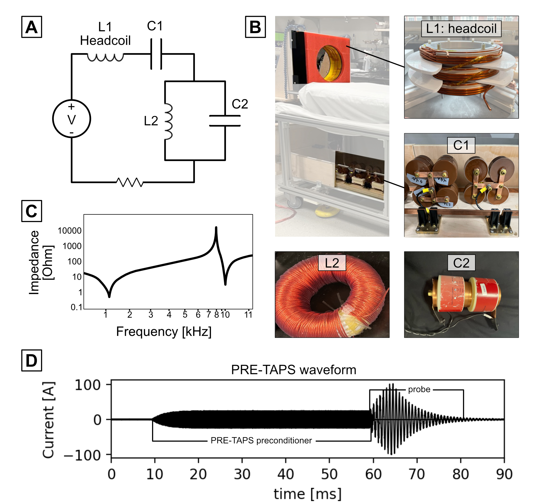
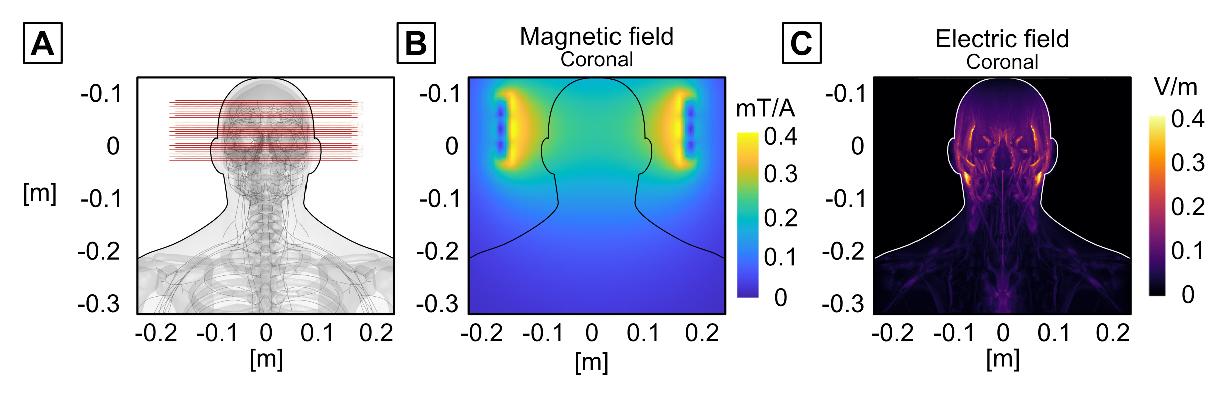
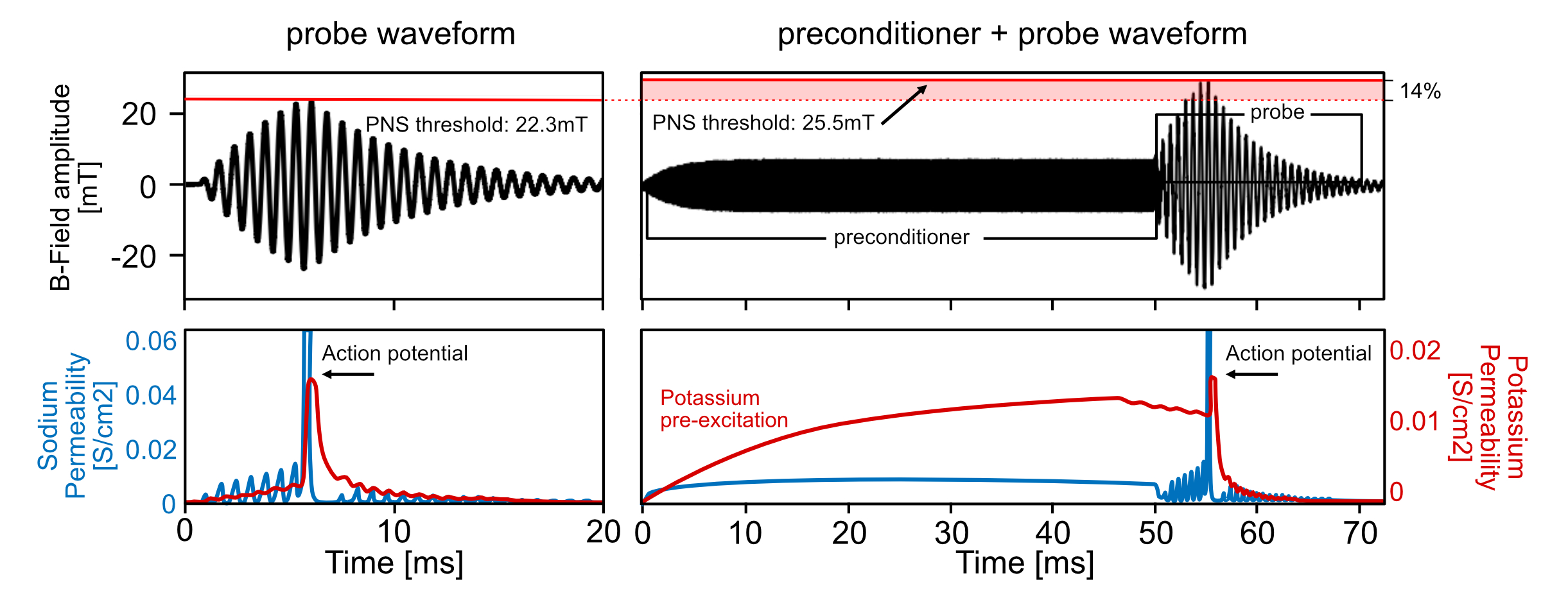
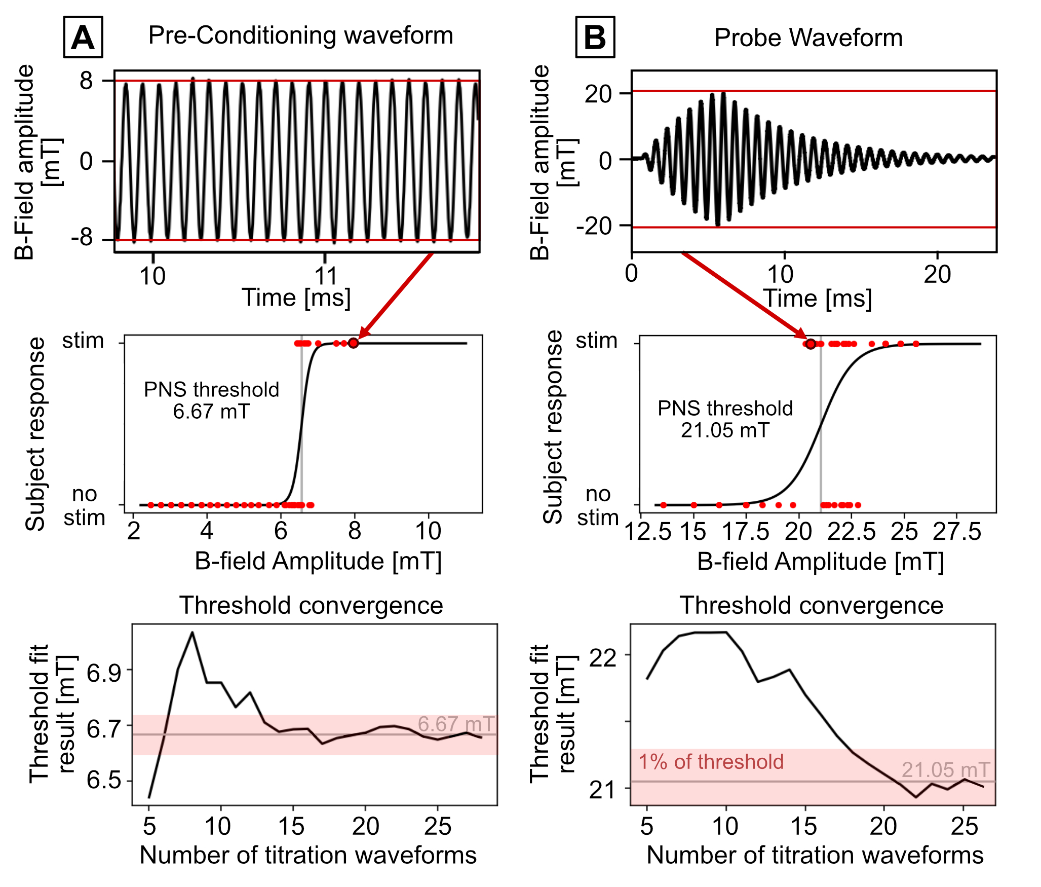
Fig 4: Experimental titration results for the preconditioner (A) and probe (B) waveforms. Titrations are performed by iteratively changing the waveform amplitude and recording binary subject responses (stim or no stim). After each titration waveform, we fit a sigmoid function to the list of binary responses to generate a new threshold estimate and track convergence of the titration (3rd row). In both cases, the threshold converged as a function of number of titration waveforms to within 1% accuracy. The final thresholds of the preconditioner and probe waveforms were 6.7 and 21.05mT.
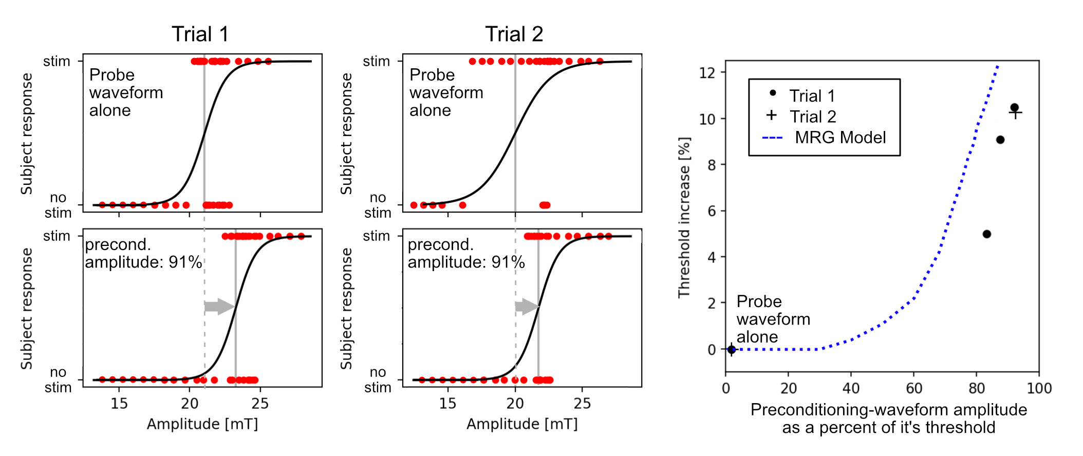
Fig 5: Titration results for two PRE-TAPS trials with the same subject. In each trial we performed a titration for the preconditioner alone, probe alone, and PRE-TAPS with the preconditioner 90% of its threshold during which only the probe amplitude was titrated. In both trials, we observed an increase in PNS threshold of the probe by 10% when the amplitude of the preconditioner was 90% of its threshold. In Trial 1, we show the PNS threshold of the probe increase with increasing preconditioner amplitude. This relationship is reproduced by our PNS model (which relies on the MRG model15).