1213
Quintuple-tuned Surface Coil Elements1Division of Medical Physics, Department of Radiology, Medical Center - University of Freiburg, University of Freiburg, Freiburg, Germany
Synopsis
Keywords: RF Arrays & Systems, RF Arrays & Systems, X Nuclei, Multinuclear coil, Flexible coil
Motivation: X-nuclear MRI is used to monitor metabolic processes but requires RF coils that are individually tuned to each resonance frequency.
Goal(s): To introduce a modular, flexible transmission line resonator array element for 5 resonance frequencies to enable multiple X-nuclear applications with a single coil.
Approach: A quintuple-tuned shielded loop resonator (SLR5) was designed consisting of three stacked shielded loop resonators with one element for 13C and 23Na, another for 31P, and a third for 19F and 1H. Switching between nuclei was realized by PIN diodes.
Results: SLR5 can be used to acquire X-nuclear signals of 5 nuclei.
Impact: The SLR5 concept might facilitate the implementation of X-nuclear MRI methods for metabolic imaging in early diagnosis and monitoring of disease. SLR5 coils can be arranged favorably in coil arrays due to the inherently low coupling of SLR coils.
Introduction
Magnetic resonance imaging (MRI) and spectroscopy (MRS) of X-nuclei is often used to detect metabolic and physiological processes. In any X-nuclear exam 1H MRI is needed for anatomical reference. Therefore, RF coil designs have been proposed to implement combined X/1H coils either as dual-resonant coils [1]–[5] or by mechanically exchanging the X and 1H coil [2], [6], [7]. If more than one X nucleus is to be imaged, coil systems with more than 2 resonances need to be realized. Recently, a quintuple-tuned coil was proposed using co-centric volume coils, LC traps and PIN-diodes to switch resonance frequencies [8]. In clinical routine an optimal coil setting for X-nuclear MRI would copy concepts developed for 1H MRI: a global transmit (Tx) volume coil, and local surface receive (Rx) coil arrays. Inspired by the quintuple-tuned volume coil [8], in this work we propose a quintuple-tuned modular and flexible surface coil array for 3T.Methods
The quintuple-tuned surface coil element design is using a stack of three shielded loop resonators (SLR). Quintuple-tuned SLR (SLR5) is realized by a capacitively tuned double-turn SLR, resonant at the 13C Larmor frequency at 3T (f13C=31 MHz), which can be tuned to f23Na=32.6 MHz via a PIN diode switch; another SLR at f31P=49.9 MHz; and a third single-turn SLR that can be switched between f19F=115.9 MHz and f1H=123.2 MHz via a PIN diode switch (Fig. 1). All 3 elements were stacked together forming a single receive coil (Fig. 2a). MRI measurements were performed at a 3T clinical MRI system (PrismaFit, Siemens) and all elements of SLR5 were interfaced to the system via high impedance on-coil preamplifiers (elcry2-u, DTU, Denmark).A phantom with 4 g/L NaPO4 was prepared in a plastic container, and two vials of PFOB and two vials 13C-enriched acetic acid were glued to the lid of the container and positioned centrally inside the phantom (Fig. 2B). For phantom measurements, SLR5 elements were fixed on a 3D-printed holder (Fig. 2A). Two prototypes were constructed to investigate coupling performance (Fig. 2C). Images of each nuclei were acquired with SLR5 and an identical mono-resonant SLR using a 2D FLASH sequence (TR/TE=8/2.5 ms, a=18°, FOV=256x256 mm2, Base resolution=128, Slice thickness=4 mm, Nslices=8, Navg=16, Total acquisition=2:20 min:s).
Results
Figure 3 shows reflection coefficient of SLR5 coil elements. Multiple modes are visible at frequencies other than the 5 nuclei. The design provided simultaneous imaging capabilities at the 1H and the 13C, 23Na, and 31P resonances without a significant resonance shift or coupling effects. When two SLR5 elements were placed next to each other, no resonance splitting was observed due to coupling, which can be attributed to the intrinsic high impedance of the SLR5 elements. Image SNR for mono-resonant SLRs and SLR5 are compared in Figure 4. SNR of SLR5 is at least 70% of the mono-resonant X-nuclear coils.Discussion
A quintuple-tuned transmission-line surface coil element, SLR5 was introduced. Image SNR of SLR5 was at least 70% of the mono-resonant X-Nuclei coils. The loss in SNR can be compensated by the time saved for a mechanical switching of coils. Modularity was demonstrated only for a 2-channel setting, yet SLR5 can be upscaled to an array of higher number of channels. Further MRI tests need to be performed to evaluate multi-channel imaging performance. SLR5 can also be used for parallel imaging in X Nuclei since signal profiles of different nuclei are very similar to 1H.Acknowledgements
This study is funded by the Deutsche Forschungsgemeinschaft (DFG) as part of the SFB1425 (#422681845) project P15 and through an individual grant (#492563001).References
[1] S. Ha, M. J. Hamamura, O. Nalcioglu, and L. T. Muftuler, “A PIN diode controlled dual-tuned MRI RF coil and phased array for multi nuclear imaging,” Phys. Med. Biol., vol. 55, no. 9, pp. 2589–2600, May 2010, doi: 10.1088/0031-9155/55/9/011.
[2] C. H. Choi, S. M. Hong, J. Felder, and N. J. Shah, “The state-of-the-art and emerging design approaches of double-tuned RF coils for X-nuclei, brain MR imaging and spectroscopy: A review,” Magn. Reson. Imaging, vol. 72, no. April, pp. 103–116, 2020, doi: 10.1016/j.mri.2020.07.003.
[3] A. Maunder, M. Rao, F. Robb, and J. M. Wild, “Comparison of MEMS switches and PIN diodes for switched dual tuned RF coils,” Magn. Reson. Med., vol. 80, no. 4, pp. 1746–1753, 2018, doi: 10.1002/mrm.27156.
[4] A. Maunder, M. Rao, F. Robb, and J. M. Wild, “An 8‐element Tx/Rx array utilizing MEMS detuning combined with 6 Rx loops for 19 F and 1 H lung imaging at 1.5T,” Magn. Reson. Med., vol. 84, no. 4, pp. 2262–2277, Oct. 2020, doi: 10.1002/mrm.28260.
[5] L. T. Muftuler, G. Gulsen, K. D. Sezen, and O. Nalcioglu, “Automatic Tuned MRI RF Coil for Multinuclear Imaging of Small Animals at 3T,” J. Magn. Reson., vol. 155, no. 1, pp. 39–44, Mar. 2002, doi: 10.1006/jmre.2002.2510.
[6] A. C. Özen et al., “Scalable and modular <scp>8‐channel</scp> transmit and <scp>8‐channel</scp> flexible receive coil array for <scp> 19 F MRI </scp> of large animals,” Magn. Reson. Med., vol. 89, no. 3, pp. 1237–1250, Mar. 2023, doi: 10.1002/mrm.29490.
[7] B. Akin and A. C. Özen, “Microstrip Array Insert for Head Coils: Towards Layer fMRI at High Fields,” in Proc. Intl. Soc. Mag. Reson. Med. 27, 2019, p. 0371.
[8] J. Dai, M. Gosselink, T. A. van der
Velden, E. F. Meliadò, A. J. E. Raaijmakers, and D. W. J. Klomp, “An
<scp>RF</scp> coil design to enable quintuple nuclear whole‐brain
<scp>MRI</scp>,” Magn. Reson. Med., vol. 89, no. 5, pp.
2131–2141, May 2023, doi: 10.1002/mrm.29577.
Figures
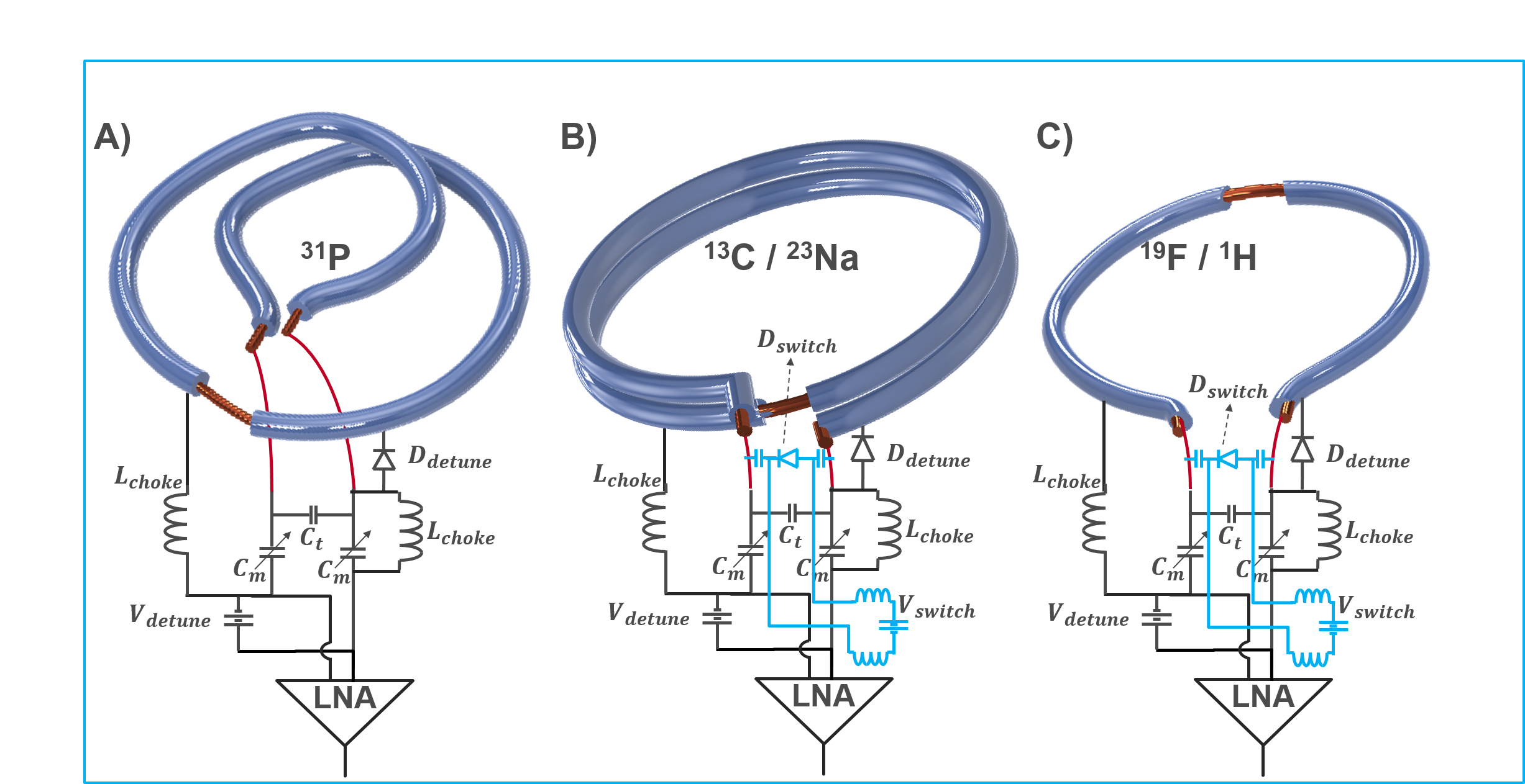
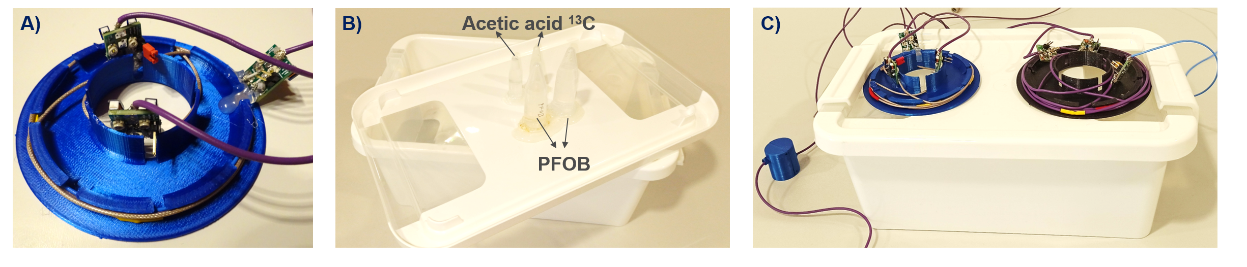
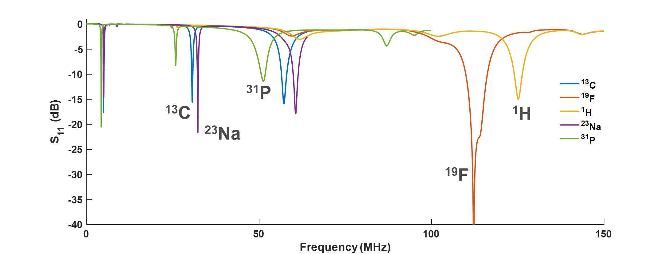
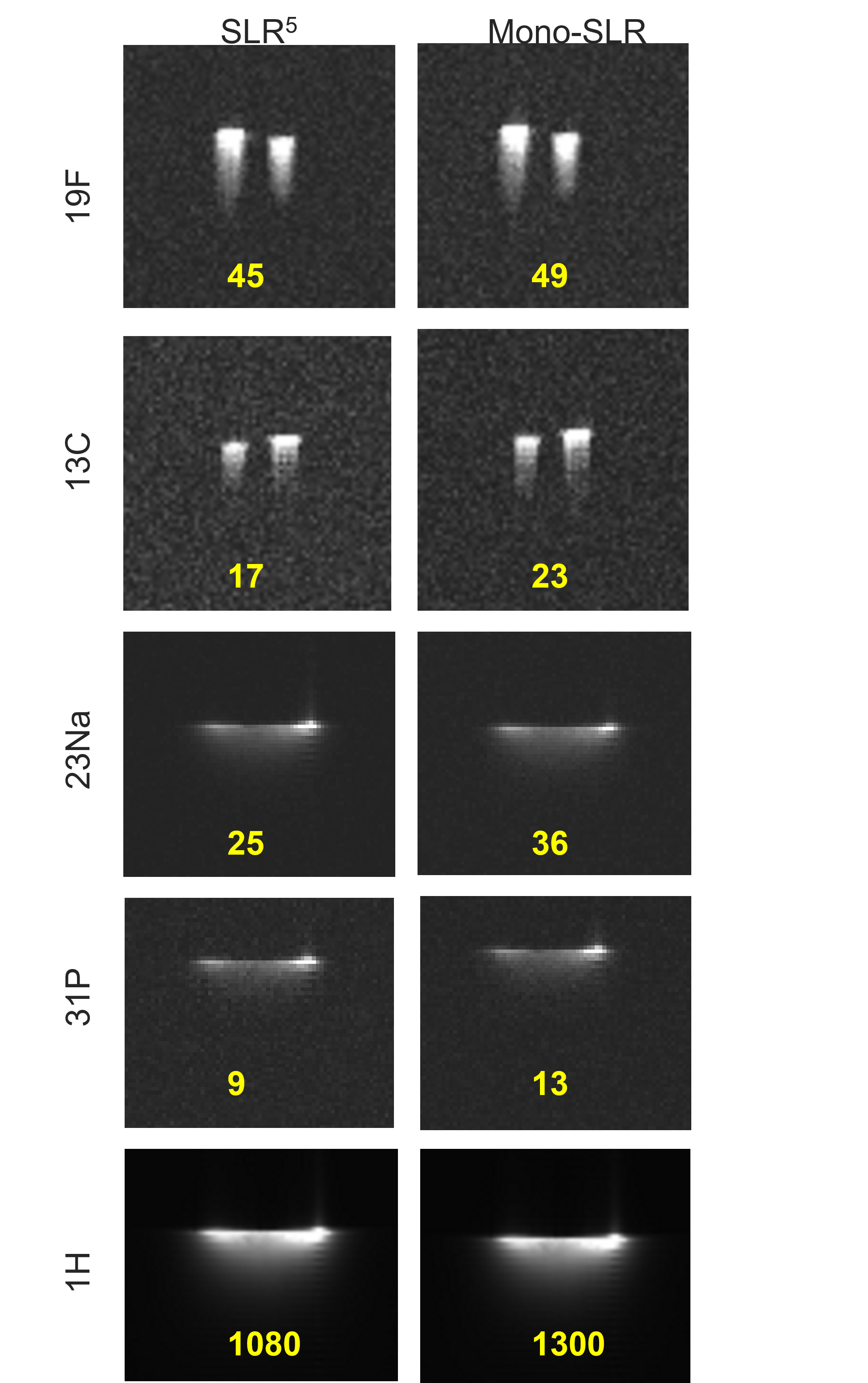
Fig.4: Phantom images acquired using SLR5 and SNR comparison to the identical mono-resonant SLR.