1212
Subwavelength dielectric waveguide for human head travelling-wave MRI at 7T1Hangzhou Institute of Technology, Xidian University, Hangzhou, China, 2National Key Laboratory of Antennas and Microwave Technology, School of Electronic Engineering, Xidian University, Xian, China, 3College of Electrical Engineering, Zhejiang University, Hangzhou, China, 4Second Affiliated Hospital of Zhejiang University School of Medicine, Zhejiang University, Hangzhou, China, 5MOE Frontier Science Center for Brain Science and Brain-machine Integration, Zhejiang University, Hangzhou, China, 6Interdisciplinary Institute of Neuroscience and Technology, School of Medicine, Zhejiang University, Hangzhou, China
Synopsis
Keywords: Non-Array RF Coils, Antennas & Waveguides, Non-Array RF Coils, Antennas & Waveguides
Motivation: Radiative excitation (e.g., travelling-wave, TW) has strength in large coverage and low SAR. It’s well compatibility with single-channel (clinical-mode) makes it promising as whole-body excitation solution at UHF. But it has apparent weakness in efficiency.
Goal(s): Improve excitation efficiency as well as homogeneity of TW MRI under clinical-mode.
Approach: Subwavelength dielectric waveguide was designed to enhance excitation efficiency as well as homogeneity through mode conversion, power-focusing, wave-impedance-matching and phase-velocity-matching.
Results: The excitation efficiency was improved by 114% over brain compared to classic TW . SAR efficiency was 10.8% higher than birdcage. The B1+ RMSE in brain was reduced by 21.9% compared to birdcage.
Impact: Our results offered insights into the design of new generation TW MRI excitation systems at UHF. The improved TW MRI systems operated under single channel may hold promises to whole-body imaging at UHF in clinical scenarios.
INTRODUCTION
Whilst human body becomes electrically large at UHF, RF coils designed under quasi-static conditions proved to be inadequate to produce uniform B1+ [1]. The SAR of RF coils placed at reactive near region is also higher than antennas used at radiative region [2]. Multi-channel transmission helps to alleviate above issues but increases system complexity. Uniform excitation under single-channel mode has been an unmet need, which is of vital importance and necessity in body coil design.Although radiative excitation (e.g., travelling-wave, TW) has shown promise at UHF under single-channel mode, it has apparent weakness in excitation efficiency [3, 4]. In the present study, we propose a novel approach to enhance efficiency as well as B1+ homogeneity by using subwavelength dielectric waveguide insert [5].METHODS
Numerical electromagnetic simulations were conducted in CST (Dassault Systèmes, France). Cubic array dielectric waveguide as shown in Figs. 1&4 were modelled by lossless materials with relative permittivity of 52. Its prototype was made up of containers filled with distilled water (mixed with sucrose to lower permittivity). MRI experiments were conducted on a 7T research scanner (MAGNETOM 7T, Siemens Healthcare, Erlangen, Germany). A quad-driven circular patch antenna placed at the service end of the bore was used for RF transmission and reception. The dielectric waveguide was placed on the patient table and positioned at isocenter. The Nova 1Tx/32Rx head coil (Nova Medical, MA, USA) was used for comparison. A head phantom was imaged with B1 mapping (AFI) (TR1/TR2: 20ms/50ms; TE: 2.53ms). In vivo study was conducted with GRE T2* images (TR: 1,000 ms, TE: 3.54 ms, nominal flip angle: 60) to qualitative evaluate the excitation homogeneity.RESULTS and DISCUSSIONS
The zero-cut-off HE11 of circular dielectric waveguide guarantees below-cut-off TE11-to-TM11 mode conversion in a circular metallic waveguide. Elevated proportion of transverse magnetic field led to efficient production of B1+ as shown in Fig. 1. The electromagnetic field distorted by subwavelength dielectric materials led to a focused power delivering as seen in Fig. 2. The wave impedance match led to maximized power transmission. Up to 70% stimulated power was delivered to the load under well-matched condition as seen in Fig. 2. B1+ homogeneity can be improved through phase-velocity match as illustrated in Figs. 3&4.In Fig. 5, the B1+ efficiency of TW MRI was improved by 114% across the entire brain region compared to classic TW. SAR efficiency of the modified TW method was 10.8% higher than the birdcage coil. Inhomogeneity in the brain measured by normalized RMSE was reduced by 21.9% through phase-velocity match.CONCLUSIONS
A novel solution for improved TW MRI excitation in electrically large human subjects with subwavelength dielectric waveguide has been presented. Its characteristics in mode conversion, power focusing, wave impedance match, as well as phase velocity match have been explored. The dielectric waveguide structure has shown advantage in improving TW excitation efficiency and homogeneity. The improved TW MRI systems driven by single transmission channel may hold promises to whole-body imaging at UHF in clinical scenarios.Acknowledgements
No acknowledgement found.References
[1] Ugurbil, K. Magnetic resonance imaging at ultrahigh fields. IEEE Trans. Biomed. Eng. 61, 1364–1379 (2014).
[2] Solomakha, G. et al. A self-matched leaky-wave antenna for ultrahigh-field magnetic resonance imaging with low specific absorption rate. Nat. Commun. 12, (2021).
[3] Brunner, D. O., De Zanche, N., Fröhlich, J., Paska, J. & Pruessmann, K. P. Travelling-wave nuclear magnetic resonance. Nature 457, 994–998 (2009).
[4] Zhang, B. et al. Whole body traveling wave magnetic resonance imaging at high field strength: Homogeneity, efficiency, and energy deposition as compared with traditional excitation mechanisms. Magn. Reson. Med. 67, 1183–1193 (2012).
[5] Yeh, C. & Shimabukuro, F. I. The essence of dielectric waveguides. Springer New York, NY (2008).
Figures
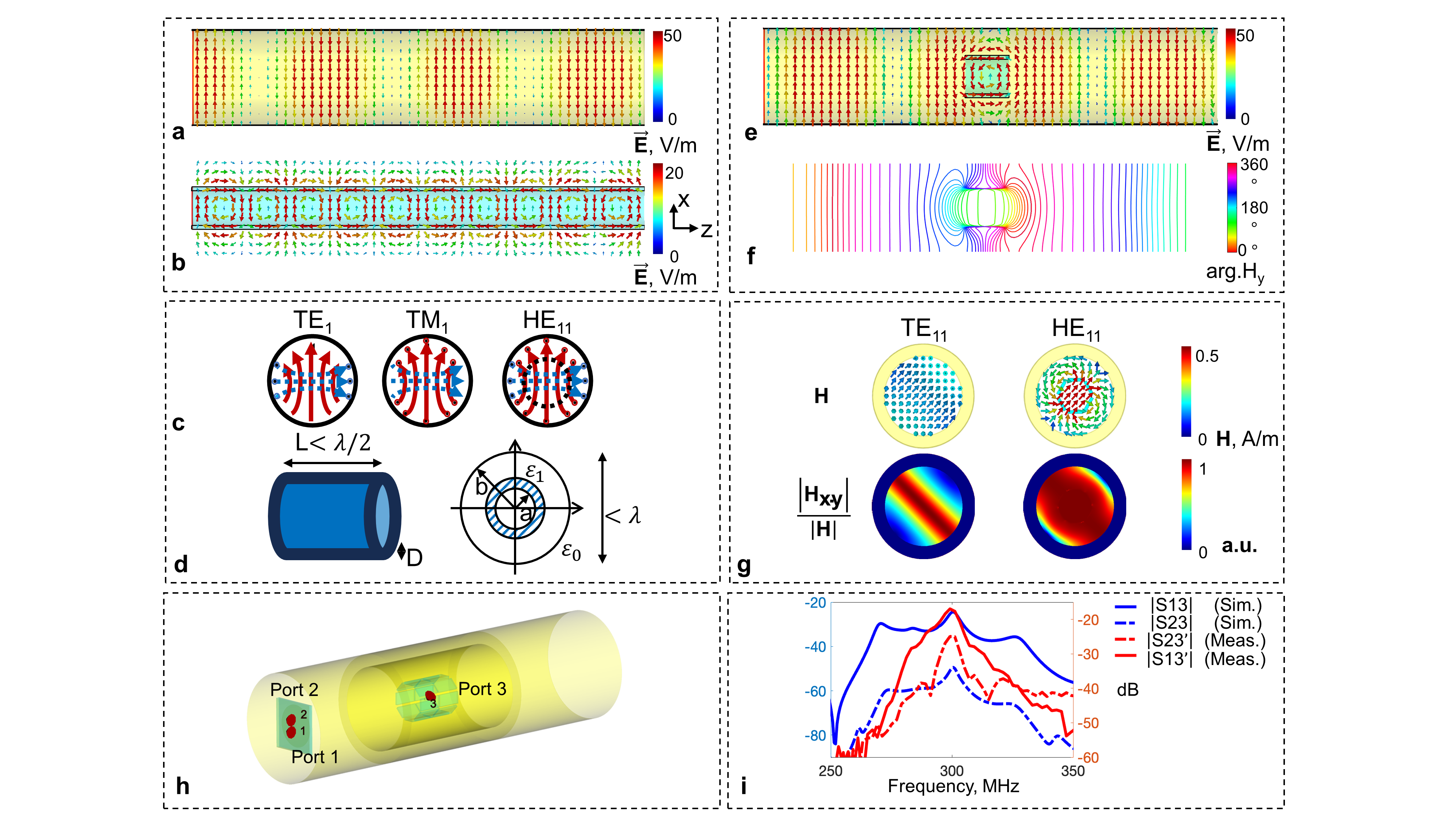
Fig. 1 Mode conversion to provide enhanced transverse magnetic field. Different modes in a circular metallic waveguide (TM11) (a) and in a dielectric waveguide (HE11) (b); The schematic field plot of TE11, TM11 modes, and hybrid mode HE11 (c). The red plot indicates the E field, while the blue plot indicates the H field; The schematic of the subwavelength dielectric waveguide (d). TE11-to-TM11 mode conversion indicated by E field (e). The waveguide discontinuity (f). The enhancement of Hxy (g). The modified TW MRI waveguide system (h). The frequency response of mode converter (i).
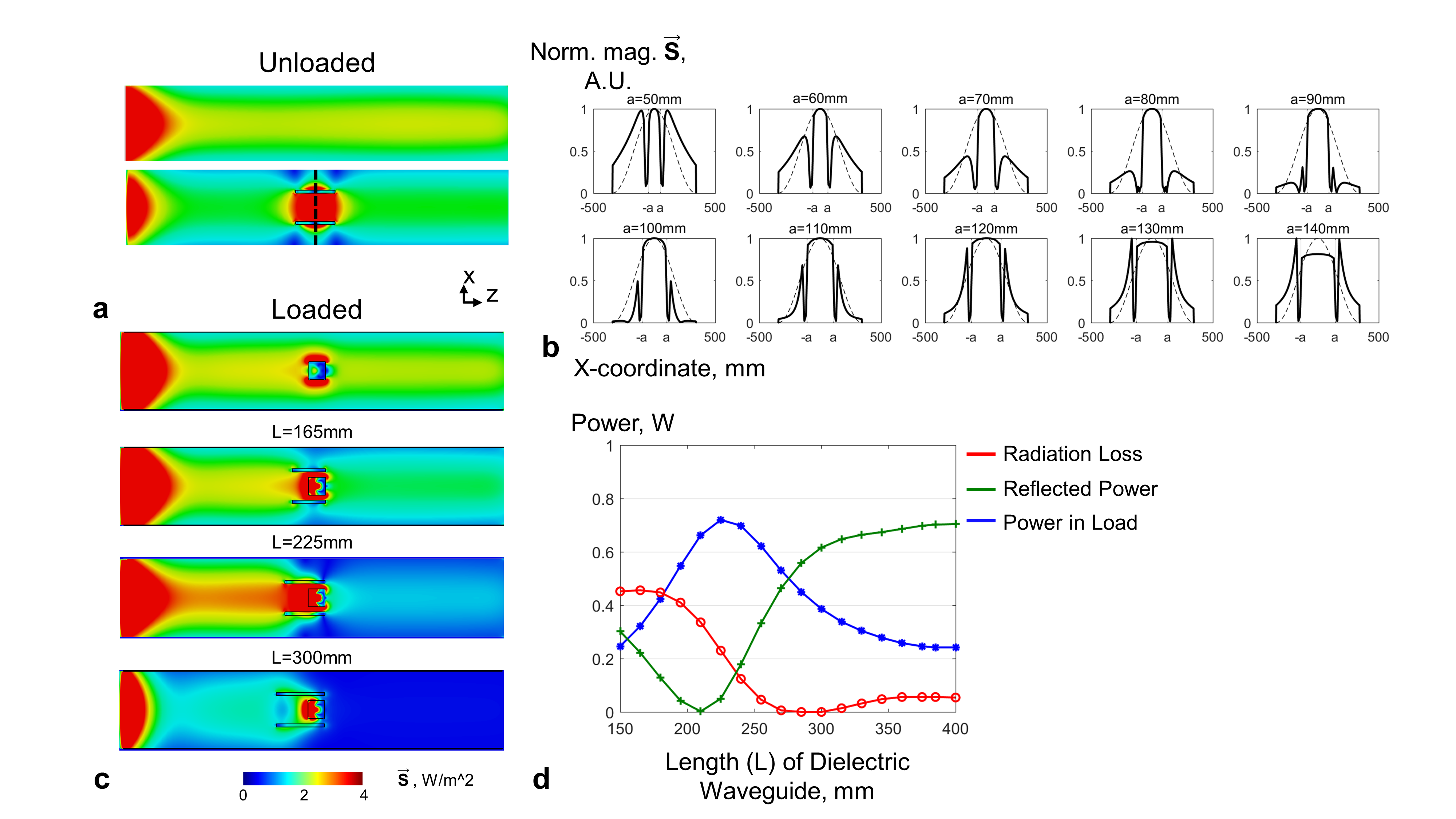
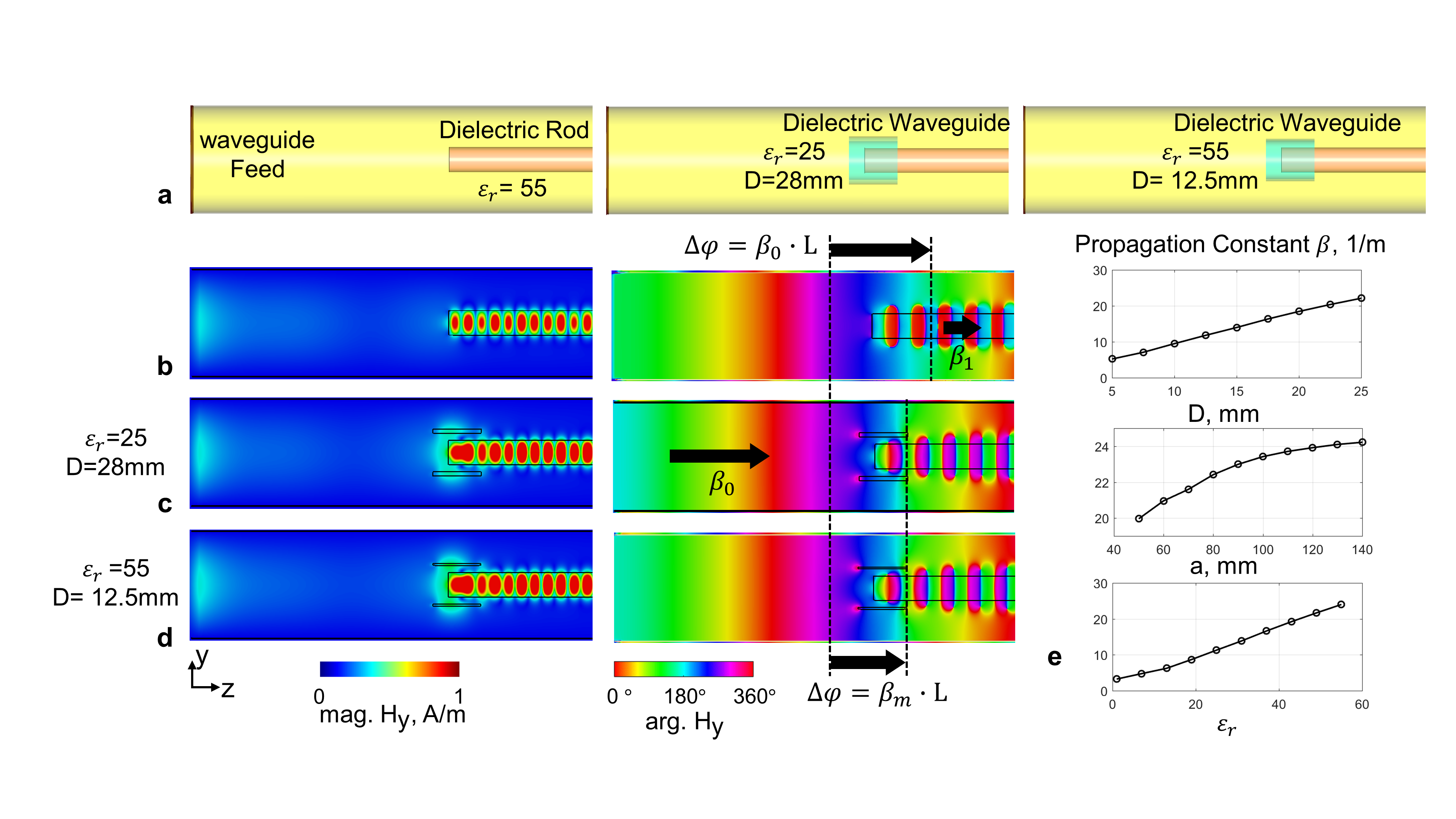
Fig. 3 Phase velocity match with dielectric waveguide. A lossless dielectric rod (160mm in diameter, εr= 55) was inserted inside a circular metallic waveguide to demonstrate the standing wave (SW) effect due to phase velocity mismatch. Two types of dielectric waveguide inserts were used to achieve phase velocity match (a); The magnitude and phase of Hy under unmatched condition (b), and well-matched condition with dielectric waveguide insert (c, d); Propagation constant β varies with thickness (D), radius (a) and dielectric constant (εr) of dielectric waveguide (e).
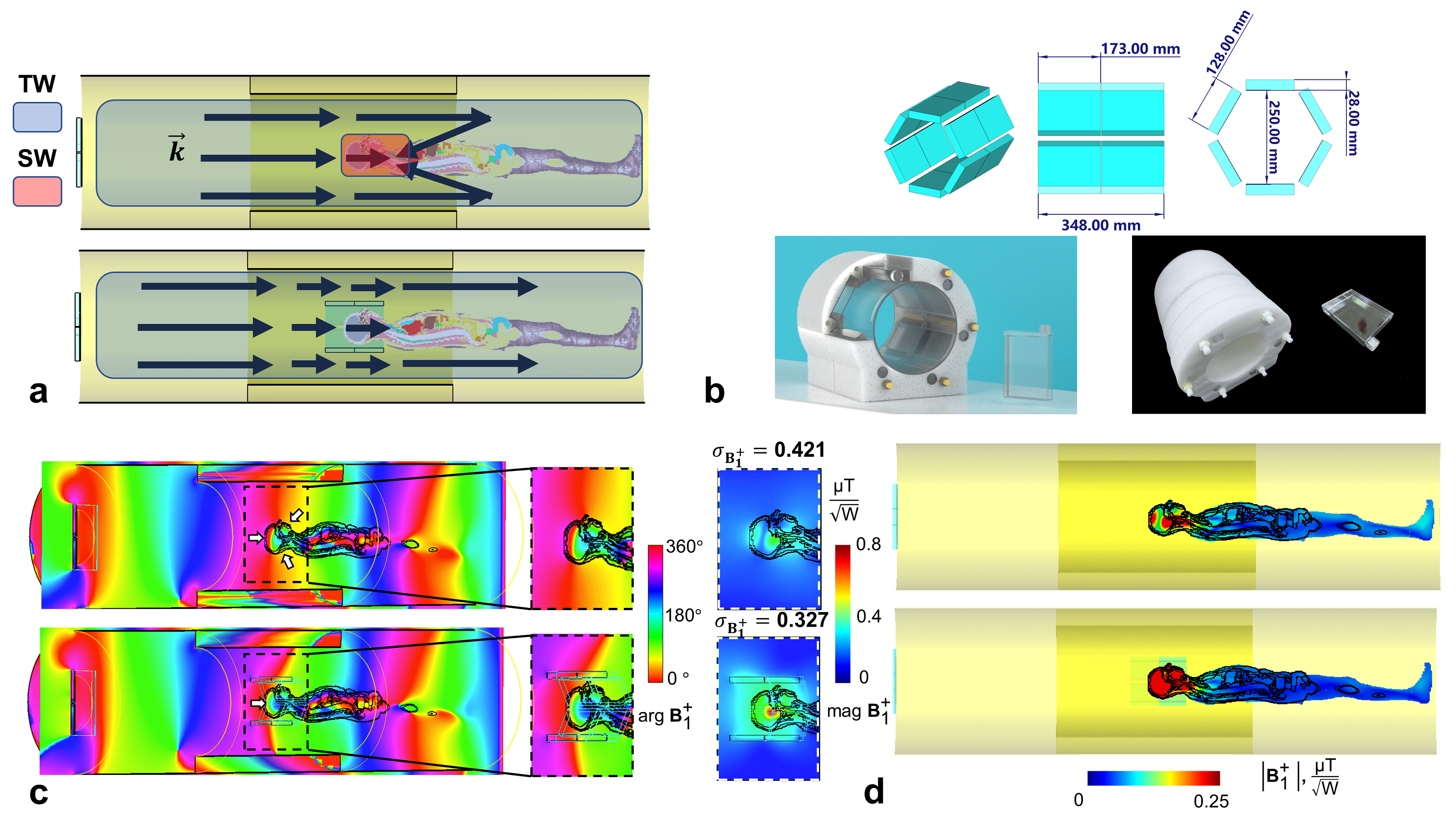
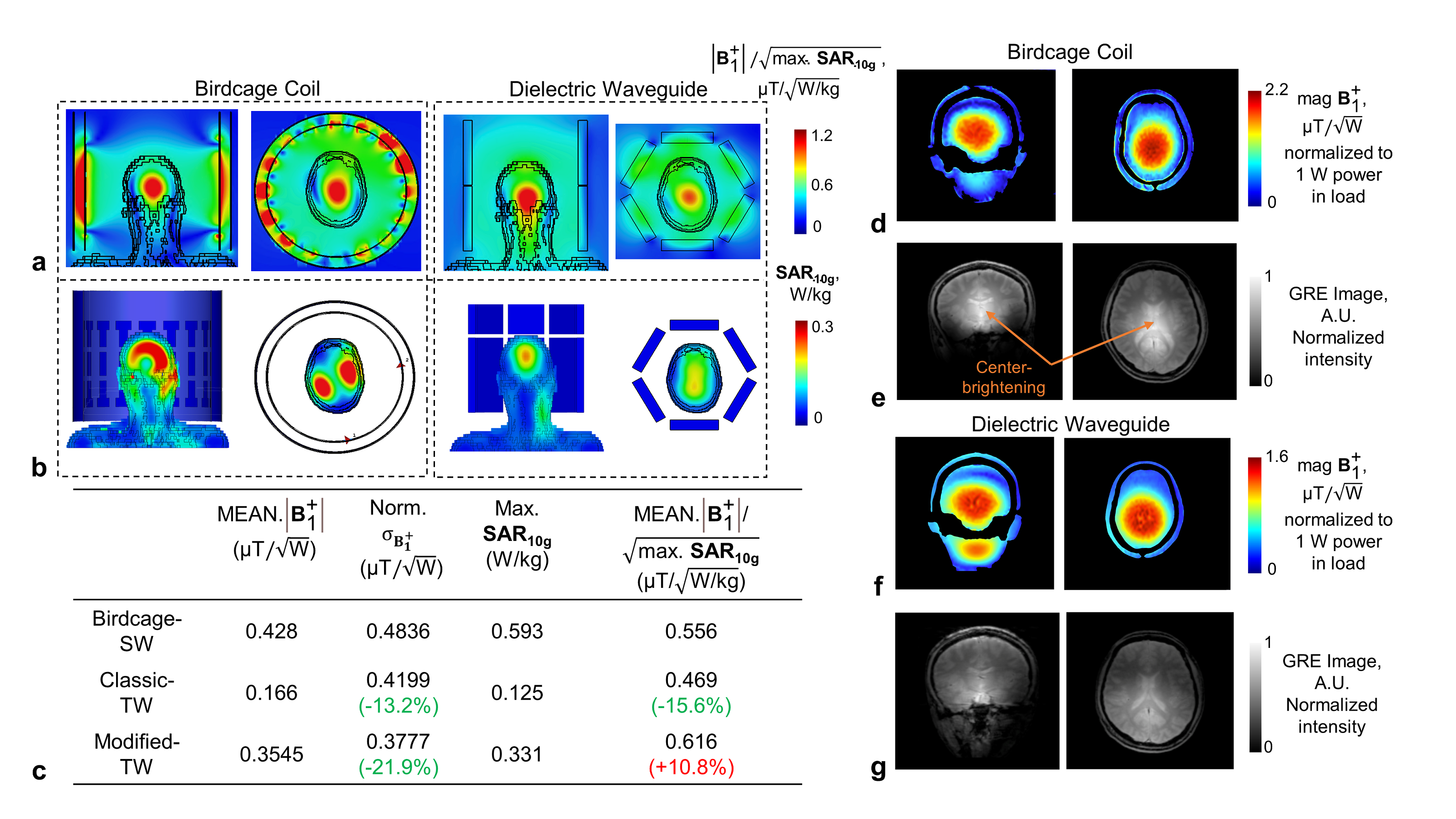
Fig. 5 The comparison of modified TW MRI with birdcage coil in human head imaging at 7T. The SAR normalized B1+ efficiency (a) and the SAR10g over the human head (b); Statistics of excitation performance (c); MRI experiments results of B1+ maps acquired from an anthropomorphic head phantom (d, f), and proton-density weighted GRE images acquired from an in-vivo human subject (e, g).