1197
A deep-learning model for effective ringing artifact removal by developing a novel multi-frequency Gibbs generator algorithm1Institute of Diagnostic and Interventional Radiology, Shanghai Sixth People's Hospital Affiliated to Shanghai Jiao Tong University School of Medicine, Shanghai, China, 2MRI R&D, Neusoft Medical Systems Co. Ltd., Shanghai, China, 3Institute of Research and Clinical Innovations, Neusoft Medical Systems Co., Ltd, Shanghai, China
Synopsis
Keywords: Analysis/Processing, Machine Learning/Artificial Intelligence, multi-frequency Gibbs artifact, deep learning, model training, artifact removal
Motivation: Gibbs artifact generated by zero-padding k-space data for model training poses a huge challenge for the model to learn different severity and manifestation of Gibbs artifact in the image domain.
Goal(s): Our goal was to effectively remove ringing artifact with a deep-learning model by developing a novel multi-frequency Gibbs generator algorithm.
Approach: We introduced Gibbs artifact generator (GAG) algorithm to create Gibbs artifacts with different truncation ratios as the input and tested the performance with a proposed deep-learning model.
Results: The images processed using the proposed approach demonstrated higher image quality score than the original images (all P < 0.05).
Impact: The images generated by our new GAG algorithm with pronounced multi-frequency Gibbs artifacts could be used as a reliable training set for deep-learning model training, enabling the model to effectively identify and eliminate Gibbs artifacts in spinal MR imaging.
INTRODUCTION
Gibbs ringing artifact is omnipresent in magnetic resonance (MR) imaging scans and manifests itself at the boundaries of the tissues. These artifacts can degrade image quality and may lead to misinterpretations[1]. In spinal MR imaging, the presence of Gibbs artifacts can result in a decline in image quality and be misidentified as syrinx, producing a false-positive diagnosis[2,3]. The traditional solution for Gibbs artifact has been achieved by applying standard filters, which, however, may reduce spatial resolution and lead to image blurring. Alternatively, more advanced approaches[4,5] have been developed without sacrificing fine image details, such as Gegenbauer reconstruction and a local subvoxel shifting method. However, such approaches either require an edge detection or do not perform well on partial Fourier acquisitions. Recently, deep learning(DL)methods have been intensively explored and employed in medical imaging field for image processing and analysis, including Gibbs ringing artifacts removal[6–9]. Zhang et al.[9] proposed a CNN model termed GRA-CNN to estimate and subtract the Gibbs artifact map from the original image. Although the cited work outperformed other approaches for reducing Gibbs artifacts, the training data were generated by simply zero-padding the k-space data, which, however, does not compensate the loss of frequency content per se. To address such an issue, a novel Gibbs artifact generator (GAG) algorithm was developed to artificially create multi-frequency Gibbs artifacts.METHODS
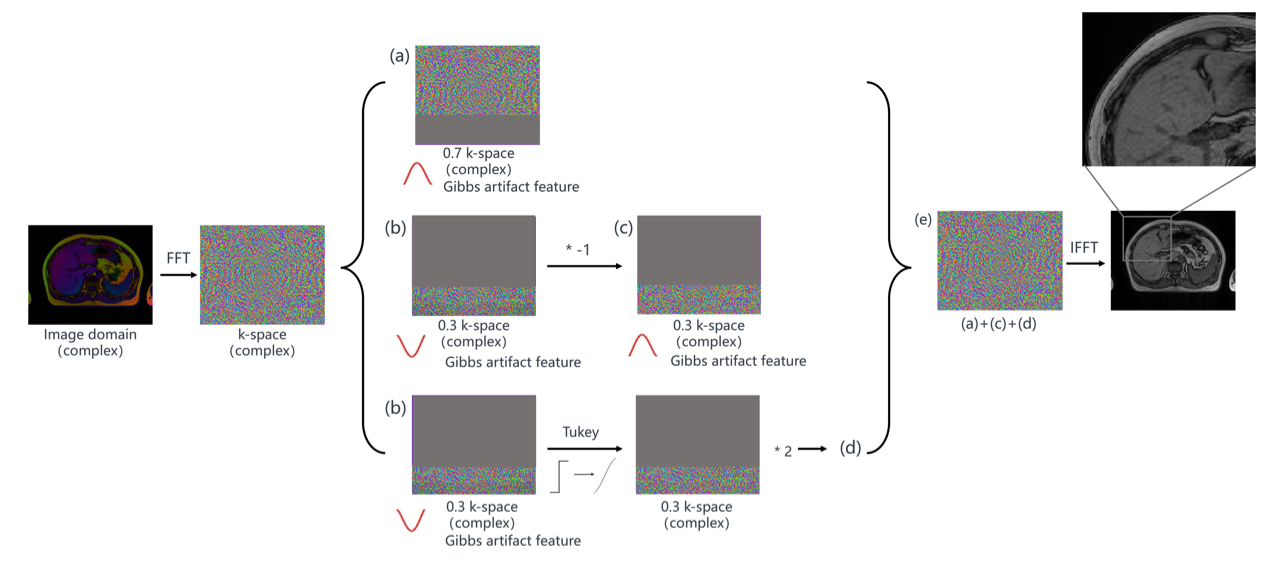
Gibbs artifact generatorThe Gibbs artifact generator (GAG), a k-space signal truncation algorithm that artificially creates truncation effects at specific frequencies in the k-space. The Gibbs artifact enhancement algorithm was then used to enhance the artifacts on the image, further improving the Gibbs artifact features, which allowed the network to learn the multi-frequency Gibbs artifact features more effectively. The workflow of GAG for creating Gibbs artifacts at truncation ratio of 0.7, for example, was shown in Figure 1.
Model for Gibbs elimination
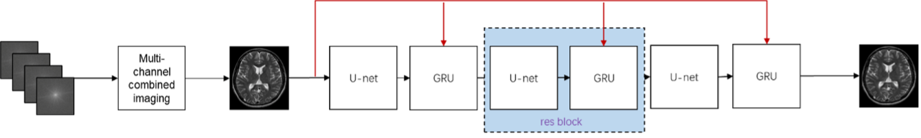
A DL network (GibbsCut) was developed to eliminate multi-frequency Gibbs artifacts, which consists of three identical blocks including a UNet [10] and a GRU[11] modules in each block, namely res block. As shown in Figure 2, the original multi-channel data is first merged into a single-channel magnitude image with Gibbs ring artifacts. Additionally, at the output layer of each res block, the output is connected to the input image, allowing the network to directly learn residual features.
Performance evaluation
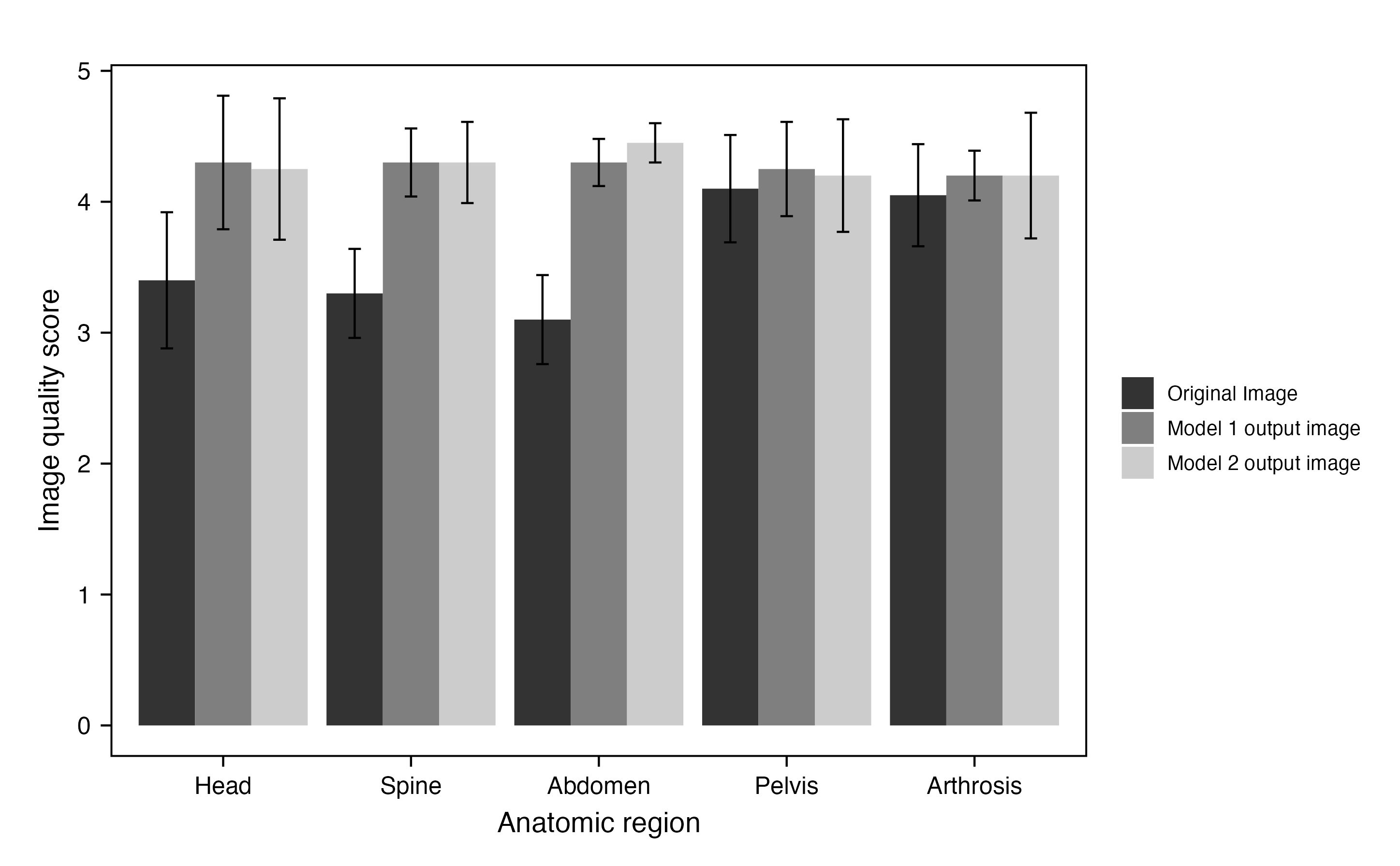
All data for model training, validation and testing were collected from a MR scanner (NeuMR Universal, Neusoft Medical Systems, Shenyang, China). A total of 290940 digital imaging and communications in medicine (DICOM) images were obtained from 4936 scans, including 67 scanning sequences. The severity of Gibbs artifact was evaluated by two radiologists with 3 years of experience in a double-blind manner and the image quality was scored according to the Likert 5-point scale. To assess the robustness on different anatomic regions, two models were trained for each anatomical region. For the case of Abdomen, model 1 utilized a combination of MR images from Head, Spine, Pelvis and Arthrosis as the training set, while model 2 employed the same anatomical region for both the training and the test sets.
Result
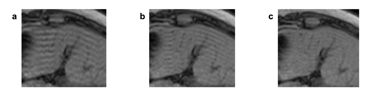
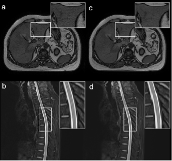
Gibbs artifacts with different frequencies and visible artifact features are shown in Figure 3, which were generated by GAG at truncation ratios of 0.675, 0.775, and 0.875, respectively. Figure 4 shows that MR images of abdomen and thoracic spine with noticeable ringing artifacts have been successfully removed by GibbsCut without visible blurring. Furthermore, no significant difference in image quality scores is observed between model 1 and 2 for all anatomical regions (all P > 0.05) in Figure 5. It is noted that both outputs of model 1 and 2 achieve much higher image quality score than the original image for Head, Spine, and Abdomen, but the difference is shortened for Pelvis and Arthrosis as the ringing artifacts in such regions are less commonly present than those in Head, Spine, and Abdomen.DISCUSSION & CONCLUSION
In routine clinical MR scanning, the application of different truncation ratios to k-space frequencies could produce different severity and manifestation of Gibbs artifacts in the image domain. Thus, the direct utilization of MR images from routine for network training would pose a huge challenge for the model to recognize and learn Gibbs artifaet features in a reliable manner. In the present study, a novel and robust algorithm was proposed to generate multi-frequency Gibbs artifacts. The generated images characterized by pronounced multi-frequency Gibbs artifacts were used as the training set for the DL model, enabling the model to effectively identify and eliminate Gibbs artifacts in an efficient manner, regardless of anatomical region.Acknowledgements
None.References
- [1] Lerch JP, van der Kouwe AJW, Raznahan A, Paus T, Johansen-Berg H, Miller KL, et al. Studying neuroanatomy using MRI. Nat Neurosci 2017;20:314–26. https://doi.org/10.1038/nn.4501.
- [2] Bronskill MJ, McVeigh ER, Kucharczyk W, Henkelman RM. Syrinx-like artifacts on MR images of the spinal cord. Radiology 1988;166:485–8. https://doi.org/10.1148/radiology.166.2.3336725.
- [3] Phillips C, Bagley B, McDonald MA, Schuster NM. Gibbs or Truncation Artifact on MRI Mimicking Degenerative Cervical Myelopathy. Pain Med 2022;23:857–9. https://doi.org/10.1093/pm/pnab346.
- [4] Archibald R, Gelb A. A method to reduce the Gibbs ringing artifact in MRI scans while keeping tissue boundary integrity. IEEE Trans Med Imaging 2002;21:305–19. https://doi.org/10.1109/TMI.2002.1000255.
- [5] Kellner E, Dhital B, Kiselev VG, Reisert M. Gibbs-ringing artifact removal based on local subvoxel-shifts: Gibbs-Ringing Artifact Removal. Magn Reson Med 2016;76:1574–81. https://doi.org/10.1002/mrm.26054.
- [6] Zhao X, Zhang H, Zhou Y, Bian W, Zhang T, Zou X. Gibbs-ringing artifact suppression with knowledge transfer from natural images to MR images. Multimed Tools Appl 2020;79:33711–33. https://doi.org/10.1007/s11042-019-08143-6.
- [7] Muckley MJ, Ades‐Aron B, Papaioannou A, Lemberskiy G, Solomon E, Lui YW, et al. Training a neural network for Gibbs and noise removal in diffusion MRI. Magn Reson Med 2021;85:413–28. https://doi.org/10.1002/mrm.28395.
- [8] Xiao L, Liu Y, Yi Z, Zhao Y, Xie L, Cao P, et al. Partial Fourier reconstruction of complex MR images using complex‐valued convolutional neural networks. Magnetic Resonance in Med 2022;87:999–1014. https://doi.org/10.1002/mrm.29033.
- [9] Zhang Q, Ruan G, Yang W, Liu Y, Zhao K, Feng Q, et al. MRI Gibbs‐ringing artifact reduction by means of machine learning using convolutional neural networks. Magn Reson Med 2019;82:2133–45. https://doi.org/10.1002/mrm.27894.
- [10] Ronneberger O, Fischer P, Brox T. U-Net: Convolutional Networks for Biomedical Image Segmentation 2015.
- [11] Cho K, van Merrienboer B, Gulcehre C, Bahdanau D, Bougares F, Schwenk H, et al. Learning Phrase Representations using RNN Encoder-Decoder for Statistical Machine Translation 2014.
Figures