1196
Contrast neutralization as a strategy to achieve generalizability in MR deep learning applications1GE HealthCare Research, Niskayuna, NY, United States, 2GE HealthCare, Bengaluru, India
Synopsis
Keywords: Analysis/Processing, Spinal Cord, Contrast Neutralization
Motivation: Provide flexibility to clinicians to fine-tune protocols/contrasts while still leveraging existing Deep-learning (DL) applications trained with limited set of MR contrasts.
Goal(s): Develop task-specific contrast neutralization pre-processing step to handle multiple imaging contrasts, that are different from the contrasts in the trainset.
Approach: Investigate Simple Contrast Neutralization (SCNe) approach that leverages Fourier domain filtering to neutralize contrast from objects of desired sizes, and demonstrate its impact on generalization of cervical foramina plane determination.
Results: Statistically significant improvements in prediction of planes when SCNe is used on new MERGE T2* contrast with DL-model that was trained only with Ax-T2 images.
Impact: Use of our Simple Contrast Neutralization (SCNe) approach as pre-processing step was effective in making DL-model trained only with Ax-T2 images robust to unseen new contrast MERGE T2* MR dataset for Spine cervical foramina (CF) plane determination.
Introduction
As MRI is inherently a multi-contrast imaging modality, deep learning (DL) models can be biased towards contrast(s) in trainset. Transfer learning1 is prudent methodology to overcome this but requires curation and labeling of each new contrasts. In this work, we investigate Simple Contrast Neutralization (SCNe) approach using object-size aware filtering in frequency domain2 to make DL models robust to contrast changes. We describe methodology using digital phantoms and demonstrate its impact by generalization of Spine cervical foramina (CF) plane determination model (trained using only axial T2w data) to new MERGE Ax T2* contrast.Methods
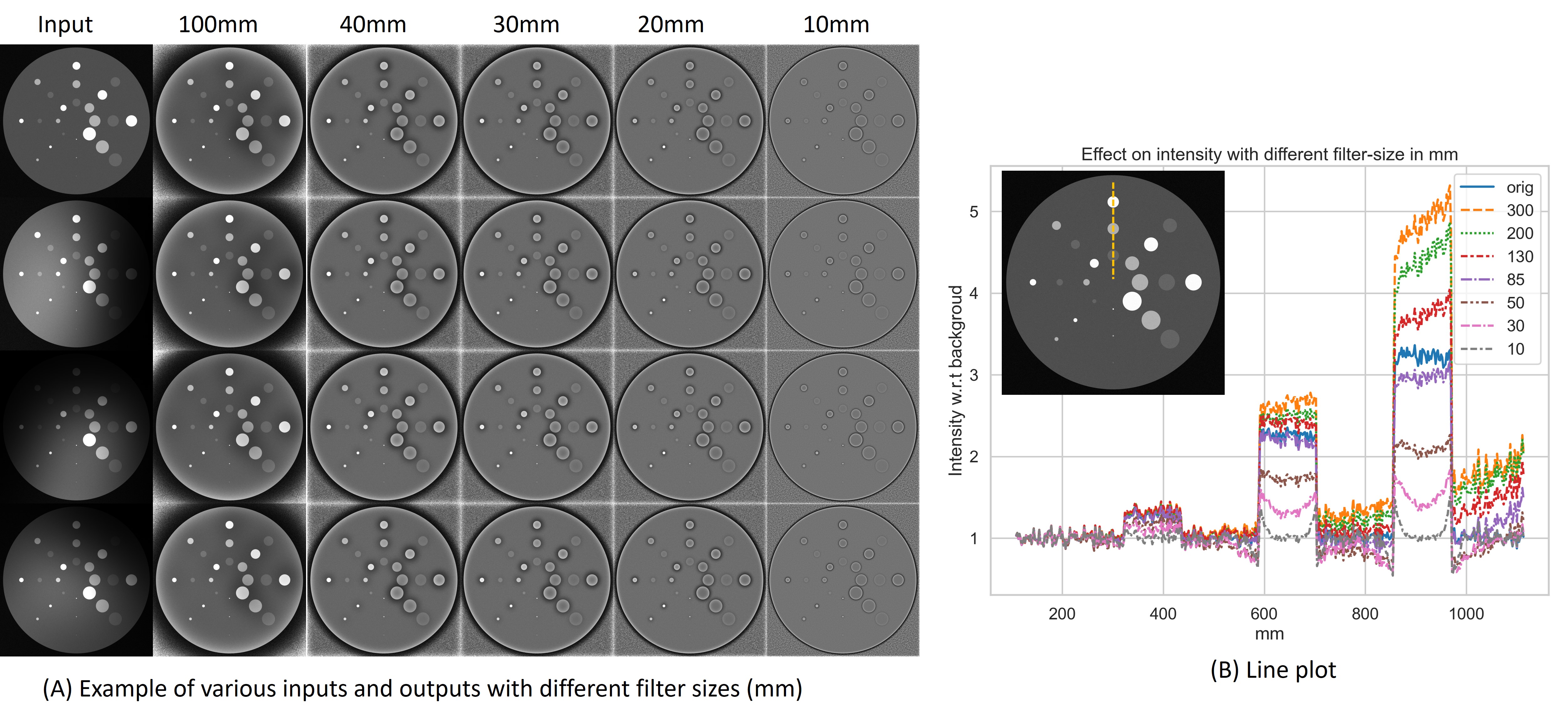
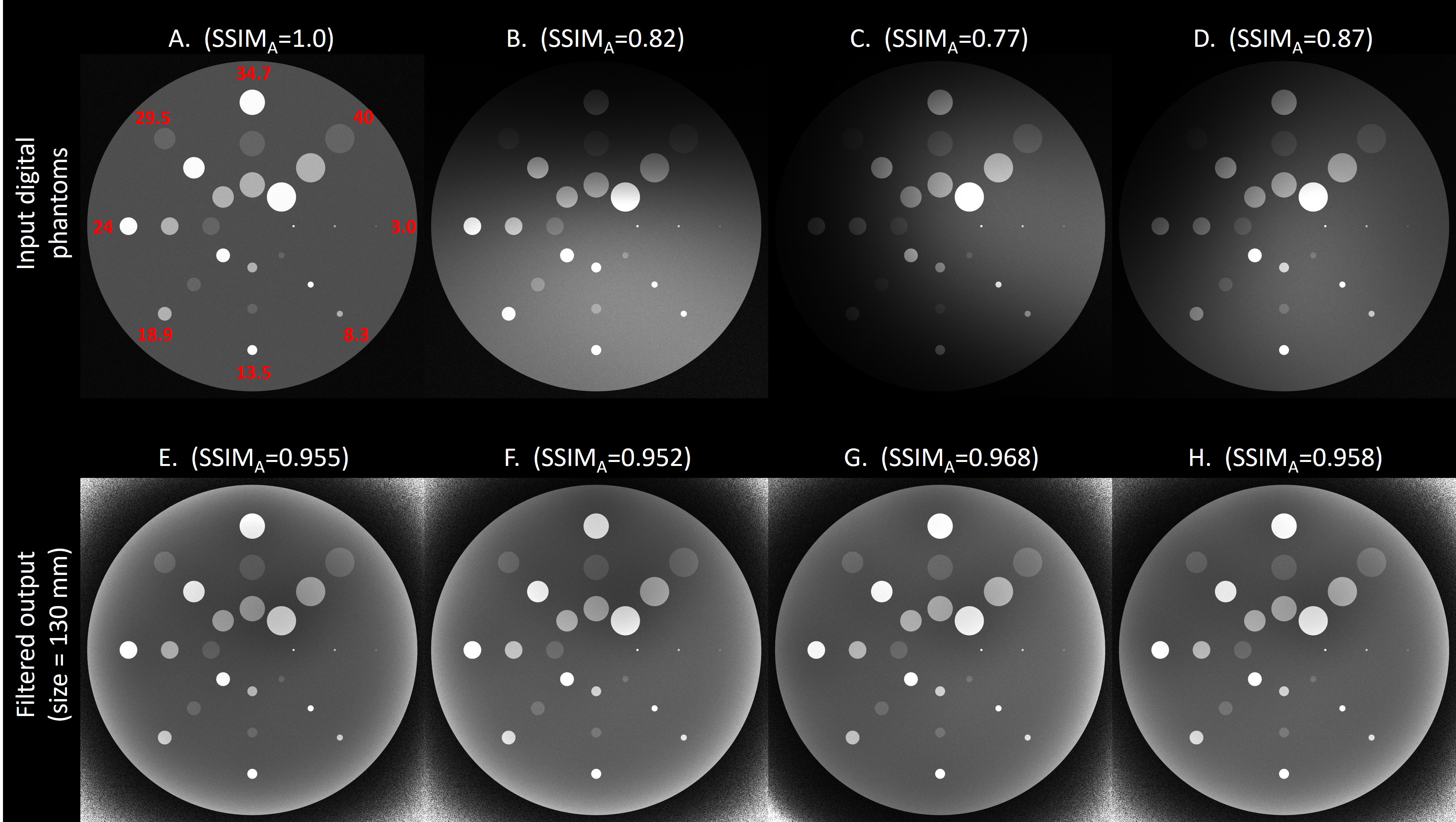
Simple Contrast Neutralization (SCNe) is a pixel-wise intensity correction approach, where pixel-intensities are governed by contrast from object(s) in a small window around it. Hence, when window size that is appropriate to underlying anatomical-object is used, SCNe neutralizes contrast from that object. Formally, given an image $$$I$$$ and window-size of $$$w$$$, we define SCNe output as $$$I_{SCNe}(w)=I/S_w$$$, where $$$S_w$$$ is smoothened version of image $$$I$$$ by applying appropriately sized symmetric four-term Blackman-Harris windowing in Fourier domain. Blackman–Harris window reduces contrast leakage across objects as it has low side-lobe levels3; although other windows can also be used. SCNe can be efficiently implemented with FFT operations for routine use in pre-processing and augmentation step for DL training.Digital Phantoms: We investigate effect of SCNe on digital phantoms that contain circular objects of different sizes and different contrasts (Fig-1, 2(A)). We used multiplicative bias-field to simulate effects of coil-sensitivity to obtain total of four digital phantoms (Fig-2), which were filtered with SCNe with varying filter sizes.
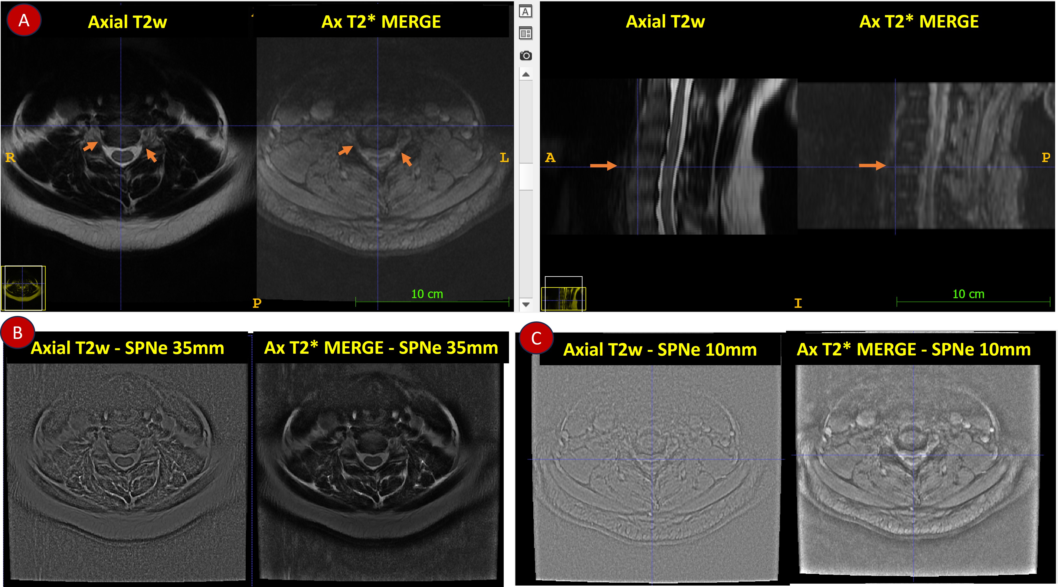
Subjects: Two sets of data from multiple clinical sites and field-strengths were used (Fig-3): Cohort A: Axial T2 spine data (N=223) and Cohort B: Axial Spoiled T2* weighted data (GE-MERGE, N=52). Different coil configurations(~38), 2D/3D acquisitions and resolutions (Axial T2w: 0.17mm to 0.78 mm in-plane, 2mm to 5mm slice thickness, MERGE: 0.35 mm to 0.93 mm in-plane , 1mm to 3mm slice thickness) were used. All studies were approved by respective IRBs.
Ground-truth (GT) marking and DL Methodology: We implemented CF plane segmentation using DL methodology4 described in Ref [4] with only axial T2w images. Briefly, a shape encoding WNET architecture was used for CF plane segmentation using z-score normalized data with dice and distance loss. With augmentation, a total of 1821 train and 242 validation Ax T2w volumes were used. As CF is around 10-mm across different vertebrae locations5, we used SCNe with window-size of 10-mm on all Axial T2w data to neutralize contrast before DL training (Fig.3). Trained DL model predicts oblique CF plane on left and right side.
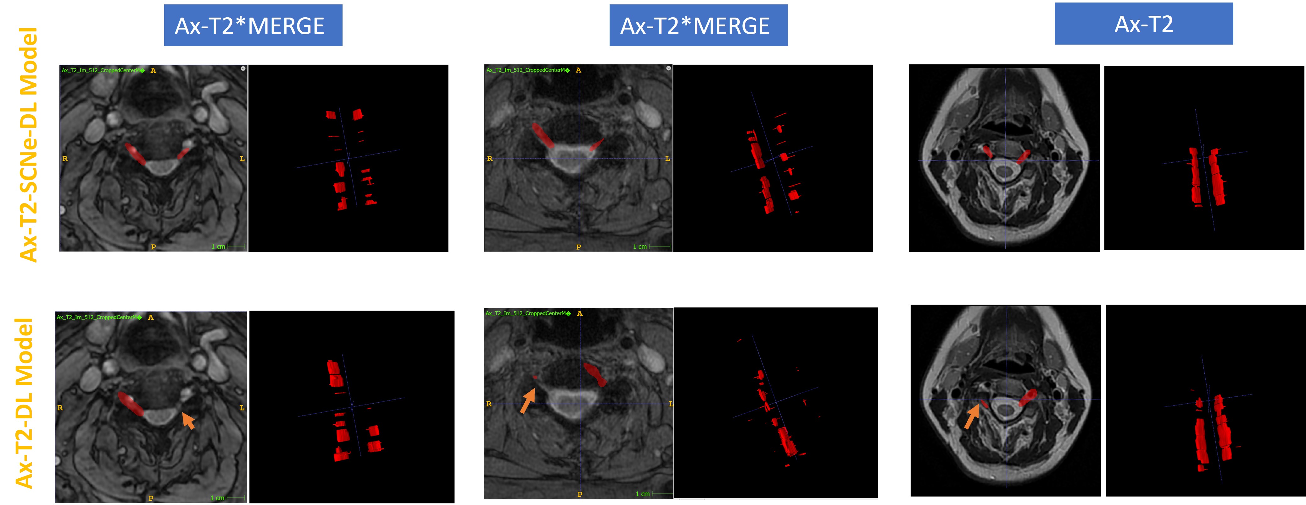
Two sets of DL models for CF plane segmentation were trained: (a) Using original data (Ax-T2-DL) and (b) using SCNe filtered data (Ax-T2-SCNe-DL). For inferencing with Ax-T2-SCNe-DL model, SCNe was applied on inputs.
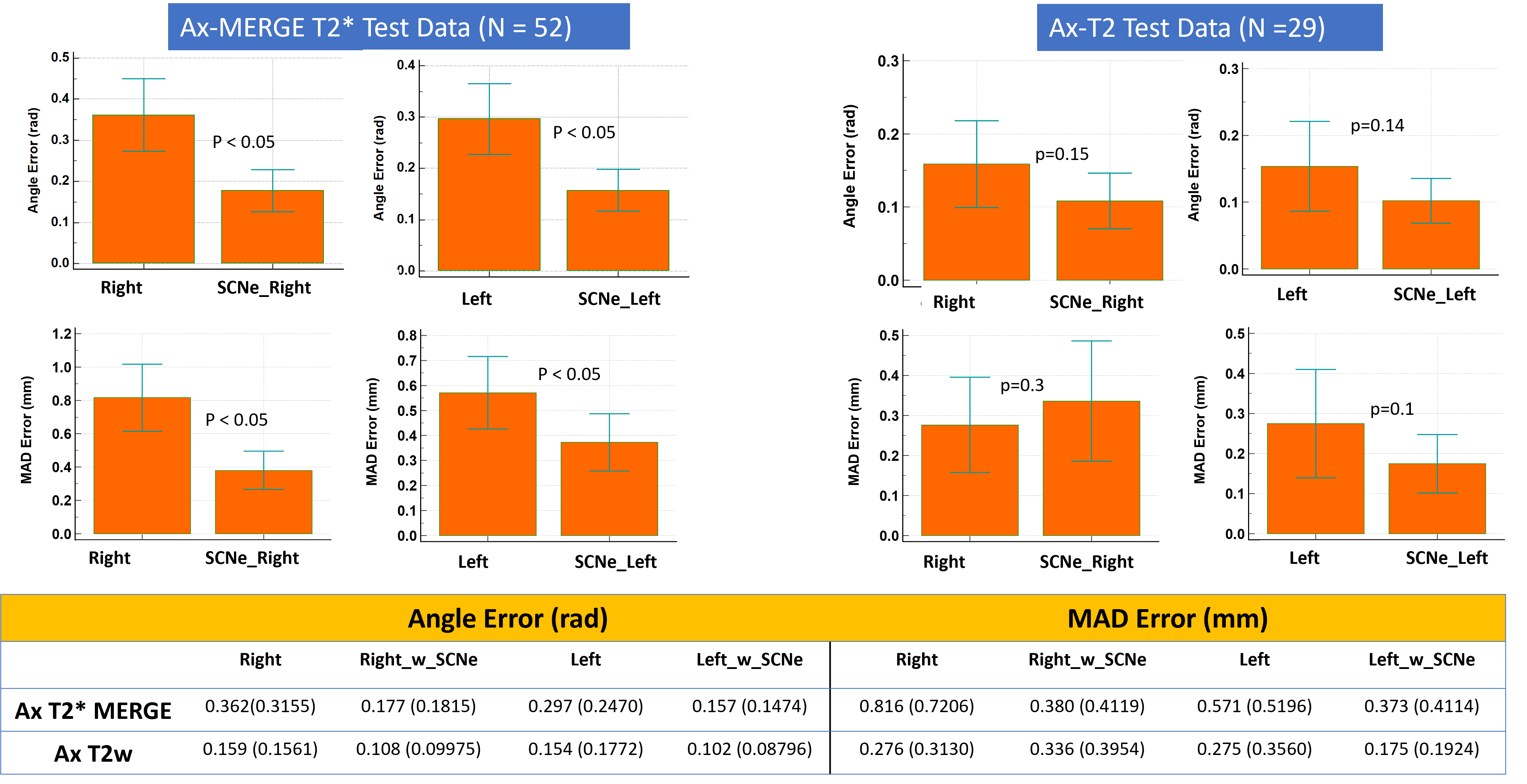
Evaluation: Evaluation was done on 29 Axial T2w datasets and 52 MERGE T2*datasets. We fit CF plane separately on left and right segments and compute angle and Mean Absolute Distance (MAD) angle error with corresponding GT plane.
Results and Discussion
In digital phantoms SCNe filtering removes contrast from objects of sizes larger than filter-size, from all contrasts (low, medium and high; Fig 1). Fig 1(B) shows that intensities inside objects are comparable to background when small filter-size of 10mm is used. When large SCNe filter size (=130mm) is used, all filtered outputs look very similar irrespective of strong & different bias field present in source images (Fig.2).In in-vivo data, Ax-T2-SCNe-DL model demonstrates significant improvement (no missing segments, correct localization) for unseen MERGE dataset, compared to non-SCNe Ax-T2-DL model (Fig 4). Quantitatively (Fig.5), use of SCNe has improved plane metrics significantly in MERGE Ax-T2* data, while there is error reduction even in Axial T2W data, which suggests that SCNe pre-processing can provide effective contrast neutralization for unseen imaging contrast data. SCNe provides flexibility to clinicians to test new imaging protocols with existing GT on limited contrasts to do same task(s). Moreover, even in intra-protocol data sets (i.e. Axial T2w), SCNe improved CF plane prediction performance; mostly neutralizing intra-protocol variations which occur based on site /scanner preferences.
Conclusion
We demonstrate that simple contrast neutralization (SCNe) scheme using size-based filtering can be effective in improving robustness of deep-learning models to MR protocol changes for a given task. Using such an approach can be effective in providing flexibility to technologists and clinicians to tune their protocol while ensuring robustness of downstream DL based solutions for routine measurement or planning tasks.Acknowledgements
No acknowledgement found.References
- Valverde JM, Imani V, Abdollahzadeh A, De Feo R, Prakash M, Ciszek R, Tohka J. Transfer Learning in Magnetic Resonance Brain Imaging: A Systematic Review. J Imaging. 2021 Apr 1;7(4):66. doi: 10.3390/jimaging7040066. PMID: 34460516; PMCID: PMC8321322.
- ImageJ Process/FFT/Bandpass Filter command. https://imagej.net/ij/plugins/fft-filter.html
- Harris, Fredric J. "On the use of windows for harmonic analysis with the discrete Fourier transform." Proceedings of the IEEE 66.1 (1978): 51-83.
- Chitresh Bhushan, Dattesh D Shanbhag, Uday Patil, Trevor Kolupar, and Maggie Fung,, “Deep Learning based prediction of the planes for automated planning of MRI imaging of cervical neural foramina and lumbar pars interarticularis”, Proceedings of ISMRM 2023, p. 2952
- Lentell G, Kruse M, Chock B, Wilson K, Iwamoto M, Martin R. Dimensions of the cervical neural foramina in resting and retracted positions using magnetic resonance imaging. J Orthop Sports Phys Ther. 2002 Aug;32(8):380-90. doi: 10.2519/jospt.2002.32.8.380. PMID: 12168956.
Figures