1192
Protocol-aware unsupervised retrospective T1 and T2 mapping with diverse imaging parameters1Biomedical Imaging Research Institute, Cedars-Sinai Medical Center, Los Angeles, CA, United States, 2Department of Bioengineering, UCLA, Los Angeles, CA, United States, 3Department of Radiological Sciences, David Geffen School of Medicine at UCLA, Los Angeles, CA, United States, 4Department of Neurology, Cedars-Sinai Medical Center, Los Angeles, CA, United States, 5Department of Imaging, Cedars-Sinai Medical Center, Los Angeles, CA, United States
Synopsis
Keywords: Analysis/Processing, Relaxometry
Motivation: Quantitative MRI has the potential for improved disease characterization, but the limited accessibility impedes its application.
Goal(s): To develop a deep learning method for retrospective T1 and T2 quantification from real-world brain MRI data, with the ability to handle diverse imaging protocols.
Approach: A protocol-aware self-supervised learning framework was developed, with the imaging parameters incorporated as additional inputs to the model.
Results: Validation on volunteers showed errors within 10% for nine brain regions when compared to prospective T1/T2 mapping. Application to 376 glioblastoma patients with diverse imaging protocols revealed statistical differences in T1 and T2 among tumor sub-regions and normal-appearing tissues.
Impact: The proposed method may allow retrospective T1 and T2 mapping in large real-world MRI datasets, enabling analysis of them regardless of the difference in protocols and scanners. This will facilitate the large-scale investigation of quantitative MRI as biomarkers for diseases.
Introduction
Quantitative MRI has major advantages in objectivity and reproducibility compared to conventional weighted MRI. Nevertheless, the clinical application of quantitative MRI faces challenges rooted in limited availability, primarily resulting from the infrequent acquisition of such data due to extended scan times and the necessity for specialized sequences. Recently, supervised and self-supervised deep learning (DL) methods were developed to retrospectively estimate relaxation parameter maps from conventional weighted MRI, thereby potentially broadening the accessibility of quantitative MR data without the need for prospective acquisition.1-9 However, these methods are trained on a specific set of imaging protocols, thus limiting their applicability to real-world MRI data which are typically acquired with different imaging protocols. To address this limitation, we developed a protocol-aware self-supervised learning framework for retrospective T1 and T2 mapping from weighted brain MR images. This method accommodates diverse imaging protocols by directly incorporating the acquisition parameters into the network. The approach was validated on volunteers with prospective maps as references and then applied to glioblastoma (GBM) patients in the UPenn GBM dataset10 to evaluate its clinical utility.Methods
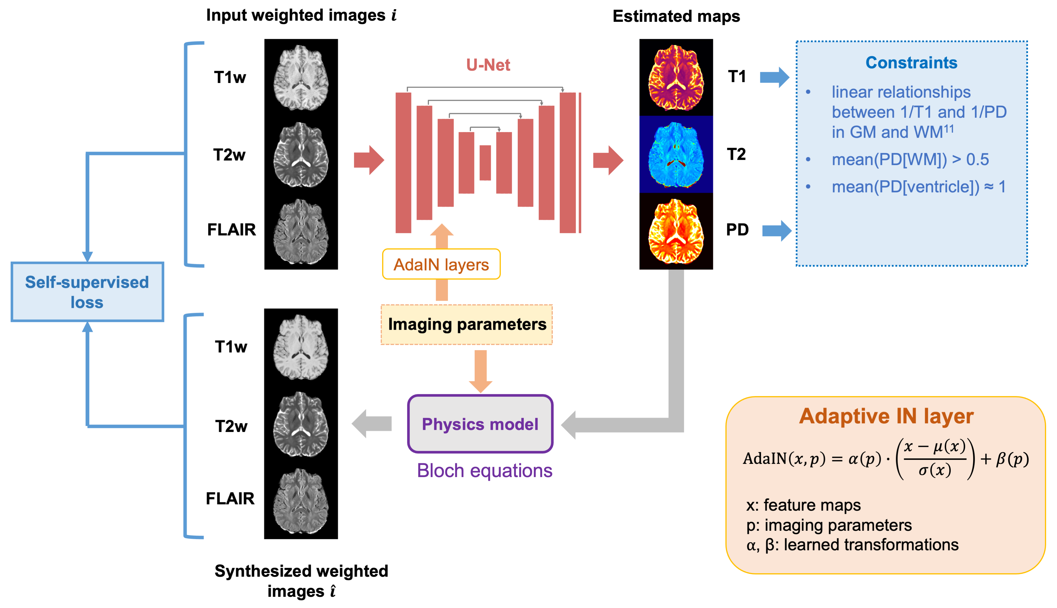
DL modelThe Unet-based model takes three weighted images (T1-weighted, T2-weighted, FLAIR) as input and outputs T1, T2, and proton density (PD) maps. Unsupervised learning was achieved by using MR physics models to convert the network outputs to weighted images and calculating losses between the synthesized and the input weighted images (Figure 1). Additional constraints were added based on prior knowledge, including literature PD values of 1 in the ventricle and 0.7 in white matter (WM).11-14 Reported linear relationship between 1/T1 and 1/PD in brain tissues was added as another constraint.12-17
To accommodate different protocols, key parameters (TR, TE, TI, and flip angle) were directly inputted to the network via adaptive instance normalization (AdaIN) layers18 in the encoder part. AdaIN layers scale and shift the feature maps according to these parameters to make the model protocol-aware.
Datasets
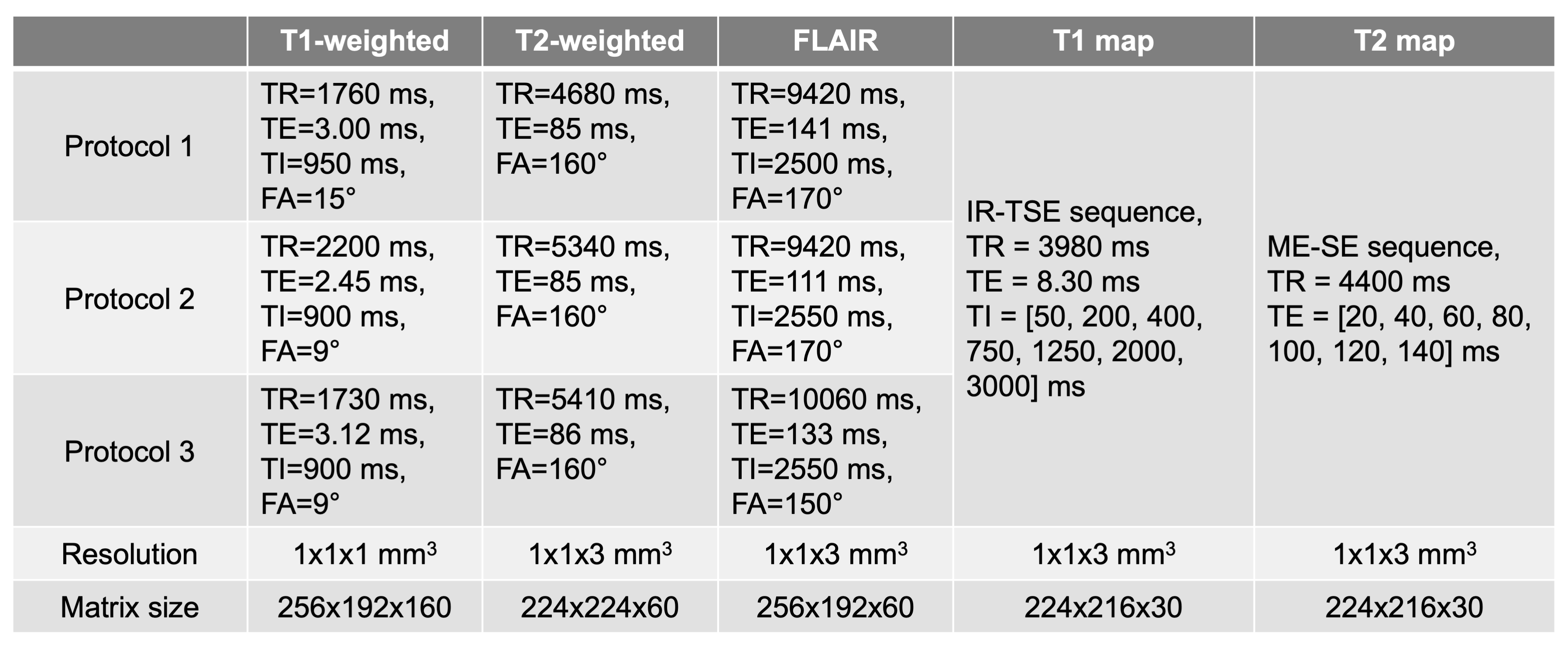
Eight healthy volunteers were scanned on a 3T Siemens Vida scanner, with T1-weighted, T2-weighted, and FLAIR images, along with conventional T1 and T2 maps. For each weighted contrast, three sets of imaging parameters were applied based on the top three protocols from the UPenn GBM dataset, resulting in 27 combinations (Table 1).
The UPenn GBM dataset was used to evaluate the method in clinical settings. 376 subjects with similar parameters to the 27 protocols were selected.
Training and evaluation
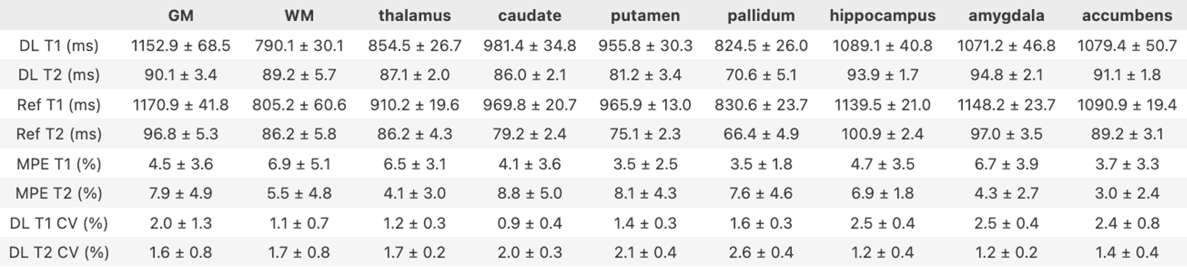
Label-free training was performed on the eight volunteers. Two subjects were used as validation data for hyperparameter tuning. Evaluation results were reported on the other six subjects (513 slices), including T1/T2 measurements and mean percentage error (MPE) in 9 brain regions. Coefficient of variance (CV) was calculated across the 27 protocols to assess robustness to protocol variations.
For the GBM dataset, the model was retrained on all subjects. Mean T1/T2 values were calculated within GM, WM, and tumor sub-regions (core, enhance, and edema). Signed-rank Wilcoxon tests were performed between regions.
Results
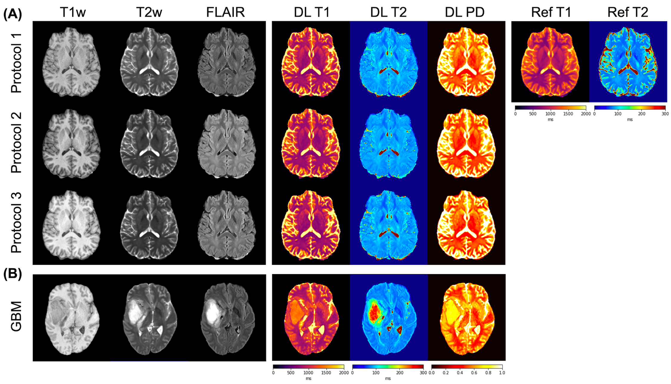
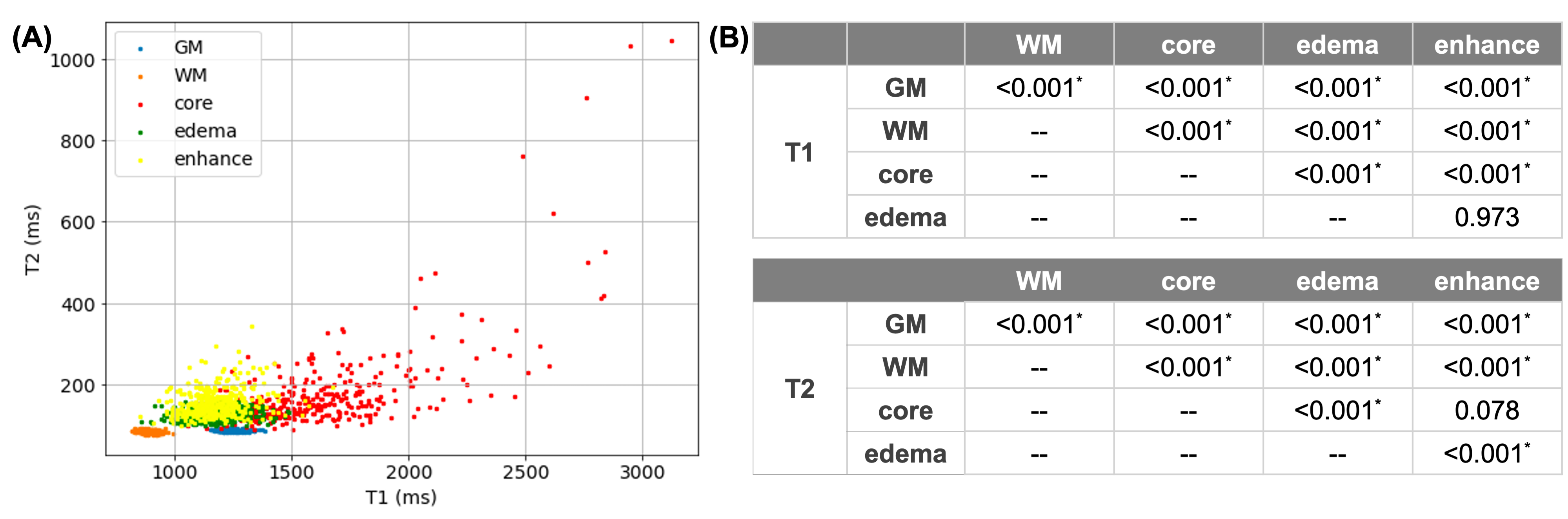
The retrospective T1 and T2 maps showed similar values and tissue contrast to the references in volunteers, regardless of variations in input image contrasts due to different imaging protocols (Figure 2 (A)). Pixel-wise evaluation (excluding CSF region) reported MPE of 9.6% and 12.1% for T1 and T2, respectively. T1/T2 measurements on nine regions showed MPE of 3.0%~8.8% (Table 2). CV across the 27 protocols was below 3%, demonstrating robustness to protocol variations.When trained with the GBM dataset, the approach also generated reasonable maps (Figure 2 (B)). Evaluation of this model on the volunteer data resulted in pixel-wise MPE of 11.7%/13.4% for T1/T2. The T1-T2 scatter plot illustrated in Figure 3 (A) showed similar clusters of tumor sub-regions and normal-appearing tissues to previous reports using prospective mapping19. Significant differences (P<0.05) were found in T1 between all pairs except for between edema and enhance, and in T2 for all comparisons but between core and enhance (Figure 3 (B)).
Discussion
The proposed protocol-aware unsupervised approach showed the feasibility of retrospective estimation of T1/T2 from real-world brain MRI with diverse imaging protocols. Validation of the DL T1/T2 maps with conventional references showed the effectiveness of the method. Initial results in a large GBM dataset demonstrated the potential value of this approach in investigating clinical applications of T1 and T2 maps.Conclusion
A protocol-aware unsupervised learning method was developed to retrospectively estimate T1 and T2 maps from conventional weighted brain MRI acquired with variable imaging protocols. The preliminary results showed the applicability and robustness of the proposed method for retrospective quantitative imaging in a large real-world MRI dataset.Acknowledgements
No acknowledgement found.References
1. Qiu S, Chen Y, Ma S, et al. Multiparametric mapping in the brain from conventional contrast-weighted images using deep learning. Magn Reson Med. 2021;87(1):488-95. https://doi.org/10.1002/mrm.28962.
2. Wu Y, Ma YJ, Du J, et al. Deciphering tissue relaxation parameters from a single MR image using deep learning. In Medical Imaging 2020: Computer-Aided Diagnosis. Proceedings of SPIE, Houston, TX, 2020. p. 113140Q. https://doi.org/10.1117/12.2546025.
3. Wu Y, Ma Y, Kee Y, et al. Quantitative Parametric Mapping of Tissues Properties from Standard Magnetic Resonance Imaging Enabled by Deep Learning. arXiv preprint arXiv:2108.04912. 2021 Aug 10.
4. Moya-Sáez E, de Luis-García R, Alberola-López C. A self-supervised deep learning approach to synthesize weighted images and T1, T2, and PD parametric maps based on MR physics priors. In Proc ISMRM, 2021. pp. 2169.
5. Qiu S, Christodoulou AG, Xie Y, et al. Hybrid supervised and self-supervised deep learning for quantitative mapping from weighted images using low-resolution labels. In Proc ISMRM, 2022. pp. 2609.
6. Qiu S, Christodoulou AG, Xie Y, et al. Physics-guided self-supervised learning for retrospective T1 and T2 mapping from conventional weighted brain MRI. In Proc ISMRM, 2023. p. 2168.
7. Moya-Sáez E, Peña-Nogales Ó, de Luis-García R, et al. A deep learning approach for synthetic MRI based on two routine sequences and training with synthetic data. Comput Methods Programs Biomed. 2021;210:106371.
8. Moya‐Sáez E, Navarro‐González R, Cepeda S, et al. Synthetic MRI improves radiomics‐based glioblastoma survival prediction. NMR in Biomedicine. 2022;35(9):e4754.
9. Varadarajan D, Bouman KL, van der Kouwe A, Fischl B, Dalca AV. Unsupervised learning of MRI tissue properties using MRI physics models. arXiv preprint arXiv:2107.02704. 2021 Jul 6.
10. Bakas S, Sako C, Akbari H, et al. The University of Pennsylvania glioblastoma (UPenn-GBM) cohort: Advanced MRI, clinical, genomics, & radiomics. Scientific data. 2022;9(1):453.
11. Neeb H, Zilles K, Shah NJ. A new method for fast quantitative mapping of absolute water content in vivo. Neuroimage. 2006;31(3):1156-68.
12. Abbas Z, Gras V, Möllenhoff K, et al. Quantitative water content mapping at clinically relevant field strengths: a comparative study at 1.5 T and 3 T. NeuroImage. 2015;106:404-13.
13. Abbas Z, Gras V, Möllenhoff K, et al. Analysis of proton‐density bias corrections based on T1 measurement for robust quantification of water content in the brain at 3 Tesla. Magn Reson Med. 2014;72(6):1735-45.
14. Volz S, Nöth U, Jurcoane A, et al. Quantitative proton density mapping: correcting the receiver sensitivity bias via pseudo proton densities. Neuroimage. 2012;63(1):540-52.
15. Mezer A, Rokem A, Berman S, et al. Evaluating quantitative proton-density-mapping methods. Hum Brain Mapp. 2016;37(10):3623-35.
16. Mezer A, Yeatman JD, Stikov N, Kay KN, Cho NJ, Dougherty RF, Perry ML, Parvizi J, Hua LH, Butts-Pauly K, Wandell BA. Quantifying the local tissue volume and composition in individual brains with magnetic resonance imaging. Nature medicine. 2013;19(12):1667-72.
17. Gelman N, Ewing JR, Gorell JM, et al. Interregional variation of longitudinal relaxation rates in human brain at 3.0 T: relation to estimated iron and water contents. Magn Reson Med. 2001;45(1):71-9.
18. Huang X, Belongie S. Arbitrary style transfer in real-time with adaptive instance normalization. In Proc IEEE ICCV, 2017. p. 1501-1510.
19. Pirkl CM, Nunez-Gonzalez L, Kofler F, et al. Accelerated 3D whole-brain T1, T2, and proton density mapping: feasibility for clinical glioma MR imaging. Neuroradiology. 2021:1-21.
Figures