1190
Fast 3D-EPI for characterization of CSF motion during the cardiac cycle.1Department of Physics, NTNU, Trondheim, Norway, 2Clinic of Radiology and Nuclear Medicine, St.Olavs University Hospital HF, Trondheim, Norway, 3DZNE, Bonn, Germany, 4Department of Physics and Astronomy, University of Bonn, Bonn, Germany
Synopsis
Keywords: Neurofluids, Brain
Motivation: Individual variation in CSF dynamics may affect the efficiency of waste clearance from the brain.
Goal(s): To develop a fast and sensitive MRI method for measurement of CSF motion during the cardiac cycle.
Approach: Whole brain 3D-EPI at 3mm resolution and volTR=187ms was acquired at 7T for 94 seconds and retrospectively sorted into 20 cardiac phases based on pulse oximeter.
Results: CSF dynamics was observed in all ventricles as well as in sub-arachnoid space, in addition to arterial pulsation. Both magnitude and phase pulsations were present in the 3 subjects acquired. Highest sensitivity to motion was observed along the phase-encoding direction.
Impact: We show that fast 3D-EPI at 7T is very sensitive to the motion of CSF during the cardiac cycle. This method may be used to characterize CSF dynamics on individual patient level and aid understanding of brain diseases.
Introduction
CSF is believed to play a key role in clearance of waste substances from the brain1. The pulsatile motion of CSF is the result of complex interplay between the driving forces such as cardiac pulsation and respiratory motion and the anatomical and mechanical properties of the brain2. Sensitive and robust methods to characterize the motion of CSF on both short and long timescales are required to advance the understanding of healthy and pathological CSF pulsation. Single-shot 3D volumetric imaging has already been applied to study brain pulsation3,4. 3D echo-planar imaging (EPI) at 7T also allows repeated acquisition of whole brain images with sub-second temporal resolution. In addition, it has intrinsic sensitivity to motion due to the non-zero first order gradient-moments at both TE and TR. This method therefore has potential for probing CSF motion.Methods
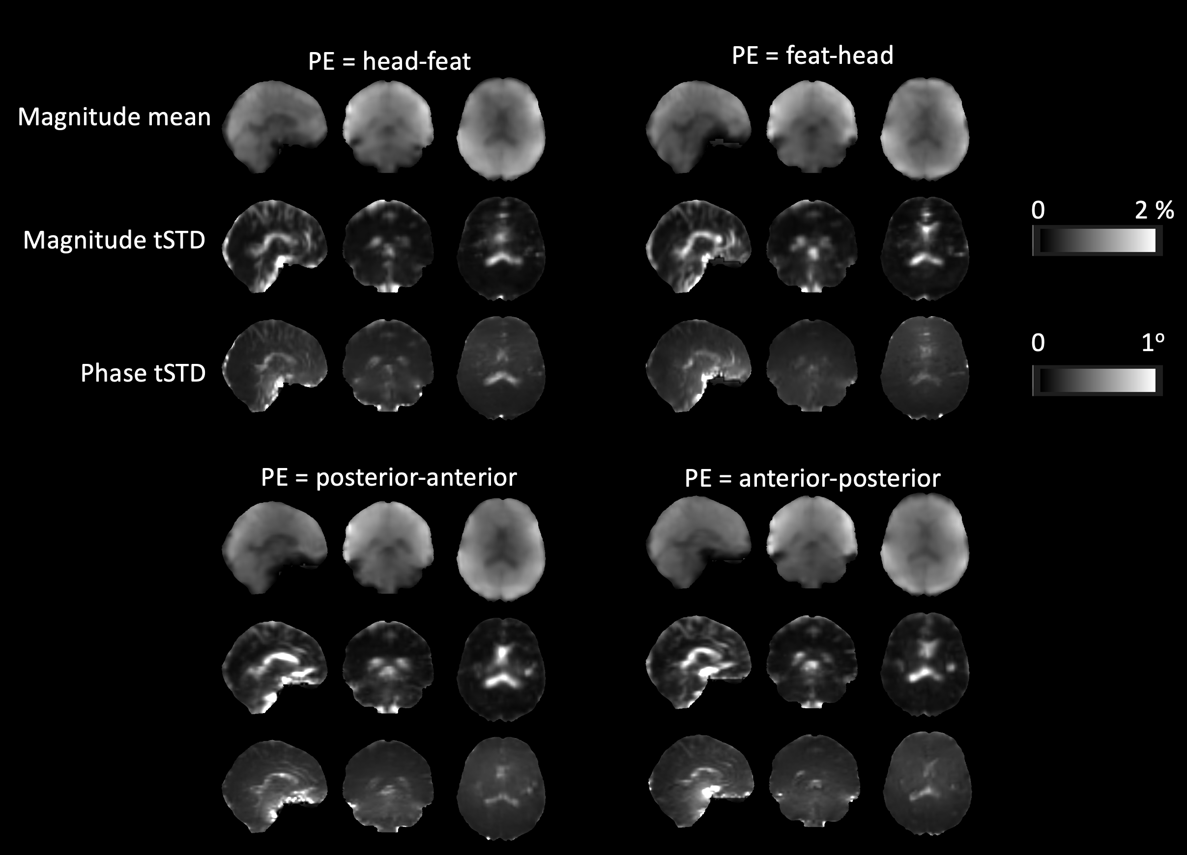
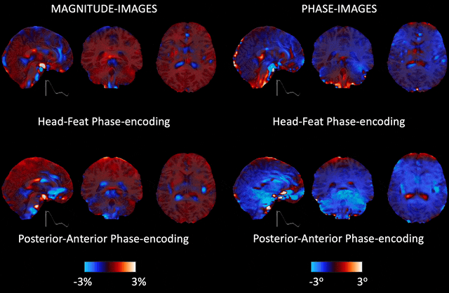
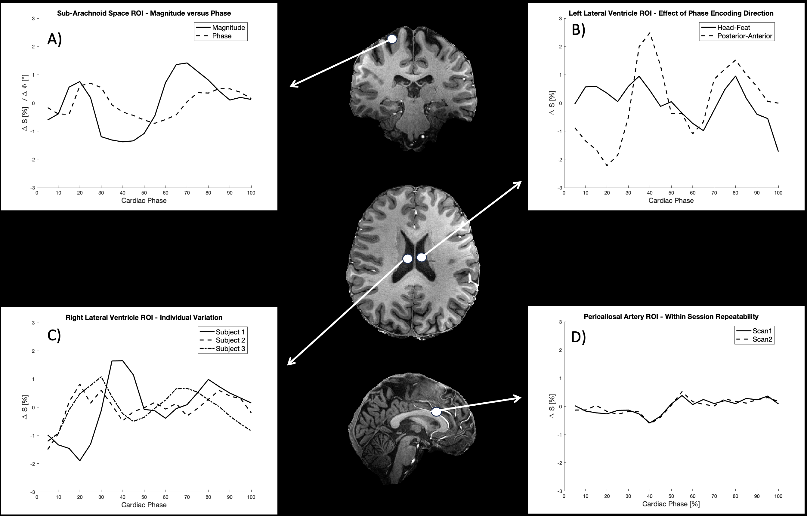
3 healthy volunteers were scanned on a Siemens Magnetom Terra 7T MR system (Siemens Healthineers, Erlangen, Germany) equipped with a 8TX/32RX head coil (Nova Medical Inc, Wilmington, Delaware) using a custom gradient-echo 3D-EPI sequence5. The protocol was optimized for minimum volume TR, with a 3D sagittally oriented matrix of 64x64x48, PF=6/8 along PE/3D and echo-spacing of 0.53 ms. The PF was placed at the end of the read-out to maximize TE within TR and maintain the first order gradient-moment at TE. 3x2 CAIPIRINHA acceleration were applied, resulting in TE/TR=6.7/11.0ms and volume TR=187ms. A binomial WE pulse at FA=10o was used for excitation. 500 volumes were acquired with a total scan time of 93.5 seconds. Physiological data were logged using a respiration belt and a pulse oximeter. The scan was repeated at different PE directions. For one subject, the same scan was repeated twice within the same session. For anatomical reference, MPRAGE6 at 0.75 mm isotropic resolution was acquired and brain-masked using SPM127 . FSL-Feat8 was used for motion correction and spatial smoothing of 3D-EPI, the first 20 volumes were deleted to ensure steady state. EPIC was used for EPI distortion correction9. For retrospective gating, the peak slope of the ascending ridge of each cardiac pulse was defined as the start of the cardiac cycle, and for each 3D volume acquired, the time delay from the latest trigger point was found. A cine-video with 20 time bins was then created by averaging across all image volumes within each time bin, resulting in around 20 images in each bin after discarding those falling outside a subject-specific predefined RR-interval . In each voxel, the magnitude and phase oscillations were calculated as percentage/degrees change from the mean across the cardiac cycle in each voxel. For ROI analysis, single voxel ROIs in the 3D-EPI images were defined, with visual guidance from MPRAGE.Results
Example cine-videos for one subject at the two different PE-directions are shown in Figures 1 (MR magnitude) and 2 (MR phase). Significant fluctuations in both magnitude and phase across the cardiac cycle is observed in CSF and blood compartments. Especially in the lateral ventricles, there is a significant difference between the Head-to-Feat versus Posterior-to-Anterior phase encoding directions, see also Figure 3B. Significant CSF dynamics are detected even in the cortical Sub-Arachnoid Space as illustrated in Figure 3A, where it can also be observed that magnitude and phase fluctuate differently. The three individuals demonstrated significant variation as illustrated for a ROI in the Right Lateral Ventricle in Figure 3C. The within-session repeatability is illustrated in Figure 3D for a ROI placed in the pericallosal artery, which also illustrates the fairly low magnitude fluctuations for this medium-sized artery.Discussion
The results from this pilot study establish that fast 3D-EPI has excellent sensitivity to fluid motion in the brain during the cardiac cycle. This sensitivity can partly be explained by the relatively high first-order gradient moment at TE along the PE direction, corresponding to a Venc of 1.0 m/s for our protocol settings. However, preliminary results from simulations using extended phase graph theory (not shown), indicate that contributions from higher order pathways might also play a role, due to the long T2 of around 2.0 sec for CSF. Further work is required to understand the contrast formation throughout the cardiac cycle, given the complex CSF dynamics in 4D. Once solved, it might be possible to quantify CSF motion with 3D-EPI.Conclusion
Dynamic 3D-EPI at low spatial resolution and short volume TR is very sensitive to CSF dynamics and has potential to be optimized for characterization of CSF motion. It represents a valuable extension of the portfolio of fast MRI methods aimed at studying brain pulsation.Acknowledgements
Staff at the Norwegian 7T MR Center, for volunteer recruitment and data acquisition. NTNU Faculty for Natural Sciences for travel support.
References
1. Bohr, T., et al., The glymphatic system: Current understanding and modeling. iScience 2022. 25(9): p. 104987.
2. Rasmussen, M.K., H. Mestre, and M. Nedergaard, Fluid transport in the brain. Physiol Rev, 2022. 102(2): p. 1025-1151.
3. Kiviniemi, V., et al., Ultra-fast magnetic resonance encephalography of physiological brain activity - Glymphatic pulsation mechanisms? J Cereb Blood Flow Metab, 2016. 36(6): p. 1033-45.
4. Raitamaa, L., et al., Spectral analysis of physiological brain pulsations affecting the BOLD signal. Hum Brain Mapp, 2021. 42(13): p. 4298-4313.
5. Stirnberg, R. and T. Stocker, Segmented K-space blipped-controlled aliasing in parallel imaging for high spatiotemporal resolution EPI. Magn Reson Med, 2021. 85(3): p. 1540-1551.
6. Mugler, J.P., 3rd and J.R. Brookeman, Three-dimensional magnetization-prepared rapid gradient-echo imaging (3D MP RAGE). Magn Reson Med, 1990. 15(1): p. 152-7.
7. Friston, K.J., Ashburner, J., Kiebel, S. J., Nichols, T. E., & Penny, W. D, Statistical Parametric Mapping (Version 12). Wellcome Trust Centre for Neuroimaging, University College London, UK.
8. Jenkinson, M., et al., Fsl. Neuroimage, 2012. 62(2): p. 782-90.
9. Holland, D., J.M. Kuperman, and A.M. Dale, Efficient correction of inhomogeneous static magnetic field-induced distortion in Echo Planar Imaging. Neuroimage, 2010. 50(1): p. 175-83.
10. Matsumae, M., et al., Changing the Currently Held Concept of Cerebrospinal Fluid Dynamics Based on Shared Findings of Cerebrospinal Fluid Motion in the Cranial Cavity Using Various Types of Magnetic Resonance Imaging Techniques. Neurol Med Chir (Tokyo), 2019. 59(4): p. 133-146.
11. Yatsushiro, S., et al., Cardiac-driven Pulsatile Motion of Intracranial Cerebrospinal Fluid Visualized Based on a Correlation Mapping Technique. Magn Reson Med Sci, 2018. 17(2): p. 151-160.
Figures