1184
Simultaneous arterial, venous, and CSF flow dynamics using interleaved multi-Venc spiral imaging1University of Wisconsin-Madison, Madison, WI, United States
Synopsis
Keywords: Neurofluids, Neurofluids
Motivation: Neurofluids dynamics are hypothesized to enable brain metabolite waste clearance pathways for healthy brain function including coupling between arterial, venous, and CSF fluid flow.
Goal(s): To enable quantification of arterial, venous and CSF flow simultaneously leveraging interleaved multi-Venc phase contrast encoding and spiral sampling.
Approach: A four-point velocity encoding scheme was integrated into a 2D golden angle spiral phase contrast sequence and used in studies of healthy subjects. The flow encoding collects multiple first moments at a fixed echo time.
Results: The multi-Venc encoding scheme allowed for simultaneous measures of cardiac-resolved arterial, venous, and CSF flow.
Impact: This work demonstrates a method for simultaneous arterial, venous, and CSF flow imaging for imaging neurofluid dynamics and their response to stimuli, challenges, and transient patient states.
Introduction:
Due to the constant space of the cranium, cerebrospinal fluid (CSF) flow oscillations are tightly coupled with arterial and venous pressure and flow dynamics partially driven by cardiac and respiratory pulsations. While neurofluid flow in each of these compartments can be independently measured using cardiac gated1 or real-time2 phase contract (PC) MRI, asynchronous measures are subject to changes in the heart rate, respiration, and repeatability of external challenges (e.g. respiratory maneuvers, exercise, neurostimulation, etc.). The main challenge limiting simultaneous neurofluid flow measures is the dramatically different velocities between arterial (<150 cm/s), venous (<40cm/s), and CSF (<10cm/s) flow. While some progress has been made on simultaneous multi-Venc using multislice (SMS) and interleaved encoding3, these techniques can be limited by restricted selection of the low Venc from signal loss due to prolonged TE, and difficulty to separate inter-Venc information due to insufficient sensitivity variations in the Venc direction. In this work, we investigate a multi-Venc imaging approach to improve the velocity dynamic range leveraging a minimal time 1st moment gradient design and accelerated golden angle spiral sampling to combat the increased acquisition time and enable both CINE and real time acquisitions.Methods:
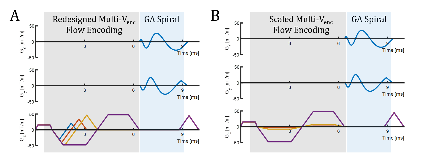
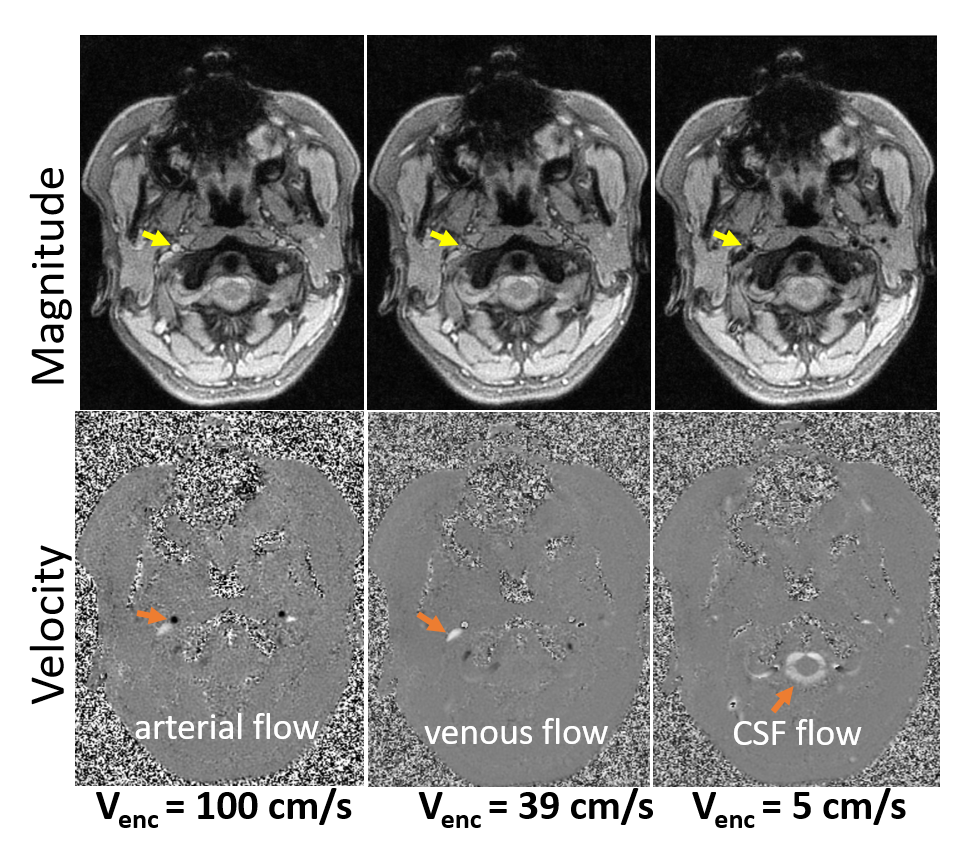
The proposed spiral multi-Venc encoding scheme is shown in Figure 1 with one sided, one directional encoding along the slice dimension. The flow encoding gradients are designed independently for each 1st encoding moment (Fig 1a) compared to naïve scaling (Fig 1b). Data sampling is performed with golden angle spiral sampling. During acquisition, pulse oximeter trigger locations and respiratory belt data were collected. Gating is entirely retrospective with corrections for pulse delay to the patients finger and rebining into cardiac- or real-time frames. Images were reconstructed utilizing multi-scale low rank image reconstruction.Healthy volunteer data (N=6) were collected on a 3T clinical scanner (Signa Premier, GE Healthcare) with a 48ch head coil (GE Healthcare). Two scans per volunteer were collected including a free breathing scan and a breath hold challenge during the second scan in which subjects were instructed to perform a 10s breath hold, 10s into the scan. Each acquisition collected 4 flow encodings with a flow compensated acquisition and images collected at Vencs of 100cm/s, 39cm/s, and 5cm/s. Relevant parameters include: flip = 4°, TE = 5.0ms, TR=9.6ms, 220mm FOV, 0.85mm in-plane resolution, 4mm slice thickness, 1,400 spiral arms collected per encode for a scan time of 54s. Two-dimensional plane placement was prescribed at the base of the skull proximal to the formen magnun to simultaneously capture signals from arteries, veins, and CSF conduits.Results:
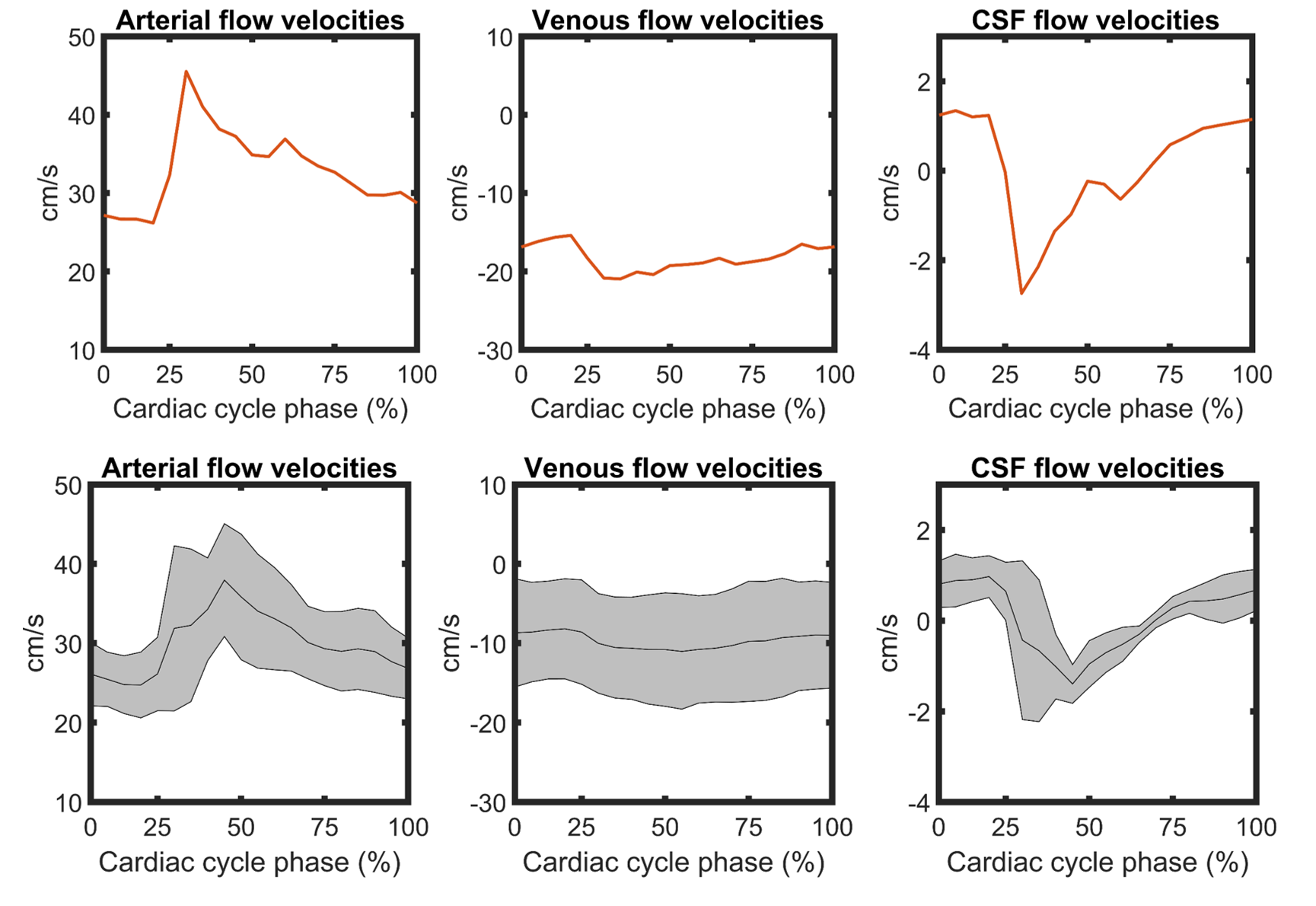
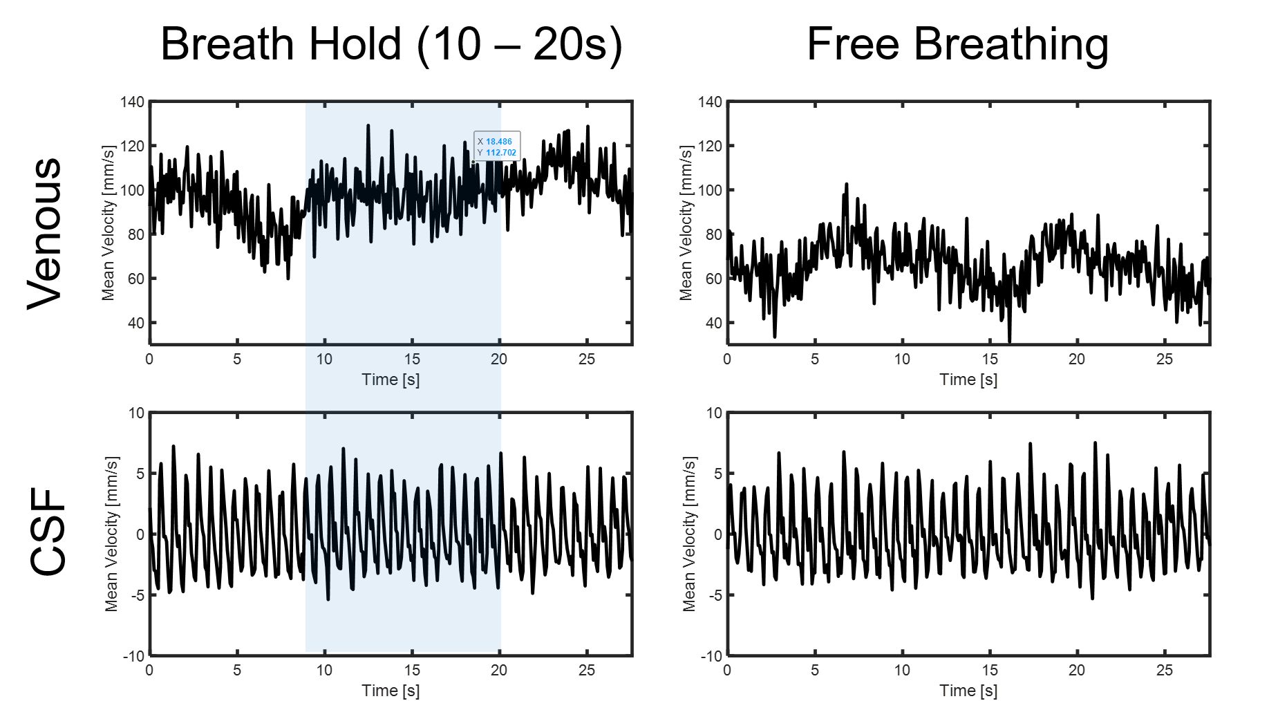
Utilizing scaled (Fig 1b) rather than redesigned gradients (Fig 1a) resulted in a 54% and 46% increase in 2nd moment values for the arterial and venous Venc’s respectively. Figure 2 shows representative magnitude and velocity images from the three Venc images. Velocity images were reconstructed separately for each velocity encode, since phase unwrapping of the low Venc images produced unreliable results in arteries and veins, presumably from intravoxel velocity distributions. Figure 3 shows cardiac-resolved velocity profiles for arterial, venous, and CSF flow across participants. Cardiac-resolved velocity profiles of arteries and CSF displayed sharp profiles including multiple features of the systolic and diastolic phases. Figure 4 shows examples of real-time venous and CSF flow velocity profiles during free breathing and breath hold conditions. Venous flow velocity variability was reduced during the breath hold compared to free breathing conditions.Discussion and Conclusions:
This work demonstrated the use of multi-Venc encoding with a 1st gradient moment minimal time optimization and fixed echo time to simultaneously measure arterial, venous, and CSF flow at a single plane. This comes with some compromise, particularly for arterial imaging. For example, the longer echo times from intermixing the velocity encoding was partially offset by the use of center out sampling; however, longer echo times still impact arterial flow imaging, including displacement artifacts. Further, the flip angle is dictated by the long T1 of CSF and reduced inflow resulting in lower arterial SNR. Work is yet needed to validate these measures in cases of real-time flow changes and against gold standard measures.Acknowledgements
We gratefully acknowledge research support from GE Healthcare, and funding support from NIH grants R01AG075788 and R21AG077337.References
1. Zarrinkoob L, Ambarki K, Wåhlin A, Birgander R, Eklund A, Malm J. Blood flow distribution in cerebral arteries. J Cereb Blood Flow Metab. 2015 Mar 31;35(4):648-54. doi: 10.1038/jcbfm.2014.241. PMID: 25564234; PMCID: PMC4420884.
2. Töger J, Andersen M, Haglund O, Kylkilahti TM, Lundgaard I, Markenroth Bloch K. Real-time imaging of respiratory effects on cerebrospinal fluid flow in small diameter passageways. Magn Reson Med. 2022;88(2):770-786. doi:10.1002/mrm.29248
3. Park S, Chen L, Townsend J, Lee H, Feinberg DA. Simultaneous Multi-VENC and Simultaneous Multi-Slice Phase Contrast Magnetic Resonance Imaging. IEEE Trans Med Imaging. 2020 Mar;39(3):742-752. doi: 10.1109/TMI.2019.2934422. Epub 2019 Aug 12.
Figures