1171
Cardiac Magnetic Resonance Cine Images derived-Radiomics for the Prediction of Event Free Survival in Patients with Acute Myocardial Infarction1Department of Cardiology, Chinese PLA General Hospital, Beijing, China, 2Chinese PLA General Hospital, Beijing, China, 3Chinese PLA General Hospital, beijing, China
Synopsis
Keywords: Myocardium, Cardiovascular
Motivation: Prognostic value of radiomic features extracted from CMR cine image remains to be investigated.
Goal(s): To evaluate the prognostic value of radiomic features derived from cine images in patients with ST-segment elevation myocardial infarction (STEMI).
Approach: Radiomic features were extracted from CMR cine images on STEMI patients, and LASSO -Cox regression used to select predictive features for MACE. Cox regression was applied to build models.
Results: RAD score provided an incremental prognostic value above baseline clinical factors and LVEF (C-index 0.78 vs 0.69; p=0.002) and outperformed the addition of CMR markers of infarct injury (C-index: 0.78 vs 0.69, p<0.001).
Impact: Radiomic features provide incremental prognostic value to clinical and infarct size in the prediction of MACE, which would promote the development of the prognostic assessment with non-contrast enhanced CMR.
Background
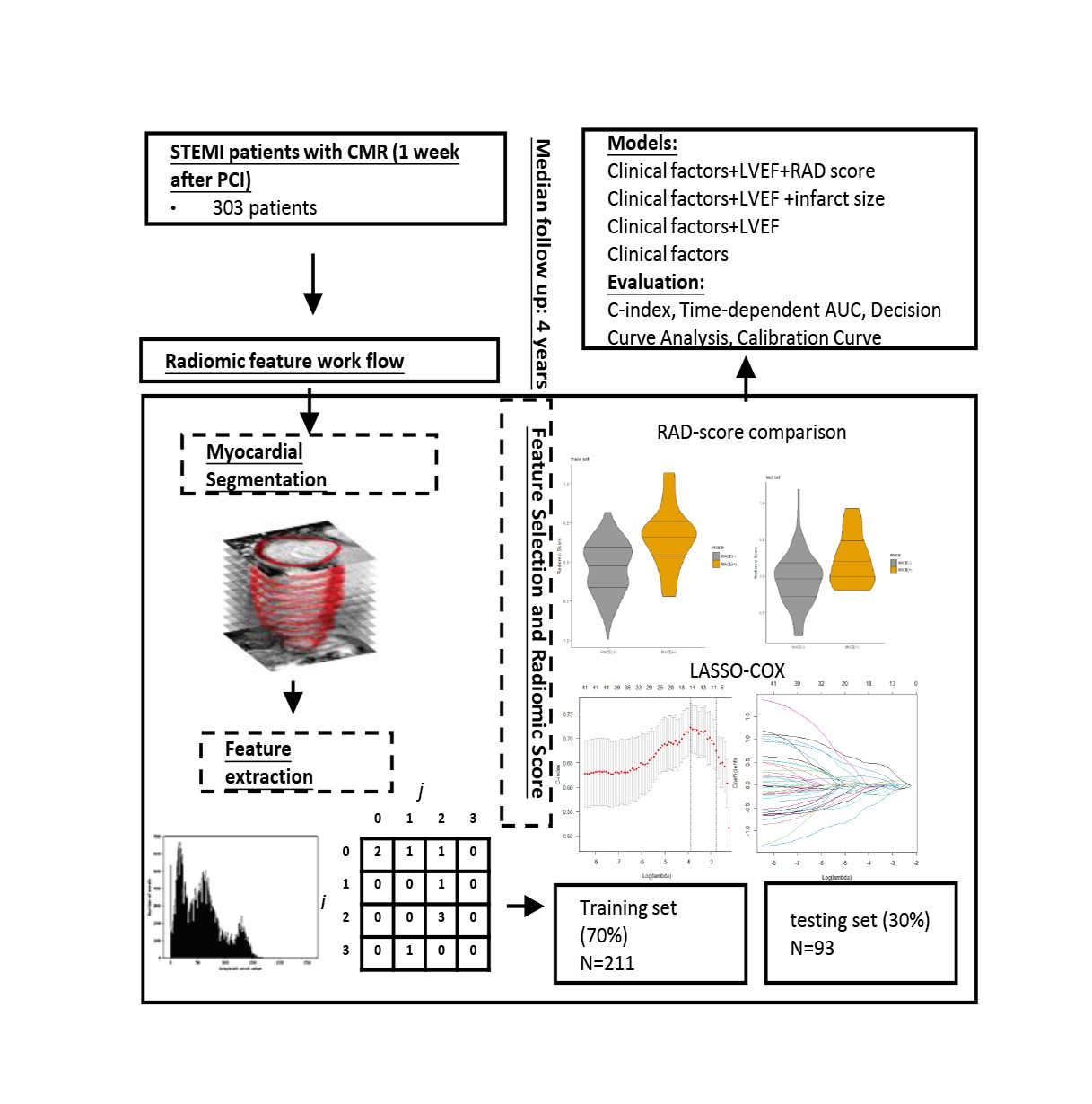
Radiomic analysis of cardiac magnetic resonance (CMR) non-contrast cine images allows quantitative analysis of myocardial tissue alterations. However, the prognostic value of radiomic features extracted from cine image remains to be investigated. The aim of this study was to evaluate the prognostic value of radiomic features derived from cine images in patients with ST-segment elevation myocardial infarction (STEMI).Methods
This prospective, multicenter observational study enrolled 303 patients with acute STEMI, who underwent CMR examination one week after percutaneous coronary intervention. The patients were randomly divided into two groups: training cohort (n=211) and validation cohort (n=92). Radiomic features were extracted from CMR cine images, and least absolute shrinkage and selection operator regression (LASSO)-Cox regression used to select predictive features for major adverse cardiac events (MACE). After univariate Cox analysis, multivariate Cox regression was applied to build combined models incorporating radiomic (RAD) score with clinical risk factors. Calibration was graphically investigated.Results
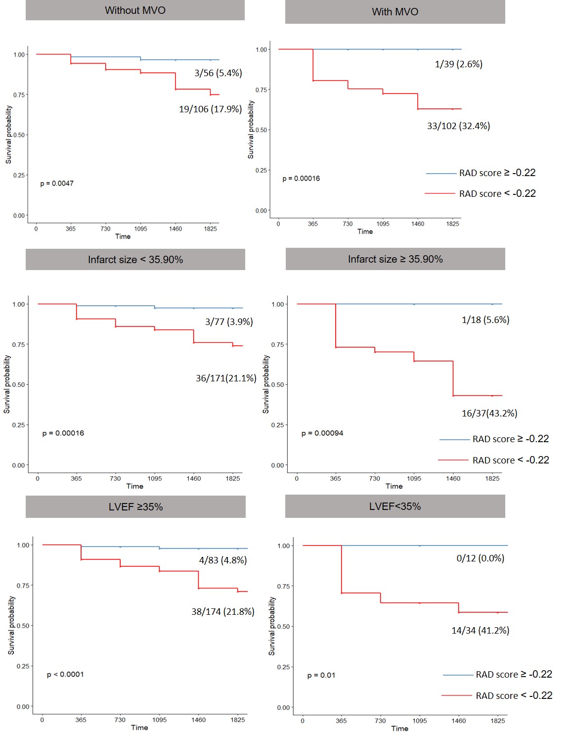
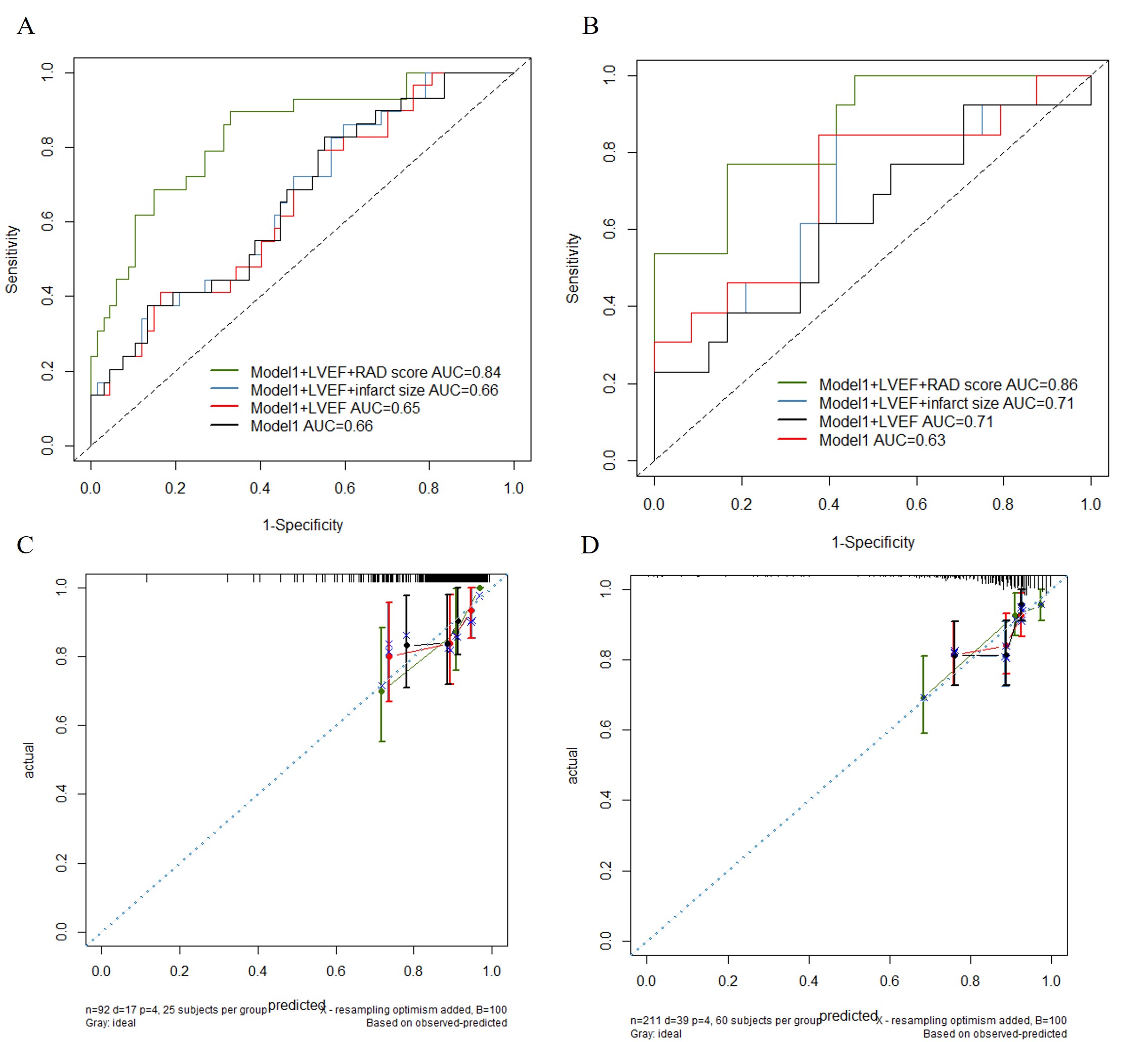
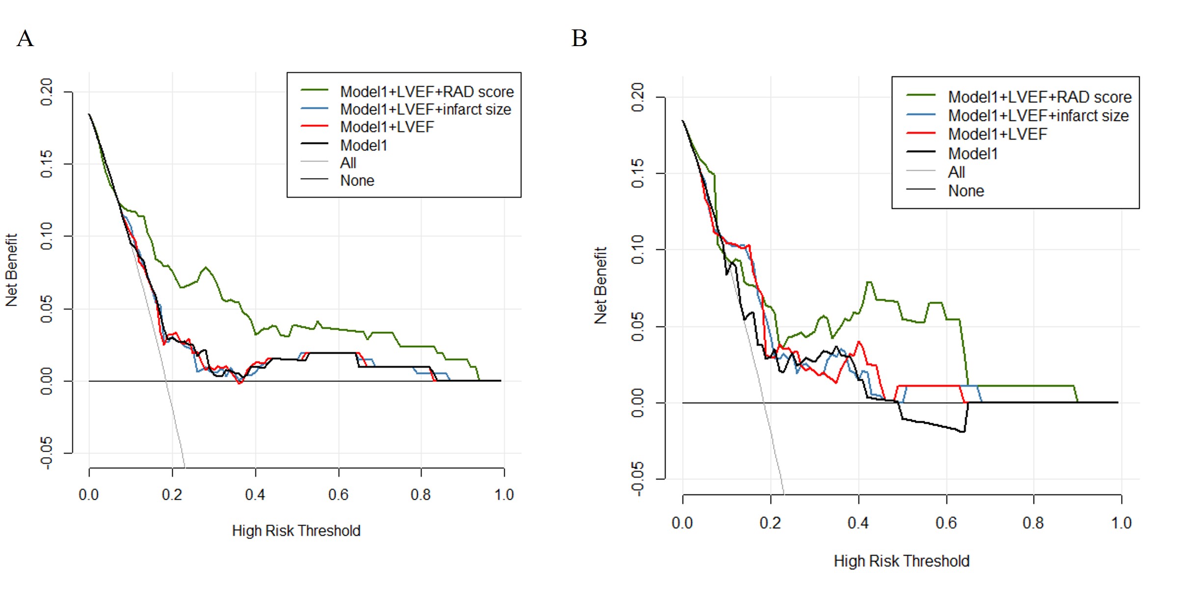
During a median follow-up of 4 years, 56 patients experienced MACE. Eight features were included in the RAD score, as selected by LASSO-Cox regression. In the multivariate analysis, RAD score remained the only significant CMR predictors in addition to clinical factors. RAD score provided an incremental prognostic value above baseline clinical factors and LVEF in both the training (C-index: 0.81vs 0.65; p<0.001) and validation cohort (C-index 0.78 vs 0.69; p=0.002) and outperformed the addition of CMR markers of infarct injury in both the training (C-index: 0.81 vs 0.61, p<0.001) and validation cohort (C-index: 0.78 vs 0.69, p<0.001). The calibration curves demonstrated a good consistency for survival prediction of the combined model.Conclusions
The radiomic signature provides important prognostic information for the development of adverse event in patients with STEMI. The prognostic value of RAD score is incremental to clinical parameters and markers of myocardial damage by contrast enhanced CMR.Acknowledgements
NoneReferences
1. Ibanez B, James S, Agewall S, Antunes MJ, Bucciarelli-Ducci C, Bueno H, et al. 2017 ESC Guidelines for the management of acute myocardial infarction in patients presenting with ST-segment elevation: The Task Force for the management of acute myocardial infarction in patients presenting with ST-segment elevation of the European Society of Cardiology (ESC). Eur Heart J. 2018;39(2):119-77. 2. Scott PA, Morgan JM, Carroll N, Murday DC, Roberts PR, Peebles CR, et al. The extent of left ventricular scar quantified by late gadolinium enhancement MRI is associated with spontaneous ventricular arrhythmias in patients with coronary artery disease and implantable cardioverter-defibrillators. Circulation Arrhythmia and electrophysiology. 2011;4(3):324-30. 3. Galea N, Dacquino GM, Ammendola RM, Coco S, Agati L, De Luca L, et al. Microvascular obstruction extent predicts major adverse cardiovascular events in patients with acute myocardial infarction and preserved ejection fraction. European radiology. 2019;29(5):2369-77. 4. Li Y, Wang G, Wang X, Li Y, Zhao Y, Gu X, et al. Prognostic significance of myocardial salvage assessed by cardiac magnetic resonance in reperfused ST-segment elevation myocardial infarction. Front Cardiovasc Med. 2022;9:924428. 5. Ibanez B, Aletras AH, Arai AE, Arheden H, Bax J, Berry C, et al. Cardiac MRI Endpoints in Myocardial Infarction Experimental and Clinical Trials: JACC Scientific Expert Panel. J Am Coll Cardiol. 2019;74(2):238-56. 6. Liu D, Borlotti A, Viliani D, Jerosch-Herold M, Alkhalil M, De Maria GL, et al. CMR Native T1 Mapping Allows Differentiation of Reversible Versus Irreversible Myocardial Damage in ST-Segment-Elevation Myocardial Infarction: An OxAMI Study (Oxford Acute Myocardial Infarction). Circulation Cardiovascular imaging. 2017;10(8). 7. Savadjiev P, Chong J, Dohan A, Agnus V, Forghani R, Reinhold C, et al. Image-based biomarkers for solid tumor quantification. European radiology. 2019;29(10):5431-40. 8. Gillies RJ, Kinahan PE, Hricak H. Radiomics: Images Are More than Pictures, They Are Data. Radiology. 2016;278(2):563-77. 9. Raisi-Estabragh Z, Izquierdo C, Campello VM, Martin-Isla C, Jaggi A, Harvey NC, et al. Cardiac magnetic resonance radiomics: basic principles and clinical perspectives. European heart journal Cardiovascular Imaging. 2020;21(4):349-56. 10. Avard E, Shiri I, Hajianfar G, Abdollahi H, Kalantari KR, Houshmand G, et al. Non-contrast Cine Cardiac Magnetic Resonance image radiomics features and machine learning algorithms for myocardial infarction detection. Comput Biol Med. 2022;141:105145. 11. Larroza A, López-Lereu MP, Monmeneu JV, Gavara J, Chorro FJ, Bodí V, et al. Texture analysis of cardiac cine magnetic resonance imaging to detect nonviable segments in patients with chronic myocardial infarction. Medical physics. 2018;45(4):1471-80. 12. Larroza A, Materka A, Lopez-Lereu MP, Monmeneu JV, Bodi V, Moratal D. Differentiation between acute and chronic myocardial infarction by means of texture analysis of late gadolinium enhancement and cine cardiac magnetic resonance imaging. European journal of radiology. 2017;92:78-83. 13. Baessler B, Mannil M, Oebel S, Maintz D, Alkadhi H, Manka R. Subacute and Chronic Left Ventricular Myocardial Scar: Accuracy of Texture Analysis on Nonenhanced Cine MR Images. Radiology. 2018;286(1):103-12. 14. Qian G, Zhang Y, Dong W, Jiang ZC, Li T, Cheng LQ, et al. Effects of Nicorandil Administration on Infarct Size in Patients With ST-Segment-Elevation Myocardial Infarction Undergoing Primary Percutaneous Coronary Intervention: The CHANGE Trial. J Am Heart Assoc. 2022;11(18):e026232. 15. van Griethuysen JJM, Fedorov A, Parmar C, Hosny A, Aucoin N, Narayan V, et al. Computational Radiomics System to Decode the Radiographic Phenotype. Cancer research. 2017;77(21):e104-e7. 16. Zhou Y, Gu HL, Zhang XL, Tian ZF, Xu XQ, Tang WW. Multiparametric magnetic resonance imaging-derived radiomics for the prediction of disease-free survival in early-stage squamous cervical cancer. European radiology. 2022;32(4):2540-51. 17. Shen W, Ning J, Yuan Y. A direct method to evaluate the time-dependent predictive accuracy for biomarkers. Biometrics. 2015;71(2):439-49. 18. Lambin P, Leijenaar RTH, Deist TM, Peerlings J, de Jong EEC, van Timmeren J, et al. Radiomics: the bridge between medical imaging and personalized medicine. Nat Rev Clin Oncol. 2017;14(12):749-62. 19. Chang S, Han K, Suh YJ, Choi BW. Quality of science and reporting for radiomics in cardiac magnetic resonance imaging studies: a systematic review. European radiology. 2022;32(7):4361-73. 20. Dastidar AG, Rodrigues JC, Baritussio A, Bucciarelli-Ducci C. MRI in the assessment of ischaemic heart disease. Heart (British Cardiac Society). 2016;102(3):239-52. 21. Symons R, Pontone G, Schwitter J, Francone M, Iglesias JF, Barison A, et al. Long-Term Incremental Prognostic Value of Cardiovascular Magnetic Resonance After ST-Segment Elevation Myocardial Infarction: A Study of the Collaborative Registry on CMR in STEMI. JACC Cardiovasc Imaging. 2018;11(6):813-25. 22. Kanda T, Fukusato T, Matsuda M, Toyoda K, Oba H, Kotoku J, et al. Gadolinium-based Contrast Agent Accumulates in the Brain Even in Subjects without Severe Renal Dysfunction: Evaluation of Autopsy Brain Specimens with Inductively Coupled Plasma Mass Spectroscopy. Radiology. 2015;276(1):228-32. 23. Chen BH, An DA, He J, Wu CW, Yue T, Wu R, et al. Myocardial extracellular volume fraction radiomics analysis for differentiation of reversible versus irreversible myocardial damage and prediction of left ventricular adverse remodeling after ST-elevation myocardial infarction. European radiology. 2021;31(1):504-14. 24. Ma Q, Ma Y, Yu T, Sun Z, Hou Y. Radiomics of Non-Contrast-Enhanced T1 Mapping: Diagnostic and Predictive Performance for Myocardial Injury in Acute ST-Segment-Elevation Myocardial Infarction. Korean J Radiol. 2021;22(4):535-46. 25. Ma Q, Ma Y, Wang X, Li S, Yu T, Duan W, et al. A radiomic nomogram for prediction of major adverse cardiac events in ST-segment elevation myocardial infarction. European radiology. 2021;31(2):1140-50. 26. Kotu LP, Engan K, Borhani R, Katsaggelos AK, Ørn S, Woie L, et al. Cardiac magnetic resonance image-based classification of the risk of arrhythmias in post-myocardial infarction patients. Artificial intelligence in medicine. 2015;64(3):205-15. 27. Baeßler B, Mannil M, Maintz D, Alkadhi H, Manka R. Texture analysis and machine learning of non-contrast T1-weighted MR images in patients with hypertrophic cardiomyopathy-Preliminary results. European journal of radiology. 2018;102:61-7. 28. Jang J, El-Rewaidy H, Ngo LH, Mancio J, Csecs I, Rodriguez J, et al. Sensitivity of Myocardial Radiomic Features to Imaging Parameters in Cardiac MR Imaging. Journal of magnetic resonance imaging : JMRI. 2021;54(3):787-94.Figures