1170
The feasibility study of multiple functional imaging modalities in the differential diagnosis of benign and malignant bone tumors1The First Affiliated Hospital of Zhengzhou University, Zhengzhou, China, 2Clinical & Technical Support, Philips Healthcare, Beijing, 100102, China, Beijing, China
Synopsis
Keywords: Cancer, Tumor
Motivation: The combination of multi-MRI techniques in the differential diagnosis of benign and malignant bone tumors represents a novel endeavor.
Goal(s): To investigate the utility of combining DWI, IVIM, DKI and APTWI in the differential diagnosis of benign and malignant bone tumors.
Approach: Relevant parameters of 45 patients were statistically compared through either the independent samples t-test or Mann-Whitney U test. Diagnostic performance was assessed using ROC curves for both individual examinations and their combined analysis in distinguishing between benign and malignant tumors.
Results: The combination of multi-MRI techniques proves to be a more effective approach in distinguishing between benign and malignant bone tumors.
Impact: Multimodal MRI provides biological and pathological information about the tumor cell microenvironment, and their combination proves to be a more effective approach in distinguishing between benign and malignant bone tumors.
Introduction
Bone tumors have a low incidence rate, a wide variety of types, diverse presentations, and complex origins, making them a long-standing challenge in clinical imaging diagnosis. The pathological diagnosis of bone tumors has always been challenging. Various biopsy locations can yield fundamentally disparate pathological findings. Therefore, the diagnosis of bone tumors relies on the collaboration of multiple disciplines, including clinical, pathological, and imaging. Selecting the suitable imaging examination method is essential for revealing the radiological features, histology, and biological traits of the tumor. This choice plays a pivotal role in guiding preoperative pathological biopsies, enabling qualitative diagnosis, evaluating postoperative effectiveness, and conducting follow-up assessments. Multimodal MRI refers to the scanning of lesion tissue or organs using various magnetic resonance sequences, thus yielding comprehensive MRI data regarding the affected tissues or organs. This study aims to investigate the efficacy of combining Diffusion-Weighted Imaging (DWI), Intravoxel Incoherent Motion (IVIM), Diffusion Kurtosis Imaging (DKI), and Amide Proton Transfer-Weighted Imaging (APTWI) in differentiating benign and malignant bone tumors.Methods
Forty-five patients (27 males and 18 females, age ranged from 7 to 70 years) with bone tumors (diagnosed based on pathological biopsy) were included in this study. According to the WHO classification of tumors of soft tissue and bone (2020) criteria, the 45 patients were categorized into two groups: 18 with benign tumors and 27 with malignant malignancies. All the patients underwent a comprehensive array of imaging examinations, including T1-weighted imaging, T2-weighted imaging, DWI, IVIM, DKI and APTWI, using a 3T MR scanner (Ingenia CX, Philips Healthcare, Best, the Netherlands). The regions of interest (ROIs) for the tumors were meticulously delineated on each parameter map. Specifically, the following parameters were recorded for each ROIs: DWI-derived apparent diffusion coefficient (ADC), IVIM-related parameters (including perfusion fraction [f], pure diffusion coefficient [D], and perfusion-related diffusion coefficient [D*]), and DKI-related parameters (including mean diffusivity [MD] and mean kurtosis [MK]). Additionally, the APTWI-derived magnetization transfer ratio asymmetry at 3.5 ppm [MTRasym (3.5 ppm)] were also documented. The independent sample t test was used to compare the differences in different parameters between benign and malignant bone tumors. Additionally, the diagnostic performance of IVIM, DKI, APTWI, and DWI, both individually and in combination, was evaluated using the Receiver Operating Characteristic (ROC) curve. Statistical analysis was conducted utilizing SPSS version 22.0.0 and MedCalc version 20.0.7 software. A significance level of p < 0.05 indicated statistically meaningful differences.Results
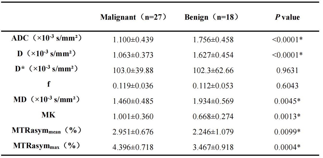
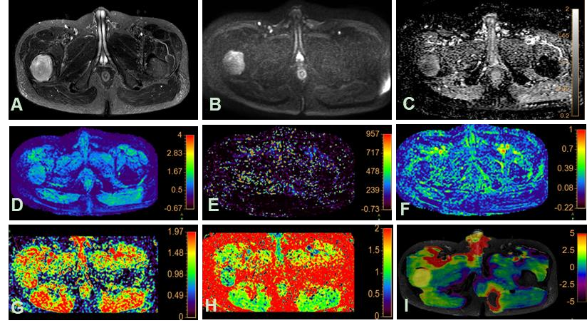
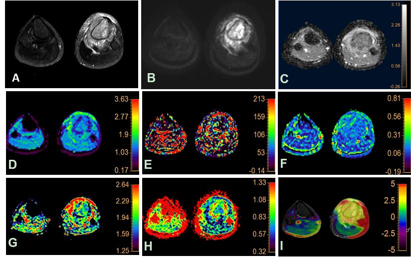
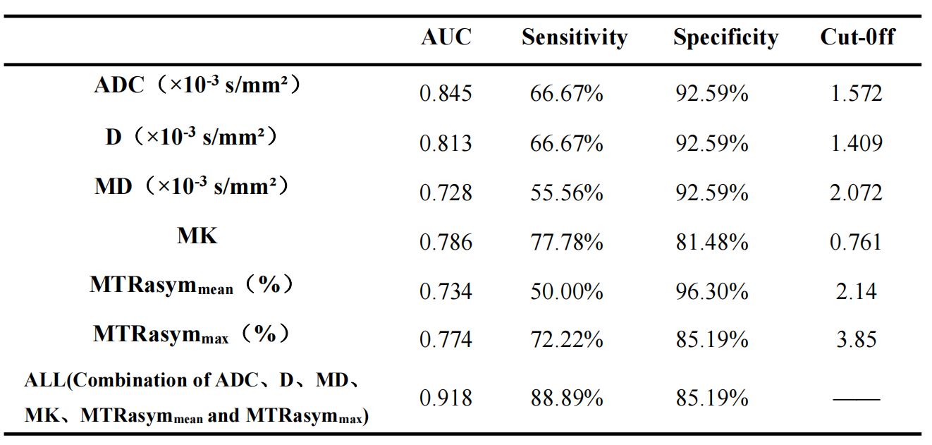
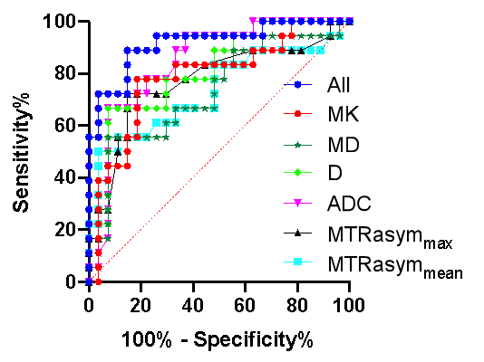
Significant differences were observed in ADC, D, MD, MK, MTRasymmean, and MTRasymmax values between benign and malignant bone tumors (Table 1). The representative images of benign and malignant tumors were shown in Figure 1 and 2. ROC analysis revealed the following AUC values: ADC (0.845), D (0.813), MD (0.728), MK (0.786), MTRasymmean (0.734), and MTRasymmax (0.774) (Table 2). Combining these parameters significantly enhanced diagnostic performance, resulting in an AUC of 0.918. When using the following cut-off values: ADC (1.57×10⁻³ mm²/s), D (1.41×10⁻³ mm²/s), MD (2.07×10⁻³ mm²/s), MK (0.76), MTRasymmean (2.14%), and MTRasymmax (3.85%), sensitivities for distinguishing benign and malignant bone tumors were as follows: 66.67%, 66.67%, 55.56%, 77.78%, 50%, and 72.22%, respectively. Specificities were 96.3%, 85.19%, 92.59%, 92.59%, and 81.48%, respectively (Figure 3). The combination of these parameters demonstrated a sensitivity of 88.89% and a specificity of 85.19%.Discussion
Traditional DWI, based on a simple single-exponential model, primarily characterizes restricted water molecule diffusion in tissues. In contrast, IVIM employs a bi-exponential model that effectively distinguishes between water molecule diffusion and microperfusion effects within tissues [1]. DKI is a diffusion imaging technique that relies on a non-Gaussian distribution single-exponential model. It assesses the extent of diffusion heterogeneity in water molecules across various directions and provides insights into the complexity of tumor cell microstructure [2]. APTWI represents an innovative non-invasive magnetic resonance molecular imaging technology, enabling the differentiation of tumor cells from healthy cells by detecting alterations in protein metabolism [3]. While IVIM, DKI, and APTWI have been explored in clinical studies for various tumors [4-6], their utilization in the context of bone tumors remains relatively limited. In our study, we found that the parameter values derived from DWI, IVIM, DKI, and APTWI parameter maps can effectively discriminate between benign and malignant bone tumors. Furthermore, the combined analysis of multiple parameters significantly enhances diagnostic performance.Conclusions
The combination of multiple functional MRI techniques provides more biological and pathological insights into the tumor cell microenvironment, enhancing the accuracy of distinguishing between benign and malignant bone tumors.Acknowledgements
No acknowledgement found.References
[1] Gang Wu, Ruyi Xie, Xuanlin Liu, et al. Intravoxel incoherent motion diffusion MR and diffusion kurtosis imaging for discriminating atypical bone metastasis from benign bone lesion[J].Br J Radiol, 2019,92(1100):20190119.
[2] Sevtap Arslan, Fatma Bilge Ergen, Güzide Burça Aydın, et al. Different Attenuation Models of Diffusion-Weighted MR Imaging for the Differentiation of Benign and Malignant Musculoskeletal Tumors[J].J Magn Reson Imaging,2022,55(2):594-607.
[3] Wu Y, Chen Y, Zhao Y, et al. Direct Radio Qin X, Mu R, Zheng W, et al.Comparison and combination of amide proton transfer magnetic resonance imaging and the apparent diffusion coefficient in differentiating the grades of prostate cancer[J].Quant Imaging Med Surg, 2023,13(2):812-824.
[4] Ma C, Tian S, Song Q, et al. Amide Proton Transfer-Weighted Imaging Combined With Intravoxel Incoherent Motion for Evaluating Microsatellite Instability in Endometrial Cancer[J]. J Magn Reson Imaging,2023,57(2):493-505.
[5] Qin X, Mu R, Zheng W, et al.Comparison and combination of amide proton transfer magnetic resonance imaging and the apparent diffusion coefficient in differentiating the grades of prostate cancer[J].Quant Imaging Med Surg,2023,13(2):812-824.
[6] Li J, Lin L, Gao X, et al.Amide Proton Transfer Weighted and Intravoxel Incoherent Motion Imaging in Evaluation of Prognostic Factors for Rectal Adenocarcinoma[J].Front Oncol, 2022,11:783544.
Figures