1165
The Impact of Pancreatic Cancer Glutamine Transporter Downregulation on Cachexia and Visceral Organ Metabolism1Department of Radiology, Division of Cancer Imaging and Reserach, The Johns Hopkins University School of Medicine, Baltimore, MD, United States., Baltimore, MD, United States
Synopsis
Keywords: Cancer, Cancer, PDAC, Pancreatic cancer, Glutamine tarnsporter
Motivation: We previously identified alterations in brain and plasma glutamine/ glutamate with cachexia that led us to downregulate the glutamine transporter, SLC1A5, in the cachexia-inducing patient derived Pa04C PDAC cells.
Goal(s): Targeting the glutamine transporter represents a promising approach to delay tumor progression and establish a novel treatment strategy in PDAC cachexia.
Approach: We performed 1H MRS to determine the metabolic changes in multiple organs of mice.
Results: We identified metabolic differences in organs of mice bearing SLC1A5 downregulated tumors compared to wild type or empty vector tumors. Our data identify SLC1A5 as a target to reduce PDAC induced cachexia and associated pathways
Impact: By detecting these visceral organ metabolic changes we identified potential metabolic pathways that can be targeted to reduce cachexia.
Introduction
PDAC induced cachexia is a major contributor to poor quality of life and poor response to treatment. We previously established that a patient-derived PDAC cell line, Pa04C, replicated the weight loss accompanying PDAC-induced cachexia in SCID mice [1]. We also found changes in mouse brain glutamine and human plasma glutamate levels with PDAC-induced cachexia [1]. These observations led us to downregulate the glutamine transporter, SLC1A5, and determine the effects on body and organ weight loss, as well as on organ metabolism using high-resolution 1H MRS.Methods:
Cachexia-inducing Pa04C pancreatic cancer cells, derived from patients, were genetically modified via lentiviral transduction to express SLC1A5 shRNA, ensuring stable downregulation of SLC1A5. This downregulation was confirmed through immunoblotting and quantitative reverse transcription-polymerase chain reaction (qRT-PCR) analyses. Wild type and empty vector (Pa04C_EVWT), and SLC1A5 downregulated Pa04C cells (Pa04C_SLC1A5_shRNA) were inoculated subcutaneously in SCID mice and body weights were monitored. Once tumors reached an approximate size of 500 mm3, the mice were euthanized, and the organs were excised and snap-frozen for further analysis. Snap frozen spleen, pancreas, lung, heart, kidney and liver from nine or more Pa04C_EVWT and Pa04C_SLC1A5_shRNA tumor bearing mice were powdered for dual phase extraction. Aqueous phase spectra were on a Bruker Avance III 750 MHz (17.6 T) MR spectrometer equipped with a 5 mm broad band inverse (BBI) probe. Spectral data processing and quantification were conducted using Topspin 3.5 software. ConsensusPathDB and metaboanalyst software was used for pathway analysis.Results:
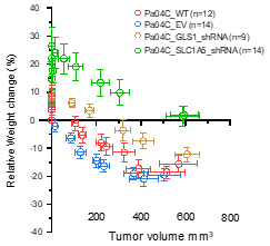
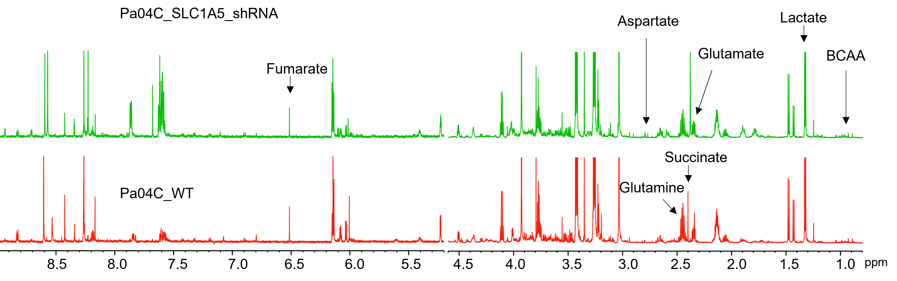
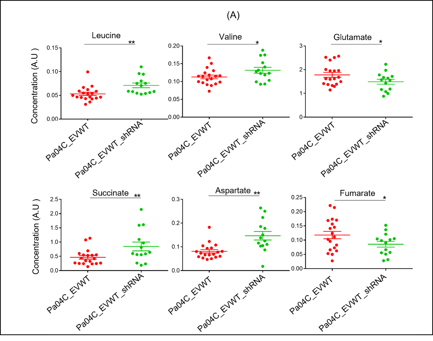
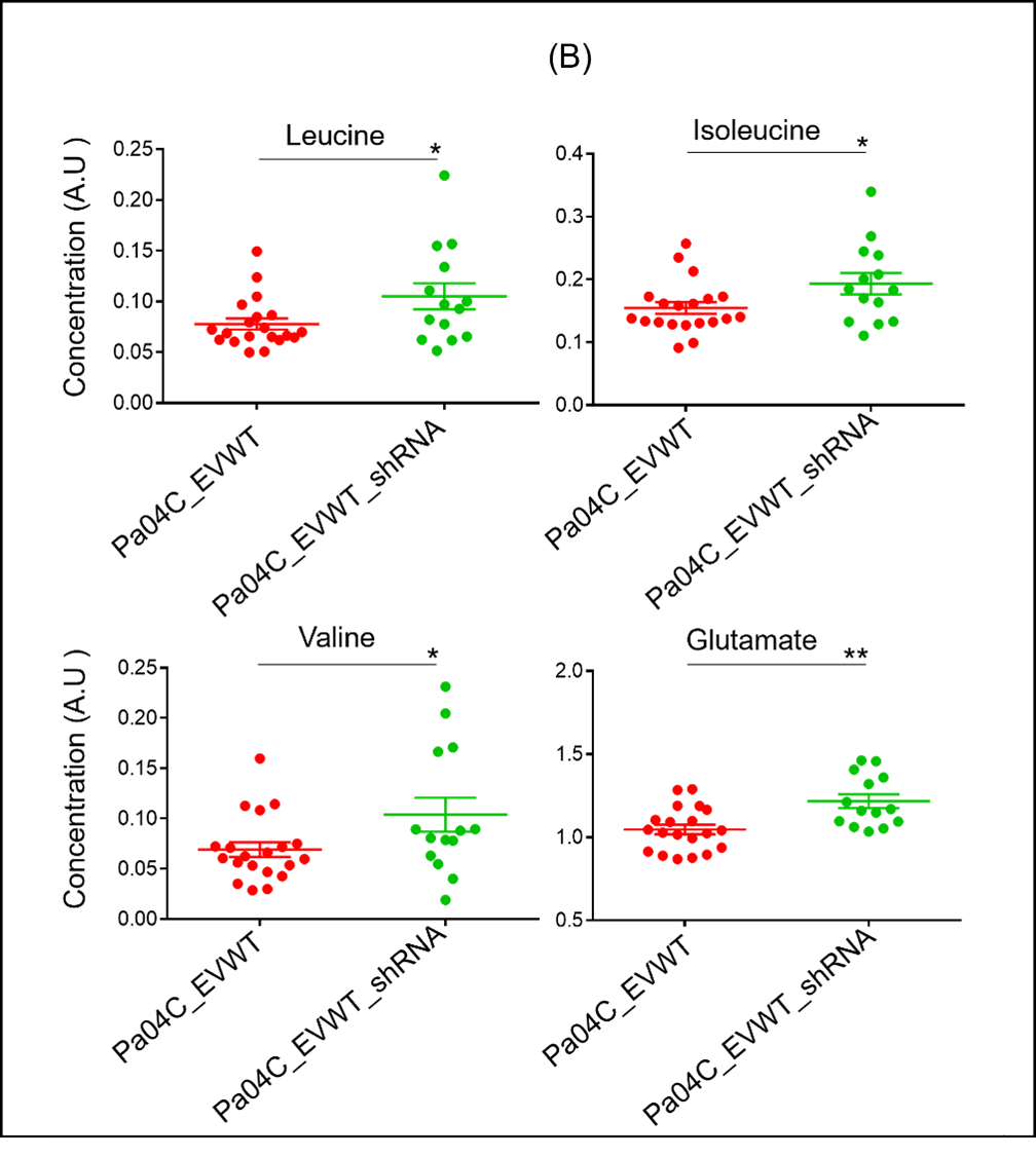
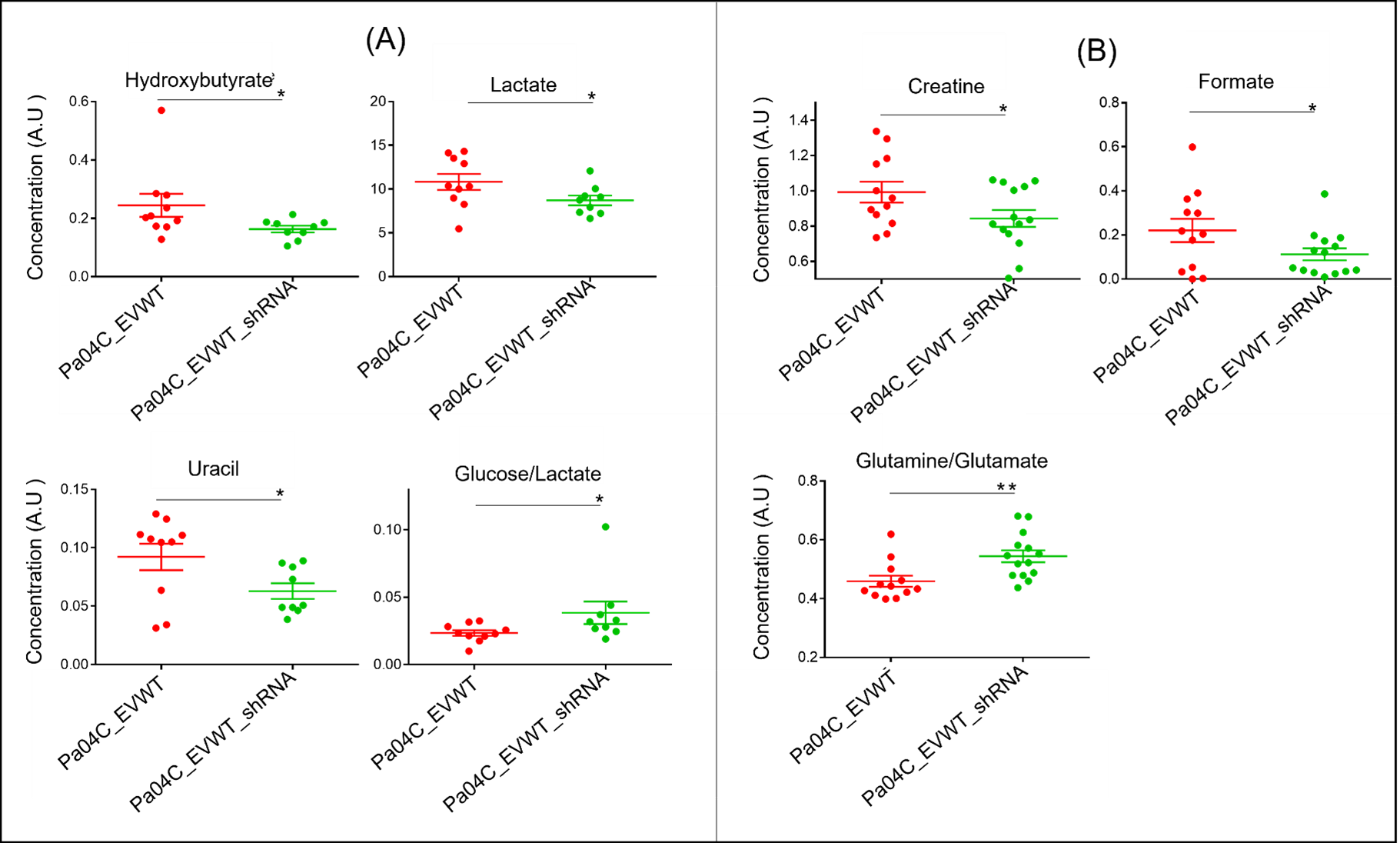
Tumors derived from SLC1A5 downregulated Pa04C cells led to a significant reduction of body weight loss compared to weight loss in mice bearing Pa04C WT and Pa04C EV tumors (Fig 1). Representative 1H MRS heart spectra from Pa04C_WT and Pa04C_SLC1A5_shRNA presented in Fig 2 reveal metabolic differences that occurred in the heart with tumor SLC1A5 downregulation compared to heart metabolites in Pa04C_EV/WT tumor bearing mice. Significant (P<0.05) increases of leucine, valine, succinate and aspartate, and a decrease of fumarate and glutamate were observed in the heart (Fig 3A) with tumor SLC1A5 downregulation. Leucine, isoleucine, valine and glutamate significantly increased in the lung (Fig. 3B); hydroxybutyrate, lactate, and uracil significantly decreased and the glucose/lactate ratio significantly increased in the kidney (Fig. 4A); creatine and formate, significantly decreased, and the glutamine/glutamate significantly increased in the spleen (Fig. 4B). In the pancreas only the glucose/lactate ratio significantly increased. Formate was significantly lower in the liver. Based on the differences, we identified commonly disturbed metabolic pathways related to glutamine and glutamate metabolism, arginine biosynthesis, alanine, aspartate and glutamate metabolism in the heart and lung.Discussion:
The heart showed the most metabolic alterations, followed by the lung, kidney and spleen. The pancreas and the liver showed the least alterations. A ConsensusPathDB Reactome analysis based on the metabolic changes revealed that the "metabolism of amino acids and derivatives" pathway was commonly disrupted in the heart, lung and spleen. Our data highlight, for the first time, the disruptive effects of tumor SLC1A5 on body weight loss and organ metabolism, and identify SLC1A5 as a potential target to reduce cancer induced cachexia. The metabolic patterns in organs hold the potential to serve as innovative targets for mitigating the associated morbidity of cachexia.Acknowledgements
Supported by NIH R01CA193365 and R35CA209960.References
Winnard, P. T, Journal of Cachexia, Sarcopenia and Muscle. 2020, 1487–1500: 11.Figures