1162
Multi-Omics Integration of MRI Habitat, Pathology, and Clinical Parameters for Predicting Platinum Resistance of HGSOC1the First People’s Hospital of Yunnan Province, Kunming, China, 2Jinshan Hospital, Fudan University, Shanghai, China, 3MR Research Collaboration Team, Siemens Healthineers Ltd., Shanghai, China
Synopsis
Keywords: Cancer, Cancer
Motivation: Platinum resistance of high-grade serous ovarian carcinoma (HGSOC) is related to tumor heterogeneity. Multi-omics integration can complement tumor heterogeneity at multiple scales and enhance the predictive power of single models.
Goal(s): We aimed to explore a range of diverse multi-omics models to predict platinum resistance of HGSOC.
Approach: Multi-omics models were developed and validated using MRI-based habitat radiomics, pathomics based on haematoxylin and eosin (H&E)-stained whole slide images (WSIs), and clinical parameters.
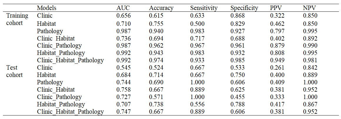
Results: Among the array of single and composite models, the Clinic_Habitat model exhibited the most promising predictive performance, with the Clinic_Habitat_Pathology model ranking as the second-best performer.
Impact: This study carries the potential to equip clinicians with treatment strategies aimed at enhancing the efficacy of individualized therapy.
Introduction
Ovarian carcinoma (OC) is the second most prevalent gynecological malignancy worldwide, following cervical cancer [1]. The majority of OC-related deaths are attributed to high-grade serous ovarian carcinoma (HGSOC) [2]. Due to response of platinum-based chemotherapy cannot currently be recognized by specific molecular biomarkers [3], it is urgently necessary to use other means to predict platinum resistance in patients with HGSOC.Habitat imaging offers a noninvasive means of evaluating intratumoral heterogeneity [4]. Previous studies suggested that multi-omics integration was conducive to complement tumor heterogeneity at multiple scales, providing complementary predictive information for each other, and enhancing the predictive power of single models [5,6].
This study aims to develop and validate diverse integrated models utilizing habitat radiomics from MRI, pathomics from H&E-stained WSIs, and clinical parameters to predict platinum resistance in HGSOC patients, providing multifaceted insights into tumor heterogeneity and treatment response.
Methods
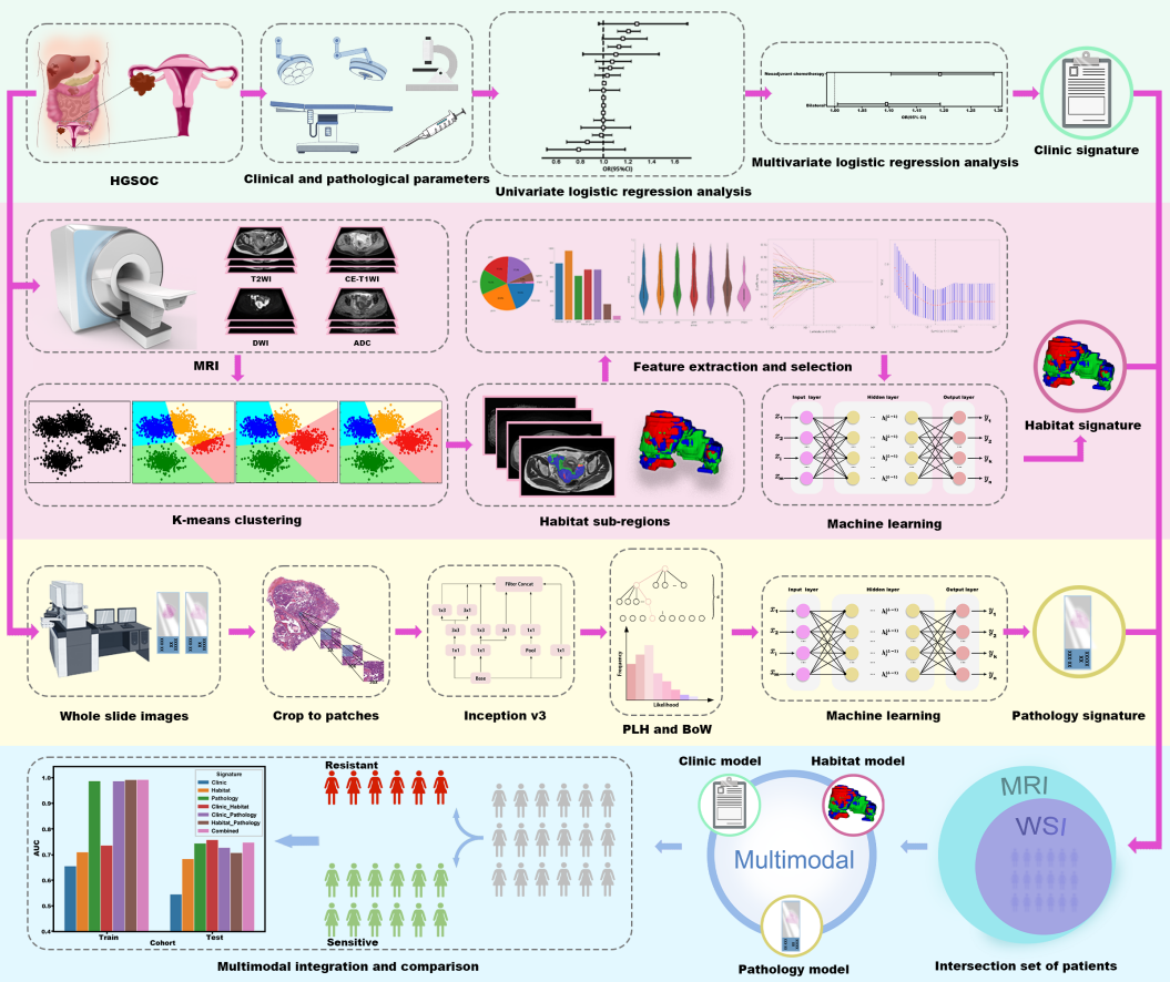
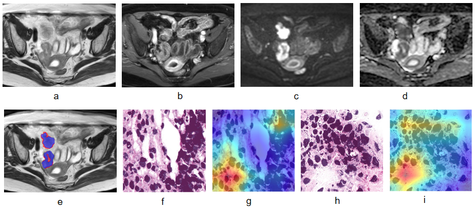
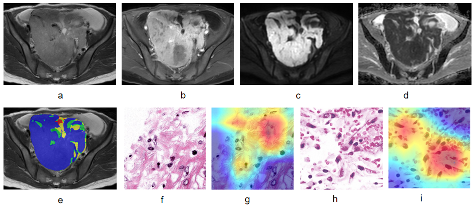
A retrospective cohort study encompassed 393 eligible patients (86 platinum-resistant and 307 platinum-sensitive) from three centres. Patients from centre A were included as the training cohort, and the eligible patients from centres B and C were designated as the test cohort. Pelvic MRI was conducted using 1.5T or 3T scanners (Siemens Magnetom Prisma, Siemens Magnetom Vida, Philips Ingenia, GE Signa Pioneer, and GE Signa HDXt). After image preprocessing and space registration, region of interest of the primary and metastatic lesions was hand-mapped slice by slice on T2WI by two radiologists who had experience in pelvic MRI of 8 and 11 years, respectively. Other sequence MRI images, operation details, and histopathological records were referenced for ensuring accurate delineation.The sequences that need to be clustered included: T2-weighted imaging (T2WI), contrast-enhanced T1-weighted imaging (CE-T1WI) on the venous phase, diffusion-weighted imaging (DWI), and apparent diffusion coefficient (ADC) maps. K-means clustering algorithm based on cohort (https://scikit-learn.org/stable/index.html) was applied to cluster MRI habitat sub-regions. The squared Euclidean distance between voxel intensities was used as the clustering cost measure. We employed Calinski-Harabasz score to determine the best number of clusters that were tested from two to ten in this study. After feature selection and extraction, the habitat radiomics model based on MRI habitat sub-regions (Habitat model) was developed for identifying platinum-resistant and platinum-sensitive patients.
H&E-stained slides of tumors were scanned to WSIs at 20x magnification. The processing of WSI involved cutting them into 512×512-pixel patches and underwent color normalization using the Macehko method. An Inception v3 deep learning model was trained for patch-level prediction. WSI-level prediction utilized multi-instance learning, encompassing Patch Likelihood Histogram (PLH) and Bag of Words (BoW) pipelines. Then, the Pathology model was developed by integrating predictive features of multi-instance learning.
Univariate and multivariate logistic regression analysis was used to select clinical predictors and construct Clinic model, respectively. The intersection of patients containing both MRI and WSI was used to validate and compare Clinic, Habitat, and Pathology models. The area under the curve (AUC) and clinical decision curve (CDC) were used to assess model performance and clinical net benefit.
Results
Four MRI habitats were generated based on Calinski-Harabasz score. Significant differences in ADC values of habitat 1, 2, and 3 were found between the platinum-resistant and platinum-sensitive patients (all P<0.05). Compared with Habitat model (0.684) and Clinic model (0.545), Pathology model had a highest AUC (0.744) in single models. Clinic_Habitat model yielded a highest AUC (0.758) among other single or compositive models, the second highest AUC (0.747) was Clinic_Habitat_Pathology model, both with good clinical net benefits.Discussion
A previous study found that habitat radiomics based on PET/CT was associated with the expression of Ki-67 and prognosis in patients with HGSOC, which implied that habitat radiomics was of certain significance for the assessment of HGSOC [7]. This study further confirmed the potential of MRI based habitat radiomics (Habitat model) to predict platinum resistance in patients with HGSOC.The combination of MRI radiomics and H&E-stained WSIs could improve the performance of predicting the pathological complete response to neoadjuvant chemoradiotherapy in patients with rectal cancer [5]. Machine learning integrated models based on CT images, clinical, pathological, and genomic features were able to effectively improve risk stratification of HGSOC [6]. These studies were consistent with our findings. Therefore, it may be promising to integrate MRI habitat and clinical information at macroscale with WSI at microscale to construct a better prediction model.
Conclusion
The Clinic_Habitat model and the Pathology model hold promise for predicting platinum sensitivity in HGSOC patients. Multimodal integration significantly enhance prediction performance.Acknowledgements
No acknowledgement found.References
1. Sung H, Ferlay J, Siegel RL, et al. Global Cancer Statistics 2020: GLOBOCAN Estimates of Incidence and Mortality Worldwide for 36 Cancers in 185 Countries. Ca-Cancer J Clin 2021; 71(3):209-249.
2. Folsom SM, Berger J, Soong TR, Rangaswamy B. Comprehensive Review of Serous Tumors of Tubo-Ovarian Origin: Clinical Behavior, Pathological Correlation, Current Molecular Updates, and Imaging Manifestations. Curr Probl Diagn Rad 2023; 52(5):425-438.
3. Gonzalez-Martin A, Harter P, Leary A, et al. Newly diagnosed and relapsed epithelial ovarian cancer: ESMO Clinical Practice Guideline for diagnosis, treatment and follow-up. Ann Oncol 2023; 34(10):833-848.
4. Napel S, Mu W, Jardim-Perassi BV, Aerts H, Gillies RJ. Quantitative imaging of cancer in the postgenomic era: Radio(geno)mics, deep learning, and habitats. Cancer-Am Cancer Soc 2018; 124(24):4633-4649.
5. Feng L, Liu Z, Li C, et al. Development and validation of a radiopathomics model to predict pathological complete response to neoadjuvant chemoradiotherapy in locally advanced rectal cancer: a multicentre observational study. Lancet Digit Health 2022; 4(1):e8-e17.
6. Boehm KM, Aherne EA, Ellenson L, et al. Multimodal data integration using machine learning improves risk stratification of high-grade serous ovarian cancer. Nat Cancer 2022; 3(6):723-733.
7. Wang X, Xu C, Grzegorzek M, Sun H. Habitat radiomics analysis of PET/CT imaging in high-grade serous ovarian cancer: application to Ki-67 status and progression-free survival. Front Physiol 2022; 13: 948767.
Figures