1158
Spatiotemporal CBF dynamics underlies emergence of limbic-sensorimotor-association cortical gradient and adaptive behavior in human infancy1Department of Radiology, Children's Hospital of Philadelphia, Philadelphia, PA, United States, 2Department of Radiology, Perelman School of Medicine, University of Pennsylvania, Philadelphia, PA, United States, 3Department of Neurology, Perelman School of Medicine, University of Pennsylvania, Philadelphia, PA, United States, 4Department of Psychiatry, Perelman School of Medicine, University of Pennsylvania, Philadelphia, PA, United States
Synopsis
Keywords: Normal Development, Perfusion, infant; cortical gradient; hierarchy; behavior; neuroscience;
Motivation: Infant cerebral blood flow (CBF) delivers nutrients to meet the brain’s energy demand for the fastest period of brain development across lifespan.
Goal(s): The presented study delineates the organizing principle of whole-brain CBF dynamics during infancy.
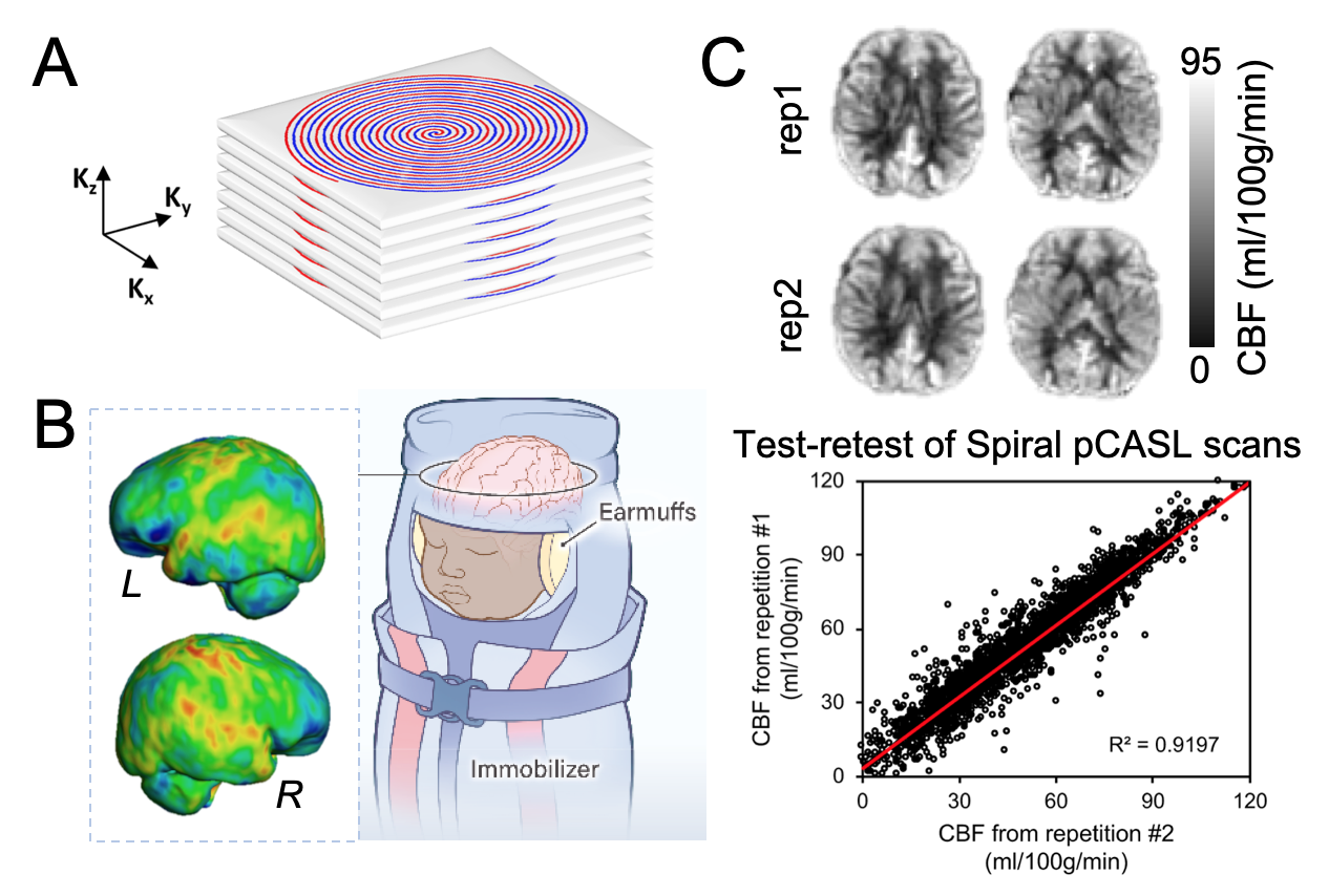
Approach: We optimized a state-of-the-art pseudo-continuous-arterial-spin-labeled (pCASL) sequence to obtain high-resolution spatiotemporal dynamics of infant CBF at isotropic 2.5mm.
Results: We revealed infant physiological heterogeneity and found the emergence of the limbic-sensorimotor-association cortical gradient based on CBF. Infant regional CBF changes were also associated with their improved real-world developmental functioning. These normative charts of infant CBF can serve as atlases for research and clinical care.
Impact: Capitalizing on a 3D multi-shot stack-of-spirals pCASL, we acquired the highest-resolution infant CBF maps available to date, discovered the emergence of the limbic-sensorimotor-association cortical gradient in infancy, and provide a standardized reference for infant CBF.
Introduction
Human infancy is characterized by striking changes in brain function and structure1,2. Infant cerebral blood flow (CBF) delivers glucose and oxygen to meet the brain’s energy demand for this fastest period of postnatal brain development across lifespan. However, the organizing principle of whole-brain CBF dynamics during infancy and its relationship with real-world developmental functioning remain unknown. Delineating infant regional CBF (rCBF) dynamics may provide insight into the physiological basis of brain and behavior development during this critical stage. The present study investigated the spatiotemporal patterns of rCBF from birth to 2 years of age, as well as associations between rCBF dynamics and infant developmental millstones. To achieve this goal, we optimized a cutting-edge pseudo-continuous arterial spin labeled (pCASL) sequence 3,4 with 3D multi-shot, stack-of-spirals readout and background suppression to obtain high-resolution rCBF at isotropic 2.5mm.Methods
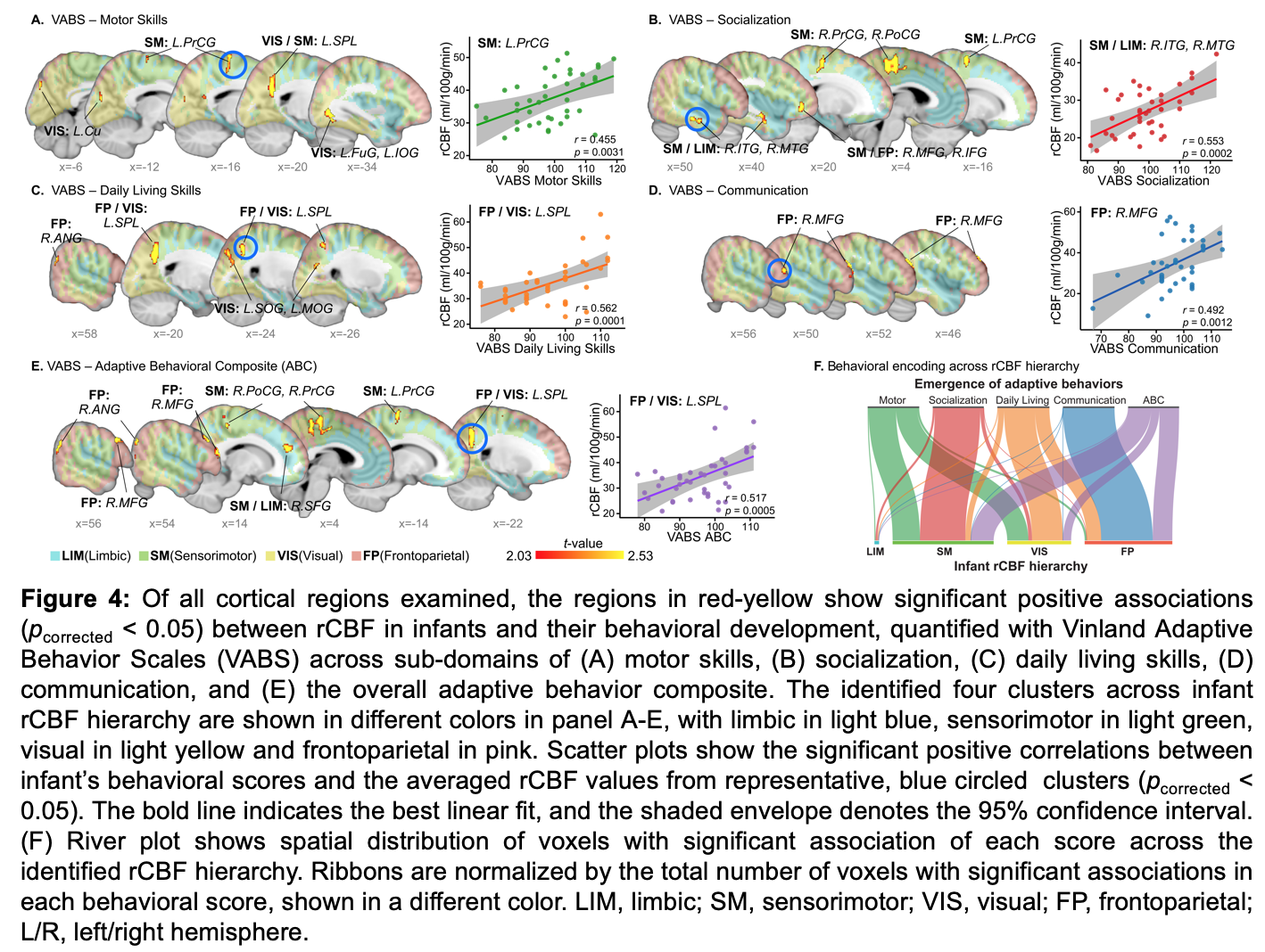
Infant subjects and acquisition of high-resolution pCASL MRI: High-resolution ASL images were acquired in fifty-two infants (17M/35F, age range: 0 to 28 months) with a 3D multi-shot, stack-of-spirals pCASL sequence (Fig. 1A) 3,4 in a 3T Siemens Prisma system. The pCASL MRI parameters were: four-shot acquisition, field-of-view = 192×192 mm2, matrix = 76×76, in-plane resolution = 2.53×2.53 mm2, 48 slices, slice thickness = 2.5 mm, no gap between slices, labeling duration=1600ms, post labeling delay = 1800ms, center of labeling slab located between cervical vertebrae C2 and C3, repetition time = 4s, echo time = 12ms, number of controls/labels = 10 pairs. High-resolution T1-weighted images were also acquired. All scans were conducted during infants’ natural sleep with earmuffs applied to reduce scanner sound (Fig. 1B). Real-world infant development: Infants’ adaptive behavior was quantified using the Vinland Adaptive Behavior Scales (VABS)5, which covers motor, socialization, daily living skills, and communication domains. rCBF quantification: The single-compartment model from the ASL white paper 6 was used to estimate rCBF from pCASL data. Developmental models of infant rCBF: To explore the effect of age on cerebral perfusion, all infants’ rCBF maps were mapped to a template space and voxel-wise age-related changes were modeled using logarithmic models with infant sex and in-scanner head motion as covariates. A data-driven clustering approach based on nonnegative matrix factorization 7 identified the cortical voxels with similar rCBF changing patterns. Associations of infant rCBF dynamics with adaptive behavior: To explore associations between infant rCBF and adaptive behavior, generalized additive models with penalized splines were used to account for linear and nonlinear effects of age, with sex and head motion as covariates.Results
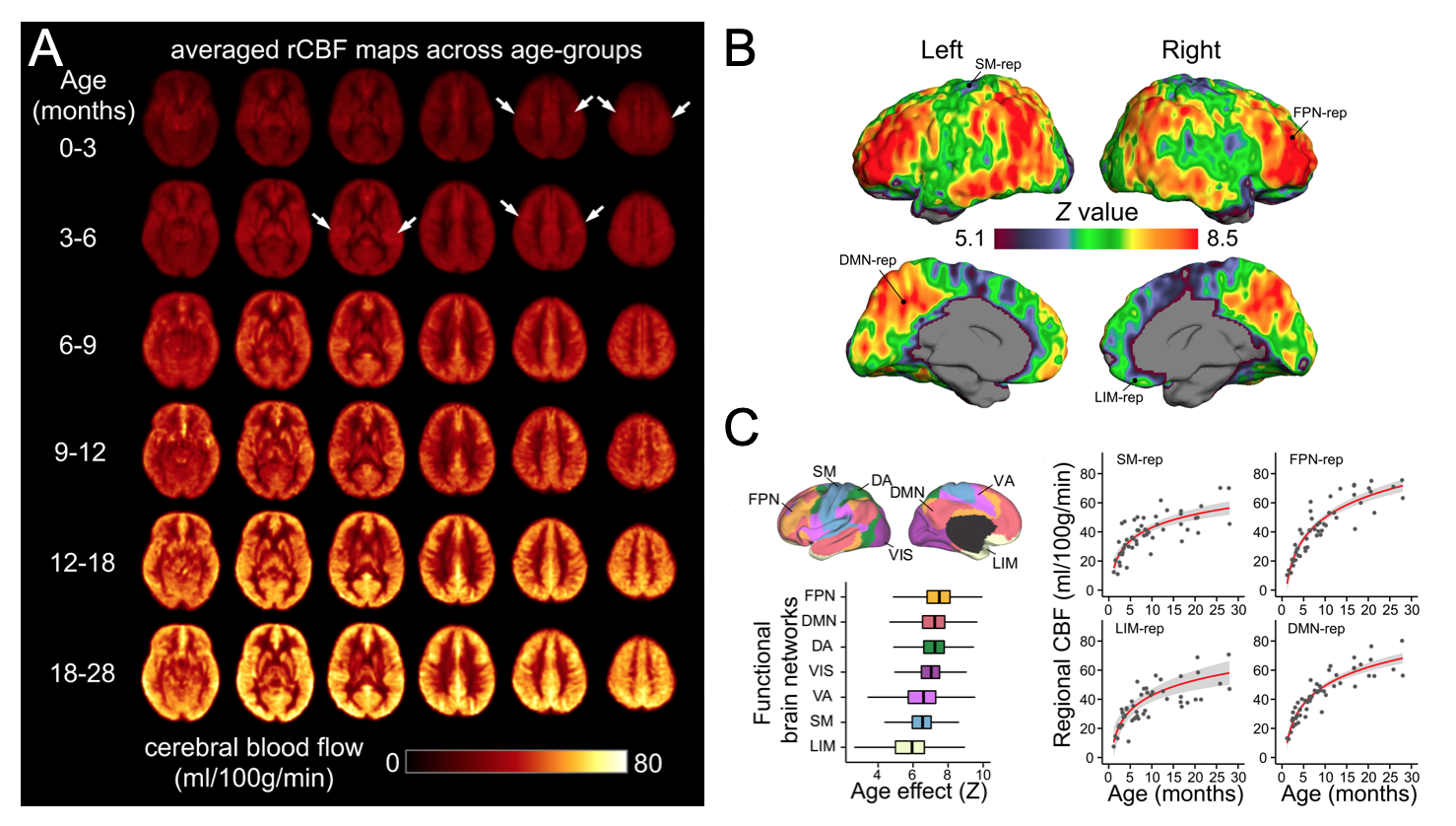
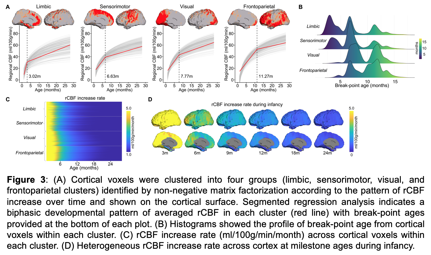
We scanned a healthy adult twice with identical pCASL protocol to examine test-retest reproducibility of rCBF (Fig. 1C). The intraclass correlation coefficient was 0.96 (Fig. 1C), indicating excellent reliability of rCBF maps obtained with the cutting-edge pCASL MRI (Fig. 1A). High-resolution rCBF maps uncover finer details of physiological heterogeneity throughout infancy (Fig. 2A). Whereas in younger infants high rCBF values are prominent in primary cortex, in older infants they are prominent in the association cortex. Significant age-related rCBF increases were nonuniform across the cortex (Fig. 2B, z > 5.1, Bonferroni corrected p < 0.05). Such age effects were unevenly distributed across the brain functional networks8 (Fig. 2C), with the largest rCBF increases occurring in frontoparietal and default-mode networks, and modest rCBF increases observed in sensorimotor and limbic networks. Furthermore, we discovered four rCBF clusters (i.e. limbic, sensorimotor, visual, frontoparietal clusters) along the hierarchical limbic-sensorimotor-association gradient with unique increase patterns (Fig. 3A). Within each cluster, averaged rCBF showed a biphasic growth pattern, with an initial rapid and then sequentially slower increases. RCBF clusters with a higher hierarchical level tended to have older break-point ages (e.g., frontoparietal cluster = 11.27months and sensorimotor cluster = 6.63months) (Fig. 3A-3B). Infant rCBF increase rates for all cortical voxels further support the limbic-sensorimotor-association gradient, with highest rates of rCBF increases observed before 6 months (Fig. 3C-3D). Finally, hierarchical rCBF changes were significantly associated with improved adaptive functioning across multiple domains, with rCBF increases in sensorimotor cortices associated with motor and socialization skills, and frontoparietal association cortices associated with communication and daily living skills (Fig. 4).Discussion and conclusion
Collectively, our findings provide the first clear view of physiological hierarchy along the limbic-sensorimotor-association gradient throughout infancy. The region-specific rCBF increases along this cortical gradient were associated with enhanced infant behaviors and development in a real-world setting. The identified infant physiological hierarchy aligns well with the pronounced regional differences in brain function e.g., 9,10, structure e.g., 1, 2,11 and metabolism e.g., 12, 13, providing crucial insights into the physiological basis of brain and behavioral development during this early developmental stage.Acknowledgements
This study is funded by NIH R01MH092535, R01MH125333, R01EB031284, R01MH129981, R21MH123930, R01EB031080, R01HD093776, R01MH107506, and P50HD105354.References
1. Ouyang M, Dubois J, Yu Q, Mukherjee P, Huang H, (2019). Delineation of early brain development from fetuses to infants with diffusion MRI and beyond. Neuroimage, 185: 836-850.
2. Huang, H, (2022). Imaging the Infant Brain. Oxford Research Encyclopedia of Psychology.
3. Chang YV, Vidorreta M, Wang Z, Detre JA, 2017. 3D-Accelerated, Stack-of-Spirals acquisitions and reconstruction of arterial spin labeling MRI. Magn Reson Med 78:1405-1419.
4. Vidorreta M, Wang Z, Chang YV, Wolk DA, Ferandez-Seara MA, Detre JA, 2017. Whole-brain background suppressed pCASL MRI with 1D-accelerated 3D RARE stack-of-spirals readout. PLoS one 12(8): e0183762.
5. Sparrow, S., Cicchetti, D. & Saulnier, C, (2016). Vineland adaptive behavior scales–third edition (Vineland-3). Circle Pines, MN: American Guidance Service.
6. Alsop DC, Detre JA, Golay X, Günther M, Hendrikse J, Hernandez-Garcia L, Lu H, et al, (2015). Recommended implementationof arterial spin-labeled perfusion MRI for clinical applications: a consensus of the ISMRM perfusion study group and the European consortium for ASL in dementia. Magn Reson Med 73:102–116.
7. Lee DD, Seung HS, (1999). Learning the parts of objects by non-negative matrix factorization. Nature 401:788-791.
8. Yeo, B. T. et al. (2011).The organization of the human cerebral cortex estimated by intrinsic functional connectivity. J Neurophysiol 106:1125-1165.
9. Cao, M. et al. (2017) Early Development of Functional Network Segregation Revealed by Connectomic Analysis of the Preterm Human Brain. Cereb Cortex 27, 1949-1963.
10. Gao, W., Alcauter, S., Smith, J. K., Gilmore, J. H. & Lin, W, (2015). Development of human brain cortical network architecture during infancy. Brain Struct Funct 220, 1173-1186.
11. Wang, F. et al. (2019) Developmental topography of cortical thickness during infancy. Proc Natl Acad Sci U S A 116, 15855-15860.
12. Chugani, H. T. & Phelps, M. E. (1986) Maturational changes in cerebral function in infants determined by 18FDG positron emission tomography. Science 231, 840-843.
13. Chugani, H. T., Phelps, M. E. & Mazziotta, J. C. (1987) Positron emission tomography study of human brain functional development. Ann Neurol 22, 487-497.
Figures