1150
3D/4D Ultrashort Echo Time Balanced-SSFP MR Lung Images Reconstructed Using XD-GRASP-Pro1Biomedical Engineering, University of Virginia, Charlottesville, VA, United States, 2Radiology and Medical Imaging, University of Virginia, Charlottesville, VA, United States, 3Health Technology and Informatics, Hong Kong Polytechnic University, Kowloon, Hong Kong, 4Physics, University of Virginia, Charlottesville, VA, United States
Synopsis
Keywords: Image Reconstruction, Sparse & Low-Rank Models
Motivation: Obtaining high-quality images of the lung using proton MRI is challenging due to breathing motion and the short T2* and low proton density of lung parenchyma.
Goal(s): Our goal was to demonstrate a free-breathing proton lung MRI approach that maximizes parenchyma and vessel signal in the lungs.
Approach: This method combines a 3D ultra-short echo time (UTE) balanced steady-state free precession (bSSFP) pulse sequence with a GRASP-Pro-based reconstruction algorithm applied to respiratory phase-binned data.
Results: Image quality was markedly better for SSFP images than for spoiled images, and end-of-exhalation frames reconstructed from 4D images compared favorably with respiratory-triggered images.
Impact: A UTE bSSFP radial pulse sequence combined with temporally-constrained reconstruction produces high-signal, high-resolution lung images at end-of-exhalation collected during free breathing. While non-end-of-exhalation reconstruction was less effective, a similar reconstruction algorithm that incorporates motion fields could improve results.
Introduction
Obtaining high-quality images of the lung using proton MRI is a significant technical challenge, due to the short T2* and low proton density of lung parenchyma and the constant motion of the lungs during breathing. We demonstrate a free-breathing proton lung MRI approach that combines a 3D ultra-short echo time (UTE) balanced steady-state free precession (bSSFP) spoke-radial pulse sequence with a GRASP-Pro-based reconstruction algorithm applied to respiratory phase-binned data. This pulse sequence takes advantage of the high signal even for short TR inherent in bSSFP techniques, as well as the ability to read out at low-frequency k-space locations immediately after excitation inherent in radial UTE techniques. The reconstruction approach exploits the natural sparsity of the sorted respiratory phase-binned image series in the respiratory-phase domain to de-noise highly under-sampled images at each phase.Methods
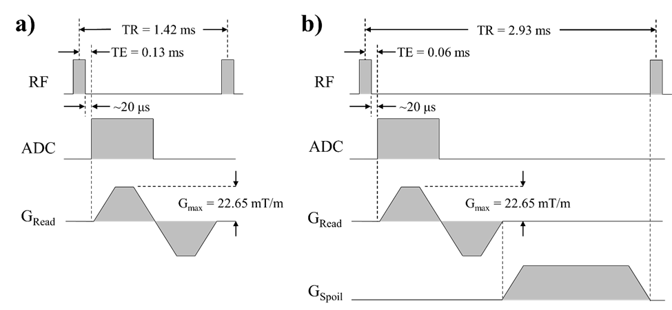
MR imaging was performed in 27 healthy subjects using a 1.5T scanner (Avanto; Siemens; Malvern, PA). Data was collected during free breathing using a 3D spoke-radial UTE bSSFP sequence1,2 (Fig. 1). A 3D spoke-radial UTE spoiled sequence identical to the UTE bSSFP sequence was used to collect separate images for comparison in 11 subjects. Spokes were organized into a spiral phyllotaxis pattern3,4, consisting of 898 passes that each included 304 individual spokes. Each pass was rotated azimuthally by a golden-angle increment of 137.51° with respect to the previous pass. Prior to the start of each pass, a 2D coronal navigator consisting of 51-101 spokes was collected. Pulse sequence parameters for bSSFP (spoiled) were as follows: TR = 1.42 (2.93) ms, TE = 0.13 (0.06) ms, flip angle = 25° (5°), matrix size = 256×256×256, resolution = 1.5 mm isotropic. Fully-sampled 3D images using identical pulse sequence parameters were also collected using prospective respiratory triggering for comparison with images collected during free breathing in 19 subjects.Coil sensitivity maps were retrospectively generated from the individual coil images using an iterative approach5. Passes collected during free breathing were retrospectively sorted into 25 respiratory phases by selecting a region of interest surrounding the diaphragm from the 2D navigator images and clustering these images via k-means6,7. Initial images corresponding to each respiratory phase were reconstructed using a multi-coil NUFFT8.
Denoised respiratory-resolved images were reconstructed in MATLAB (Mathworks; Natick, MA) using the XD-GRASP-Pro9,10 technique. Briefly, a low-resolution image series was reconstructed using XD-GRASP11 with a temporal total-variation constraint, and used as the basis to construct a lower-dimensional temporal subspace using the first 6 dominant PCA components. After obtaining this subspace from the low-resolution reconstruction, the subspace coefficients that represent the full-resolution image series under the subspace are found by performing an XD-GRASP-like optimization, using a temporal total-variation constraint applied to the subspace image representation and a spatial total-variation constraint applied to the subspace coefficient matrix.
Results
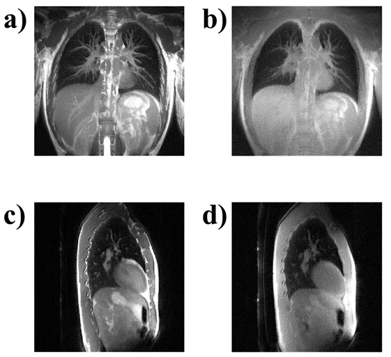
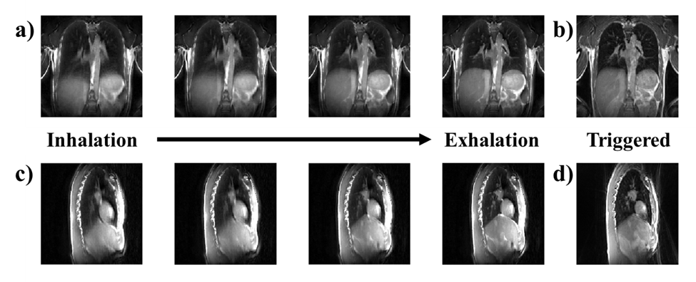
Fig. 2 depicts a comparison of UTE bSSFP and UTE spoiled images collected in the same individual. Higher vessel and parenchyma signal is observed for the UTE bSSFP than the UTE spoiled images, with vessel signal particularly improved in the UTE bSSFP images.Fig. 3 shows end-of-exhalation frames from a free-breathing UTE bSSFP compared with images collected using the same pulse sequence but with respiratory triggering. Similar image quality and feature resolution is observed between the two images.
Discussion
The UTE bSSFP pulse sequence displayed higher signal and feature contrast than the UTE spoiled sequence, particularly for pulmonary blood vessels. Banding artifacts characteristic of bSSFP12 are absent due to the short TR, avoiding one of the key downsides of bSSFP.MRI of the lung using prospective respiratory triggering is generally effective at freezing lung motion, but prolongs scan times significantly and only permits visualization of the lung at one respiratory phase. Images reconstructed from free-breathing data using a method that takes advantage of temporal sparsity can closely replicate fully-sampled images collected using respiratory triggering, as demonstrated here, permitting three-fold shorter scan times and allowing the possibility of reconstructing high-quality 4D images at several respiratory phases.
The images shown here were reconstructed using only k-space samples collected while moving from the k-space center to the periphery. A future version of this approach might seek to incorporate data sampled during the retracing of each ray from the k-space periphery to the center. Additionally, reconstruction of frames not taken at the end of exhalation might be improved by direct incorporation of frame-to-frame motion estimates into the reconstruction13,14.
Conclusion
The combined UTE bSSFP radial pulse sequence and XD-GRASP-Pro reconstruction approach demonstrated here produces high-quality images of the lung collected during free breathing. Future work will focus on improving reconstruction of non-end-of-exhalation respiratory phases.Acknowledgements
Research reported in this abstract was supported by the National Cancer Institute of the National Institutes of Health under award number R01 CA226899.References
1. Miller GW, Mugler III JP, Sá RC, Altes TA, Prisk GK, Hopkins SR. Advances in functional and structural imaging of the human lung using proton MRI. NMR Biomed. 2014;27(12):1542-1556.
2. Bauman G, Bieri O. Balanced steady-state free precession thoracic imaging with half-radial dual-echo readout on smoothly interleaved archimedean spirals. Magn Reson Med. 2020;84(1):237-246.
3. Piccini D, Littmann A, Nielles-Vallespin S, Zenge MO. Spiral phyllotaxis: The natural way to construct a 3D radial trajectory in MRI. Magn Reson Med. 2011;66(4):1049-1056.
4. Delacoste J, Chaptinel J, Beigelman-Aubry C, Piccini D, Sauty A, Stuber M. A double echo ultra short echo time (UTE) acquisition for respiratory motion-suppressed high resolution imaging of the lung. Magn Reson Med. 2018;79(4):2297-2305.
5. Inati SJ, Hansen MS, Kellman P. A fast optimal method for coil sensitivity estimation and adaptive coil combination for complex images. Proc Intl Soc Mag Reson Med. 2014;22:4407.
6. Lloyd S. Least squares quantization in PCM. IEEE Trans Inf Theory. 1982;28(2):129-137.
7. Arthur D, Vassilvitskii S. k-means++: the advantages of careful seeding. Proc Annu ACM-SIAM Symp Discrete Algorithms. 2007;18:1027-1035.
8. Fessler JA, Sutton BP. Nonuniform fast Fourier transforms using min-max interpolation. IEEE Trans Signal Process. 2003;51(2):560-574.
9. Feng L, Wen Q, Huang C, Tong A, Liu F, Chandarana H. GRASP-Pro: imProving GRASP DCE‐MRI through self-calibrating subspace-modeling and contrast phase automation. Magn Reson Med. 2020;83(1):94-108.
10. Feng L, Liu F. High spatiotemporal resolution motion-resolved MRI using XD-GRASP-Pro. In: Proc Intl Soc Mag Reson Med 28. Vol 28. ; 2020:597.
11. Feng L, Axel L, Chandarana H, Block KT, Sodickson DK, Otazo R. XD-GRASP: Golden-angle radial MRI with reconstruction of extra motion-state dimensions using compressed sensing. Magn Reson Med. 2016;75(2):775-788.
12. Scheffler K, Lehnhardt S. Principles and applications of balanced SSFP techniques. Eur Radiol. 2003;13(11):2409-2418.
13. Zhu X, Chan M, Lustig M, Johnson K, Larson P. Iterative motion compensation reconstruction ultra-short TE (iMoCo UTE) for high resolution free breathing pulmonary MRI. Magn Reson Med. 2020;83(4):1208-1221.
14. Miller Z, Johnson KM. Motion compensated self supervised deep learning for highly accelerated 3D ultrashort echo time pulmonary MRI. Magn Reson Med. 2023;89(6):2361-2375.
Figures