1148
High SNR rapid T1-weighted MP-RAGE and MP-FISP using a 3D stack-of-spirals trajectory at 0.55 T1Department of Biomedical Engineering, University of Southern California, Los Angeles, CA, United States, 2Ming Hsieh Department of Electrical and Computer Engineering, University of Southern California, Los Angeles, CA, United States
Synopsis
Keywords: Pulse Sequence Design, Low-Field MRI, structural brain imaging
Motivation: Acquiring High SNR T1-weighted MP-RAGE at low field strengths, such as 0.55T, often requires multiple averages due to reduced equilibrium polarization (~30 min for 3 averages).
Goal(s): Provide high SNR MP-RAGE (and MP-FISP) within a reasonable scan time (~15 min) using an SNR-efficient readout while mitigating spatial blurring caused by static off-resonance and concomitant fields.
Approach: MP-RAGE and MP-FISP sequences with a stack-of-spirals trajectory were implemented with the Pulseq framework. Spatial blurring was mitigated using the MaxGIRF framework implemented in BART.
Results: Spiral MP-RAGE achieves comparable image quality and higher SNR relative to Cartesian MP-RAGE given only half of the scan time.
Impact: This work demonstrates the feasibility of acquiring high SNR T1-weighted structural brain imaging at 0.55T within a reasonable scan time (~15 min). This opens opportunities for structural neuroimaging and harmonized multi-site studies via code sharing with open-source frameworks.
Introduction
3D T1-weighted magnetization-prepared rapid gradient-echo (MP-RAGE)1 is used to capture brain anatomy with excellent gray/white contrast and isotropic spatial resolution. It is part of nearly every neuroimaging protocol. However, acquiring high SNR T1-weighted MP-RAGE at low field strengths, such as 0.55T, often requires multiple averages due to reduced equilibrium polarization (~30 min for 3 averages), making it prone to motion artifacts. Wang et al2 recently demonstrated the benefits of spiral-based MP-RAGE, which are increased SNR and shorter scan time. Schäper et al3 recently demonstrated that magnetization-prepared fast imaging with steady-state free precession (MP-FISP) is a viable alternative to MP-RAGE at 0.55T, with improved gray/white matter contrast. Inspired by these studies, we investigate the feasibility of providing high SNR T1-weighted MP-RAGE and MP-FISP within a reasonable scan time (~15 min) using SNR-efficient long spiral readouts. The MaxGIRF reconstruction framework4 is used to mitigate spatial blurring caused by static off-resonance and concomitant fields at 0.55T. In the spirit of reproducible research, both pulse sequence and image reconstruction were implemented using open-source frameworks. We used the Pulseq framework5 to implement spiral MP-RAGE and MP-FISP sequences and the BART6,7 for MaxGIRF reconstruction.Methods
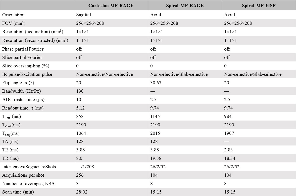
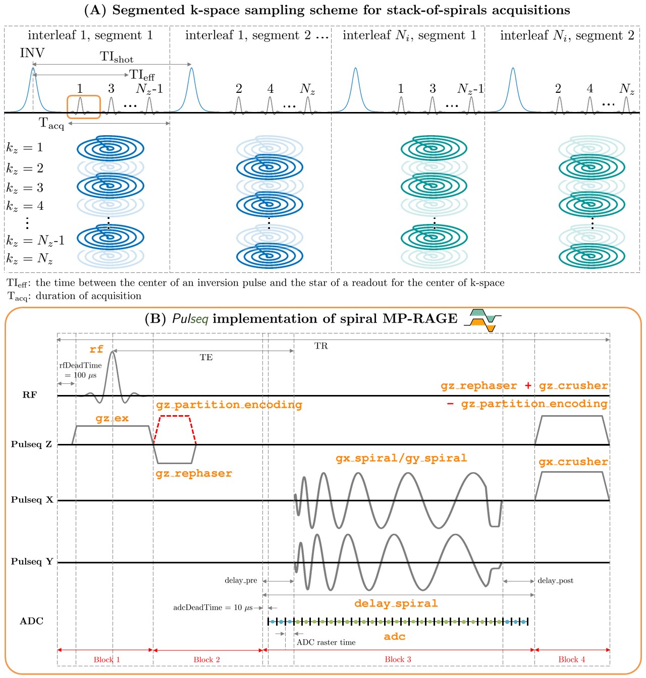
Pulse sequence: Spiral MP-RAGE and MP-FISP sequences were implemented with the Pulseq framework5. Figure 1 illustrates the segmented sampling scheme for stack-of-spirals acquisitions. A base 2D spiral gradient waveform was designed using a numerical algorithm8. A time-optimal 2D gradient rewinder waveform was designed9. Spiral imaging parameters were: FOV=256x256mm2, isotropic resolution=1.0x1.0mm2, Gmax=20mT/m, Smax=180 mT/m, and number of partition-encoding steps=208. Table 1 summarizes imaging parameters for all sequences.Spiral MP-RAGE: A gradient preparation scheme10 (6 cycles) was used for stabilization of eddy currents. An RF spoiled GRE (FLASH) was used. The flip angle of TR of spiral MP-RAGE was modified2 to match the T1 contrast of Cartesian MP-RAGE. A slab-selective excitation was used. Crusher gradients with 2π dephasing were added along the readout and slice-selection directions. An RF-spoiling phase increment of 117° was used.
Spiral MP-FISP: The (α/2-TR/2) preparation was used to reduce signal oscillations11. A phase increment of 180° per TR was used. A crusher with 4π dephasing was added along the slice-selection direction.
Experiments: All imaging experiments were performed on a whole-body 0.55T scanner (prototype MAGNETOM Aera; Siemens Healthineers, Erlangen, Germany) with gradients capable of 45mT/m amplitude and 200 T/m/s slew rate. A 16-channel head and neck array coil was used for signal reception. The “standard” shim setting was used. One healthy male volunteer was scanned under a protocol approved by our institutional review board after providing written informed consent.
Static off-resonance estimation: A single-echo 3D FLASH sequence was used to acquire datasets at different TEs (3.7,4.7,5.7,6.7,7.7 ms). Coil sensitivity maps were estimated from the first echo and applied to different TEs to perform optimal coil combination. For each voxel, phase unwrapping was performed, and linear least-squares fitting was used to estimate static off-resonance. A k-space filter based on a Sobolev norm12 was applied to enforce spatial smoothness on a static off-resonance map.
Center-frequency drift: A center frequency drift of ~60 Hz due to heavy gradient loading was compensated prior to image reconstruction13,14.
MaxGIRF image reconstruction: Density compensation factors were estimated15. A Cartesian FFT was applied along the kz dimension to enable a slice-by-slice image reconstruction. Coil sensitivity maps were estimated with ESPIRiT16 from gridded 3D k-space. A 2D MaxGIRF reconstruction4 was performed to mitigate spatial blurring caused by static off-resonance and concomitant fields17.
Results
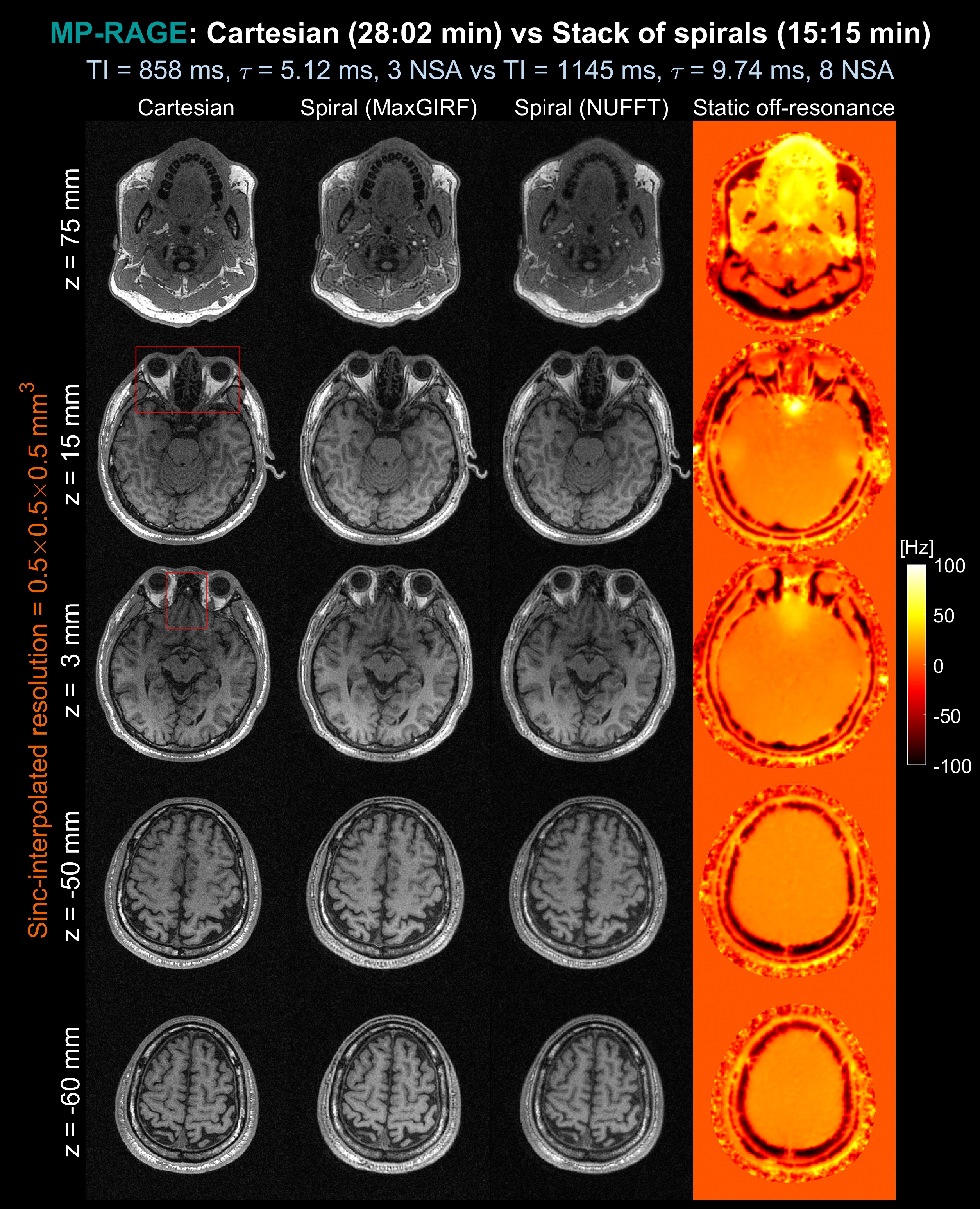
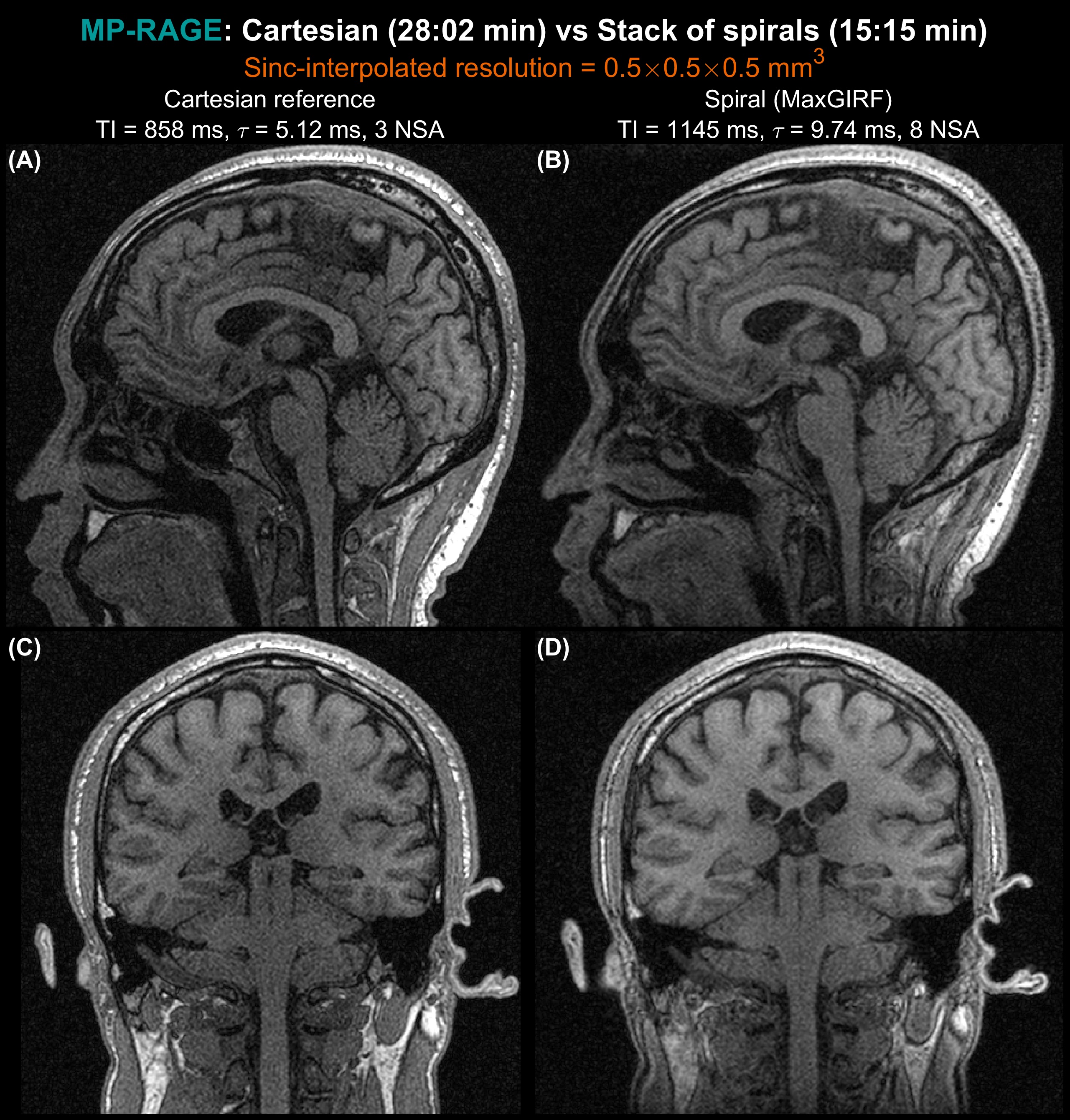
Figure 2 shows a comparison between spiral MP-RAGE (NUFFT and MaxGIRF) against Cartesian MP-RAGE. Spatial blurring due to concomitant fields increases as a distance from isocenter increases, and severely degrades image quality when a slice offset is greater than 40 mm. The MaxGIRF framework successfully mitigates spatial blurring caused by static off-resonance and concomitant fields.Figure 3 shows a comparison between spiral MP-RAGE (MaxGIRF) against Cartesian MP-RAGE using sagittal and coronal reformats. Spiral MP-RAGE achieves comparable image quality and higher SNR relative to Cartesian MP-RAGE given only half of the scan time. A slight reduction in spatial resolution was observed due to the difference in the volume of k-space coverage8,18.
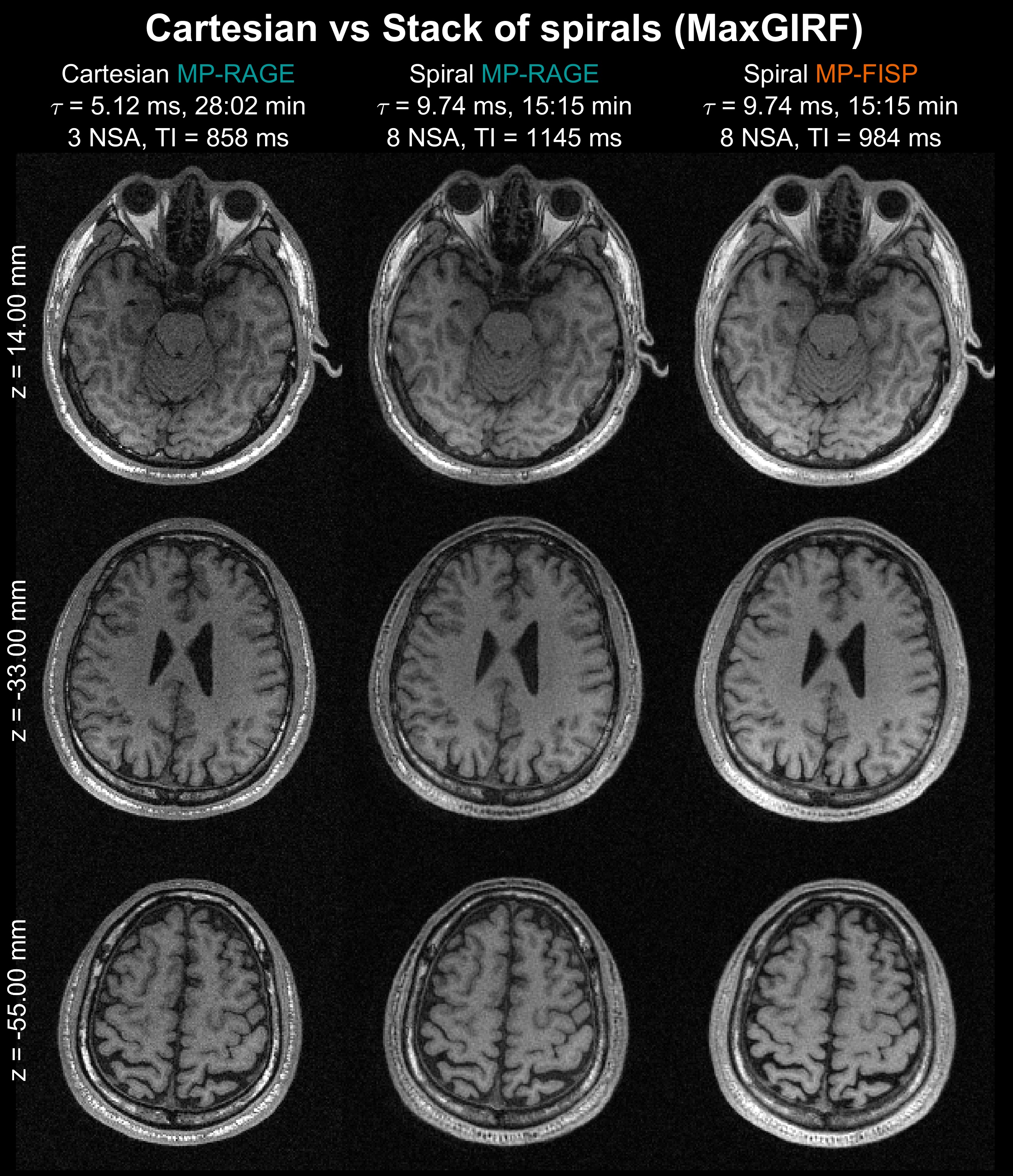
Figure 4 shows a comparison between Cartesian MP-RAGE, spiral MP-RAGE, and spiral MP-FISP. Spiral MP-FISP shows visually improved SNR without noticeable improvements in CNR at TI = 984 ms.
Discussion and Conclusion
We have successfully demonstrated high SNR T1-weighted structural brain imaging with an SNR-efficient spiral trajectory within a reasonable scan time at 0.55T. Multiple spiral acquisitions inevitably causes center-frequency drift and thus it is desirable to have a short f0 navigator14 after each NSA to track center-frequency drift.Acknowledgements
We acknowledge grant support from the National Science Foundation (#1828736) and research support from Siemens Healthineers.References
1. Mugler JP, Brookeman JR. Three-dimensional magnetization-prepared rapid gradient-echo imaging (3D MP RAGE). Magn Reson Med. 1990;15(1):152-157. doi:10.1002/MRM.1910150117
2. Wang D, Robison RK, Li Z, Pipe JG. High SNR rapid T1-weighted MPRAGE using spiral imaging with long readouts and improved deblurring. Magn Reson Med. 2023;89(3):951-963. doi:10.1002/MRM.29492
3. Schäper J, Bauman G, Bieri O. Improved gray-white matter contrast using magnetization prepared fast imaging with steady-state free precession (MP-FISP) brain imaging at 0.55 T. Magn Reson Med. 2023. doi:10.1002/MRM.29838
4. Lee NG, Ramasawmy R, Lim Y, Campbell-Washburn AE, Nayak KS. MaxGIRF: Image reconstruction incorporating concomitant field and gradient impulse response function effects. Magn Reson Med. 2022;88(2):691-710. doi:10.1002/MRM.29232
5. Layton KJ, Kroboth S, Jia F, et al. Pulseq: A rapid and hardware-independent pulse sequence prototyping framework. Magn Reson Med. 2017;77(4):1544-1552. doi:10.1002/MRM.26235
6. Berkeley Advanced Reconstruction Toolbox. Proc. Intl. Soc. Mag. Reson. Med. 23. (2015). p2486.
7. Tamir JI, Ong F, Cheng JY, Uecker M, Lustig M. Generalized Magnetic Resonance Image Reconstruction using The Berkeley Advanced Reconstruction Toolbox. doi:10.5281/zenodo.31907
8. Pipe JG, Zwart NR. Spiral trajectory design: A flexible numerical algorithm and base analytical equations. Magn Reson Med. 2014;71(1):278-285. doi:10.1002/MRM.24675
9. Hargreaves BA, Nishimura DG, Conolly SM. Time-optimal multidimensional gradient waveform design for rapid imaging. Magn Reson Med. 2004;51(1):81-92. doi:10.1002/MRM.10666
10. Mugler JP, Brookeman JR. Rapid three-dimensional T1-weighted MR imaging with the MP-RAGE sequence. J Magn Reson Imaging. 1991;1(5):561-567. doi:10.1002/JMRI.1880010509
11. Deimling M, Heid O. Magnetization prepared True FISP imaging. Proc. Intl. Soc. Mag. Reson. Med. 2 (1994). p 495.
12. Tan Z, Voit D, Kollmeier JM, Uecker M, Frahm J. Dynamic water/fat separation and [Formula: see text] inhomogeneity mapping-joint estimation using undersampled triple-echo multi-spoke radial FLASH. Magn Reson Med. 2019;82(3):1000-1011. doi:10.1002/MRM.27795
13. Intra-Scan Center Frequency Drift Correction for 3D
Spiral Exams. Proc. Intl. Soc. Mag. Reson. Med. 25 (2017). p1482.
14. Spiral Deblurring Using B0 Maps with B0 Drift
Correction. Proc. Intl. Soc. Mag. Reson. Med. 24. (2016). p1760.
15. Zwart NR, Johnson KO, Pipe JG. Efficient sample density estimation by combining gridding and an optimized kernel. Magn Reson Med. 2012;67(3):701-710. doi:10.1002/MRM.23041
16. Uecker M, Lai P, Murphy MJ, et al. ESPIRiT — An Eigenvalue Approach to Autocalibrating Parallel MRI: Where SENSE meets GRAPPA. Magn Reson Med. 2014;71(3):990. doi:10.1002/MRM.24751
17. Bernstein MA, Zhou XJ, Polzin JA, et al. Concomitant gradient terms in phase contrast MR: Analysis and correction. Magn Reson Med. 1998;39(2):300-308. doi:10.1002/MRM.1910390218
18. Van Gelderen P. Comparing True Resolution in Square versus Circular K-space Sampling. Proc. Intl. Soc. Mag. Reson. Med. 6, (1998). p1424.
Figures