1146
Ultra-Short Echo-Time Based Accelerated Four-Dimensional Rosette J-resolved Spectroscopic Imaging (4D UTE-ROS-JRESI): A pilot study1Radiological Sciences, University of California, Los Angeles, Los Angeles, CA, United States, 2College of Health and Human Sciences, Purdue University, West Lafayette, IN, United States, 3School of Nursing, University of California, Los Angeles, Los Angeles, CA, United States
Synopsis
Keywords: Pulse Sequence Design, Spectroscopy, UTE, Rosette Spectroscopic Imaging, 4D JRESI
Motivation: Two-dimensional spectrum resolves information-coupled metabolites along an additional spectral dimension. However, acquisition after adding the 2nd spectral encoding can increase the total acquisition time significantly.
Goal(s): To achieve clinically feasible runtimes for J-resolved Spectroscopic Imaging (JRESI) using Ultra-Short Echo-Time (UTE) based sequence implementation.
Approach: Implement a UTE based 4D JRESI sequence with rosette readout, which would enable shorter repetition times and use of higher undersampling factors.
Results: This study demonstrated in-vivo 4D UTE-ROSE-JRESI in less than 10 minutes as compared to a semi-laser sequence with a TR of 1.5 seconds which will take 17 minutes for the same sequence.
Impact: With higher incoherence level of sampling patterns rosette based spectroscopic imaging sequence showed the potential for highly accelerated acquisitions. Using UTE based rosette 4D J-resolved Spectroscopic Imaging sequence allowed further reduction in scan time with the help of shorter TR.
Introduction
MR spectroscopy (MRS) is an efficient biochemical tool for non-invasively analyzing metabolite and lipid concentrations in human tissues (1-4). Compared to one dimensional spectrum, two-dimensional spectral variant resolves peak information along an additional spectral dimension which helps to disperse the spectrum better (1-4). However, acquisition of MRSI after adding the 2nd spectral encoding can increase the total acquisition time significantly. Even though k-space-weighted and average-weighted schemes have been used to shorten the total duration of MRSI, echo-planar spectroscopic imaging (EPSI) showed further acceleration of the total acquisition duration (5,6). Undersampling the spatial and 2nd spectral dimension is essential to achieve clinically feasible scan times. Compressed sensing (CS) based reconstruction techniques are known to be capable of recovering the signal depending on the signal sparsity and incoherent sampling patterns (7). However, ultra-short echo-time (UTE) based sequences for non-cartesian J-resolved spectroscopic imaging has not been attempted so far. In this pilot study, we implemented a four-dimensional (4D) UTE based rosette echo-planar J-resolved spectroscopic imaging (4D UTE-ROSE-JRESI) sequence and studied the feasibility of achieving clinically feasible runtimes.Materials and Methods
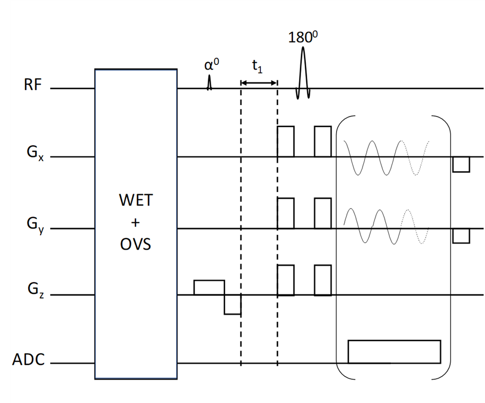
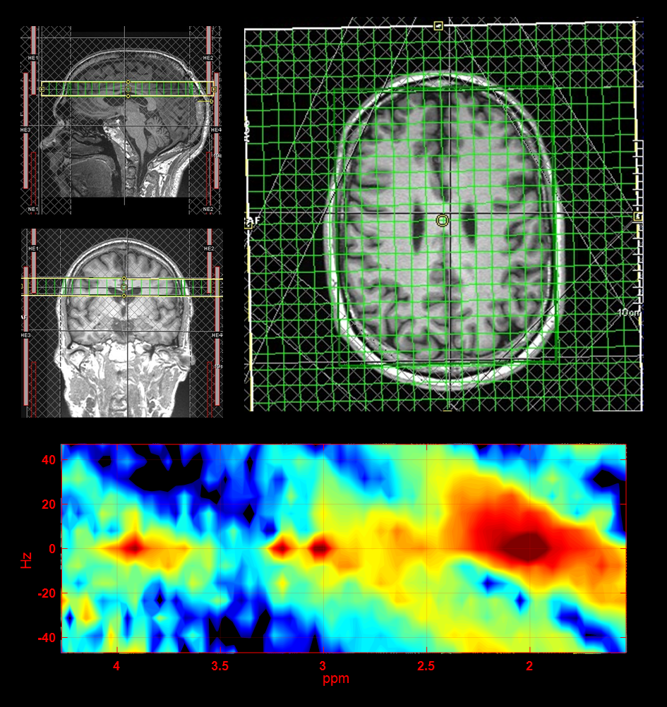
The 4D UTE-ROSE-JRESI sequence was designed for the Siemens VE11C (Prisma) platform as a low flip angle (FA) slab-selective RF pulse followed by an 1800 hard pulse and rosette (8) readout gradients. The 1800 pulse is incremented to encode the second spectral dimension (t1). Shown in Fig.1 is a schematic diagram of the 4D UTE-ROS-JRESI sequence. Outer Volume Suppression (OVS) and Water Suppression Enhanced Through T1 Effects (WET) methods (9) were used to suppress the signals from fat (outside region of interest (ROI)) and water. Data from a brain phantom containing metabolites at physiological concentrations was acquired. A 61-year-old healthy volunteer was recruited with IRB approval for the acquisition of in vivo brain data. This scan was acquired with a 32×32 matrix size, a 24×24×2 cm3 slab, TE=3.9ms, TR=800ms, FA = 48° and a spectral width of 1250 Hz, spatially interleaved 11 petals, with 512 t2 points, 32 t1 points (2x undersampled) and 2 averages. This amounts to a total of 6-7x acceleration based on the Nyquist criterion for rosette sampling. Total scan time was 9 minutes and 26 seconds. A separate non-water suppressed (NWS) data was acquired for coil combination and eddy current phase correction with only 1 t1 point in 12 seconds. CS reconstruction using Perona-Malik (PM) (10-13) and non-uniform FFT (nuFFT) (14) was used to estimate the missing samples of k-space. ProFit quantitation was used to quantify the resulting spectra (15).Results
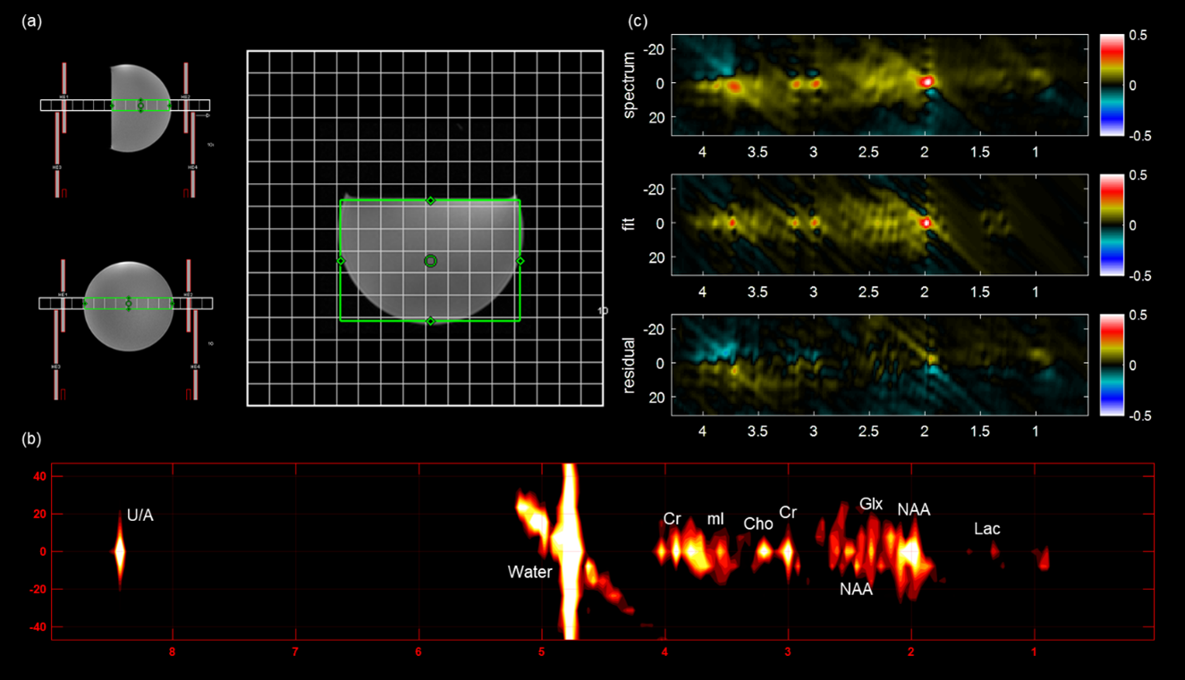
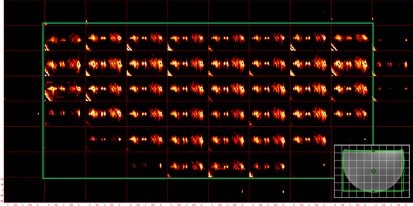
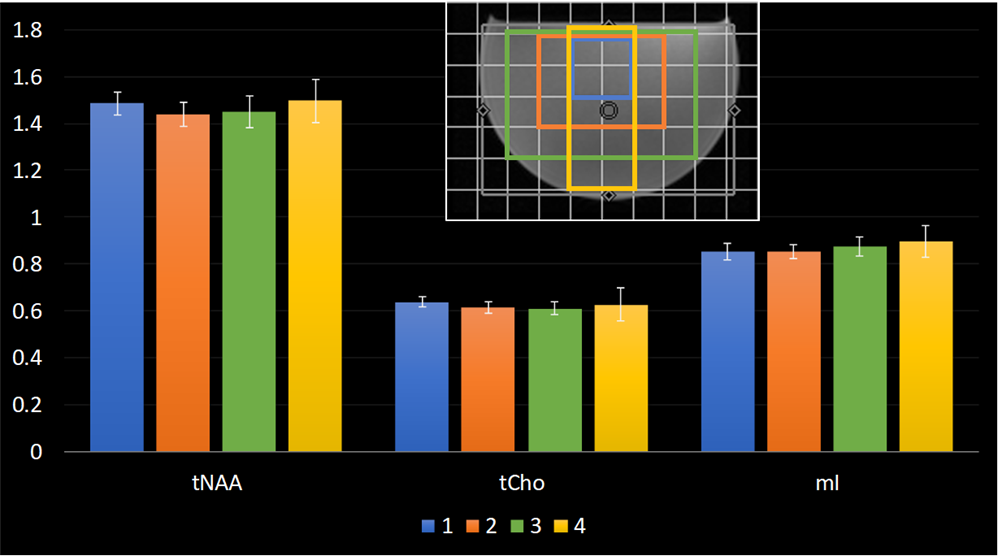
Figure 1 shows a schematic pulse sequence diagram for proposed 4D UTE-ROS-JRESI. Figure 2 shows (a) localizer images of phantom data and (b) an extracted spectrum along with (c) the results of profit quantitation. Multivoxel spectra showing all voxels in the phantom are depicted in Figure 3. A bar chart comparing metabolite ratios with respect to Creatine 3.0 (Cre3.0) across multiple voxels is shown in Figure 4. Sagittal, axial and coronal localization images, as well as an extracted spectrum from the in-vivo data is shown in Figure 5.Discussion
UTE based rosette J-resolved 4D spectroscopic imaging was implemented and tested using phantom and in-vivo datasets. UTE allows the use of shorter TR which greatly decrease the total scan time. This study demonstrated 4D UTE-ROSE-JRESI in less than 10 minutes as compared to a semi-laser sequence with a TR of 1.5 seconds which will take 17 minutes for the same sequence. Metabolite ratios stayed within relatively same range across different regions in the phantom scan. Total NAA (tNAA) ratio was slightly overestimated. OVS side bands were required to suppress the signal from Intracranial lipid signal due to lipid bleed. Another solution would be to use a higher spatial resolution at the expense of scan time or remove the lipid signal during post processing.Conclusion
Due to the higher incoherence level of sampling patterns based on rosette trajectories, rosette based spectroscopic imaging sequence has the potential for highly accelerated acquisitions. Using UTE based rosette 4D J-resolved Spectroscopic Imaging sequence allows further reduction in scan time with the help of shorter TR. Feasibility of the technique using both phantom and in-vivo scans are demonstrated.Acknowledgements
Authors acknowledge grants support from National Institute of Health (5R21MH125349-02 and 5R01HL135562-04).References
1. Thomas MA, Ryner LN, Mehta MP, Turski PA, Sorenson JA. Localized 2D J‐resolved H MR spectroscopy of human brain tumors in vivo. Journal of Magnetic Resonance Imaging. 1996 May;6(3):453-9.
2. Thomas MA, Ryner LN, Mehta MP, Turski PA, Sorenson JA. Localized 2D J‐resolved H MR spectroscopy of human brain tumors in vivo. Journal of Magnetic Resonance Imaging. 1996 May;6(3):453-9.
3. Ludwig C, Viant MR. Two‐dimensional J‐resolved NMR spectroscopy: review of a key methodology in the metabolomics toolbox. Phytochemical Analysis: An International Journal of Plant Chemical and Biochemical Techniques. 2010 Jan;21(1):22-32.
4. Schulte RF, Lange T, Beck J, Meier D, Boesiger P. Improved two‐dimensional J‐resolved spectroscopy. NMR in Biomedicine: An International Journal Devoted to the Development and Application of Magnetic Resonance In vivo. 2006 Apr;19(2):264-70.
5. Mansfield P. Spatial mapping of the chemical shift in NMR. Magn Reson Med 1984;1(3):370-86.
6. Posse S, Otazo R, Caprihan A, Bustillo J, Chen H, Henry PG, et al. Proton echo‐planar spectroscopic imaging of J‐coupled resonances in human brain at 3 and 4 Tesla. Magn Reson Med 2007;58(2):236-44.
7. Furuyama JK, Wilson NE, Burns BL, Nagarajan R, Margolis DJ, Thomas MA. Application of compressed sensing to multidimensional spectroscopic imaging in human prostate. Magn Reson Med 2012;67(6):1499-505.
8. Shen X, Özen AC, Sunjar A, Ilbey S, Sawiak S, Shi R, Chiew M, Emir U. Ultra-short T2 components imaging of the whole brain using 3D dual-echo UTE MRI with rosette k-space pattern. Magnetic Resonance in Medicine. 2022 Sep 25.
9. Ogg RJ, Kingsley RB, Taylor JS. WET, a T1-and B1-insensitive water-suppression method for in vivo localized 1H NMR spectroscopy. Journal of Magnetic Resonance, Series B. 1994 May 1;104(1):1-0.
10. Joy A, Paul JS. Multichannel compressed sensing MR image reconstruction using statistically optimized nonlinear diffusion. Magn Reson Med 2017;78(2):754-62.
11. Joy A, Paul JS. A mixed-order nonlinear diffusion compressed sensing MR image reconstruction. Magn Reson Med 2018;80(5):2215-22.
12. Joy A, Jacob M, Paul JS. Compressed sensing MRI using an interpolation‐free nonlinear diffusion model. Magn Reson Med 2021;85(3):1681-96.
13. Joy A, Jacob M, Paul JS. Directionality guided non linear diffusion compressed sensing MR image reconstruction. Magn Reson Med 2019;82(6):2326-42.
14. Fessler JA, Sutton BP. Nonuniform fast Fourier transforms using min-max interpolation. IEEE transactions on signal processing. 2003 Jan 22;51(2):560-74.
15. Schulte RF, Boesiger P. ProFit: two‐dimensional prior‐knowledge fitting of J‐resolved spectra. NMR in Biomedicine: An International Journal Devoted to the Development and Application of Magnetic Resonance In vivo. 2006 Apr;19(2):255-63.
Figures