1144
Efficient T2 and diffusion weighted imaging using the multiple-echo steady-state (MESS) sequence with a 3D PROPELLER acquisition1Department of Radiology and Nuclear Medicine, St. Olav's University Hospital, Trondheim, Norway, 2Department of Circulation and Medical Imaging, NTNU - Norwegian University of Science and Technology, Trondheim, Norway, 3Division of Medical Physics, Department of Diagnostic and Interventional Radiology, University Medical Center Freiburg, Faculty of Medicine, University of Freiburg, Freiburg, Germany
Synopsis
Keywords: Pulse Sequence Design, Pulse Sequence Design, Diffusion, New Trajectories
Motivation: The multiple-echo steady-state (MESS) sequence extends double-echo steady state and efficiently measures multiple images with different contrasts. Because diffusion contrasts are important in clinical imaging, this study aims to include strong diffusion weighting in the MESS sequence.
Goal(s): To extend the MESS sequence to provide distortion-free, high-resolution 3D T1-, T2- and diffusion-weighted imaging.
Approach: The DW-MESS sequence utilizes a 3D EPI-PROPELLER acquisition to acquire 32 echoes per repetition. An incoherent non-Cartesian k-space trajectory enables reconstruction of individual echoes.
Results: The DW-MESS images show contrasts comparable to SE-EPI with anterior-posterior diffusion encoding. Artifacts are present in other diffusion directions, and require further phase corrections.
Impact: This study enables stronger diffusion-weighting in DESS-type sequences in addition to T1- and T2-weighting. The incoherent k-space trajectory allows reconstruction of a B0-map and coil-sensitivities, and could allow T2* and susceptibility quantification. This provides new opportunities for efficient multi-parametric imaging.
Purpose
The double-echo steady-state (DESS) is a steady-state free-precession (SSFP) pulse sequence that acquires both a FID-like echo (S+) and a spin-echo-like echo (S-), which have substantially different T2 and/or diffusion weighting1-4. Measuring multiple echoes enables quantification of other parameters, such as T15 or water-fat fractions in MESS6. Diffusion-weighted DESS is distortion free, and can reach higher resolutions than EPI-based diffusion imaging7. However, DW-DESS is very sensitive to artifacts from motion and flow, which can be partially resolved with self-navigated non-Cartesian imaging5,8.Here, we propose a diffusion-weighted multiple-echo steady-state (DW-MESS) sequence with non-Cartesian sampling. In our sequence, every individual echo forms an incoherently undersampled k-space, and can be reconstructed separately to correct for B0-induced dephasing. This enables imaging with longer echo trains, which allows for longer repetition times and thus stronger T2- and diffusion-weighting, with high scan efficiency.
Methods
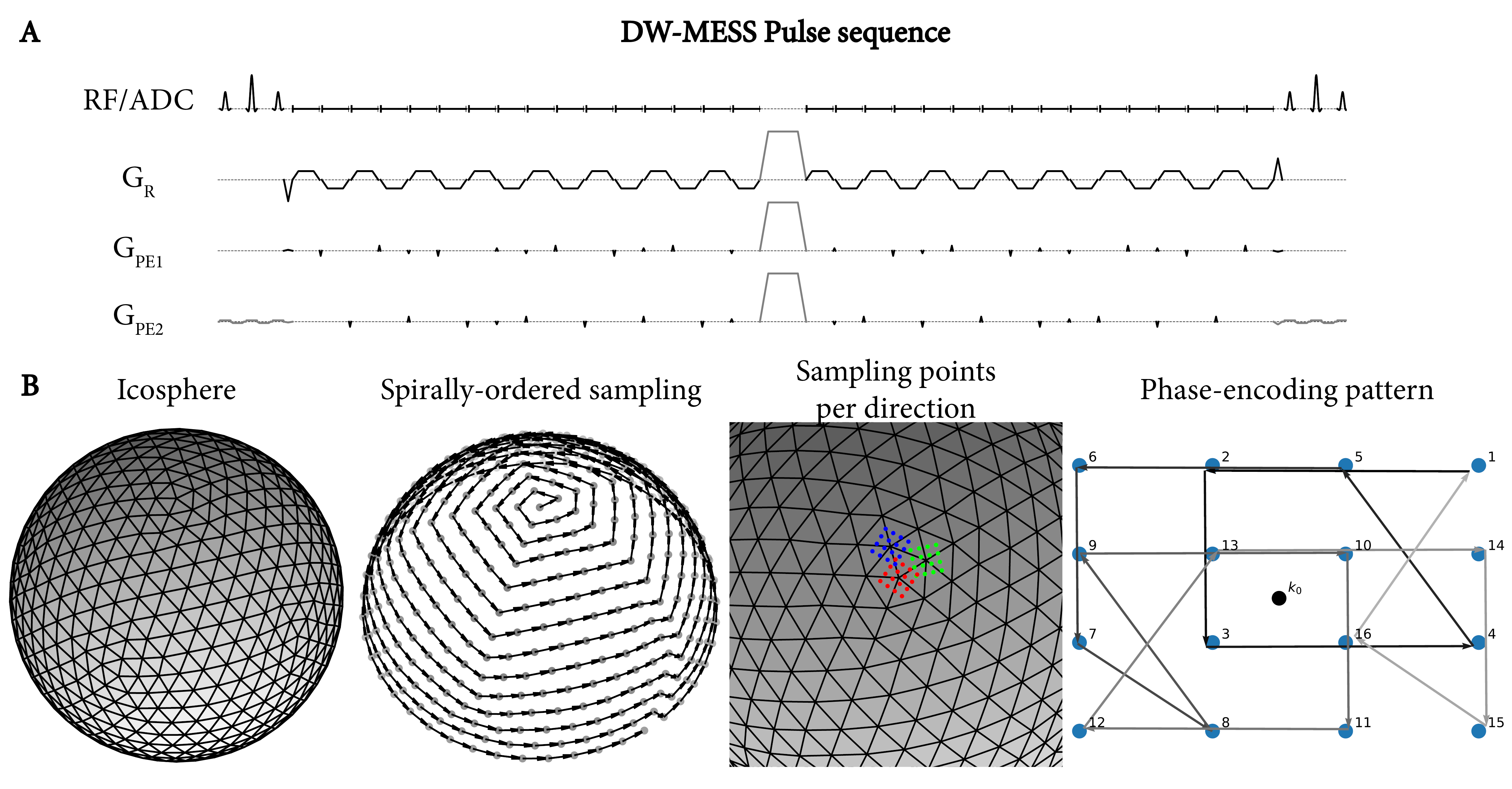
AcquisitionOur sequence extends the PROPELLER9 sequence to 3D by phase-encoding$$$\:4\times4\times\:N_x\:$$$beams, which are rotated in 3D according to a spirally-ordered subdivided icosahedron (icosphere) pattern (Figure 1B). Each beam is sampled with an EPI-DESS sequence with 16 S+ echoes and 16 S- echoes, separated by a spoiler gradient (Figure 1A). In order for each separate echo to have a uniform sampling pattern with incoherent aliasing, we rotated the phase-encoding beam around the readout direction with golden-angle increments (Figure 1B), and cycled the phase-encoding pattern to sample each phase-encode uniformly across echo times.
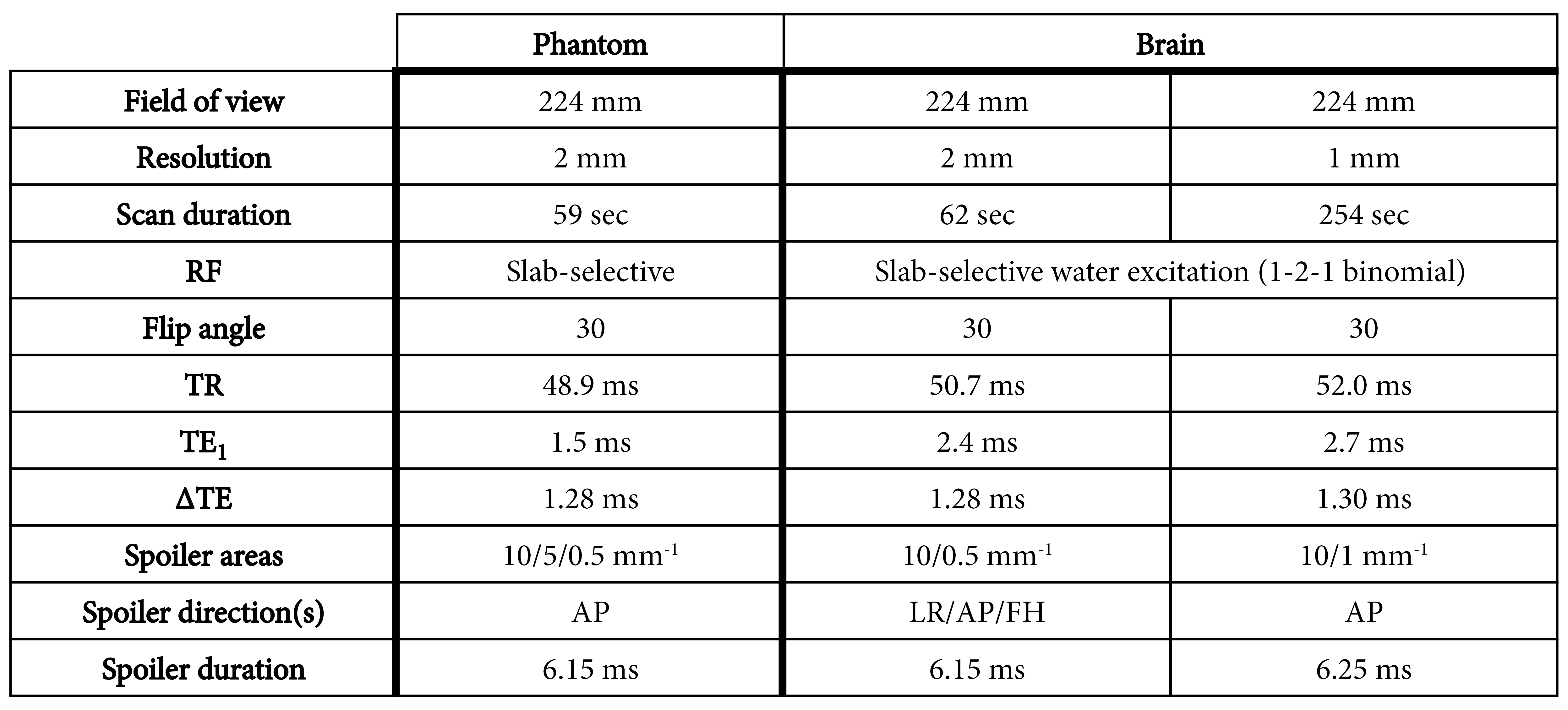
The sequence was implemented using pyPulseq10 and measurements were performed at 3 tesla (Trio, Siemens, Erlangen, Germany) using a 12-channel head coil. Both a phantom and in vivo scans of the brain of a volunteer were acquired (in accordance with institutional guidelines). Figure 2 lists all relevant scan parameters for the DW-MESS sequences. T1-weighted gradient echo and SE-EPI diffusion scans were acquired for reference.
Reconstruction
Reconstruction of the non-Cartesian 3D PROPELLER data was performed using the following steps:
- Raw data was coil-compressed11 to 4 channels.
- 1D regridding in the readout direction to remove ramp-sampling and oversampling.
- 1D Phase correction to correct for shifts between even and odd EPI readout lines.
- Removal of average phase in S- data.
- Reconstruction of all 16 S+ echoes for all receive channels separately using the adjoint NUFFT12 operator with sampling density correction13.
- Estimation of B0-induced phase from phase differences between each echo:$$b_0=exp(i\:arg\sum_{n,c}I_{n,c}\overline{I_{n-1,c}})$$(where$$$\:I_{n,c}\:$$$is the reconstruction of the$$$\:n$$$’th echo and$$$\:c$$$’th receive channel).
- B0-corrected reconstruction of the S+ image, separated by receive channel:$$R_c=\sum_{n}I_n(b_0)^{-n}$$
- Coil sensitivity calculation from R using Inati’s method14.
- B0-corrected SENSE reconstruction of both S+ and S- using the LSMR algorithm15. We solved the damped least squares problem:$${min}_x||WFSBx-Wy||+||x||$$(where$$$\:B\:$$$is the B0-induced phase,$$$\:S\:$$$is the coil sensitivity matrix,$$$\:F\:$$$is the NUFFT operator,$$$\:W\:$$$are the sampling density correction weights, and$$$\:y\:$$$is the acquired data)
Results
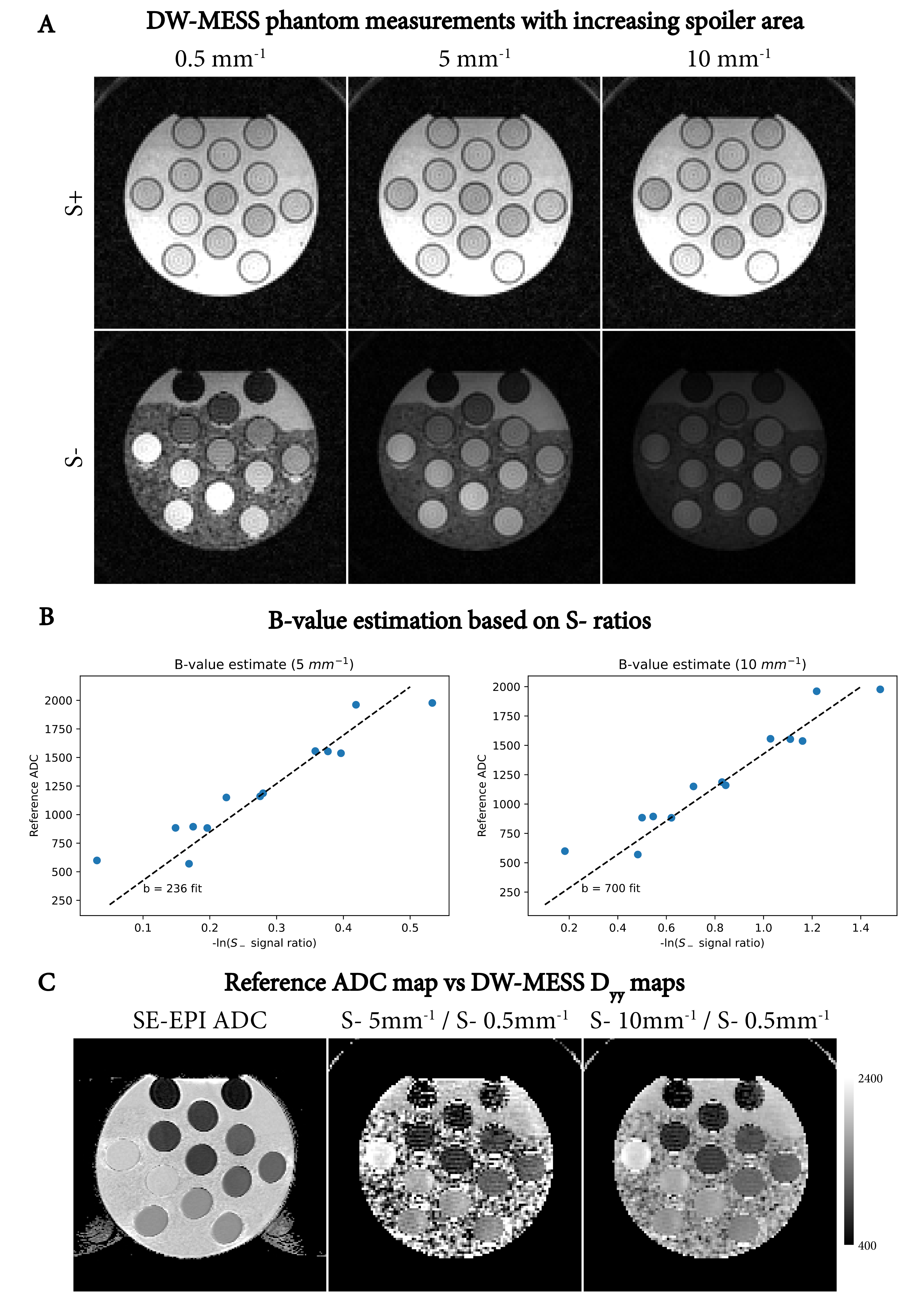
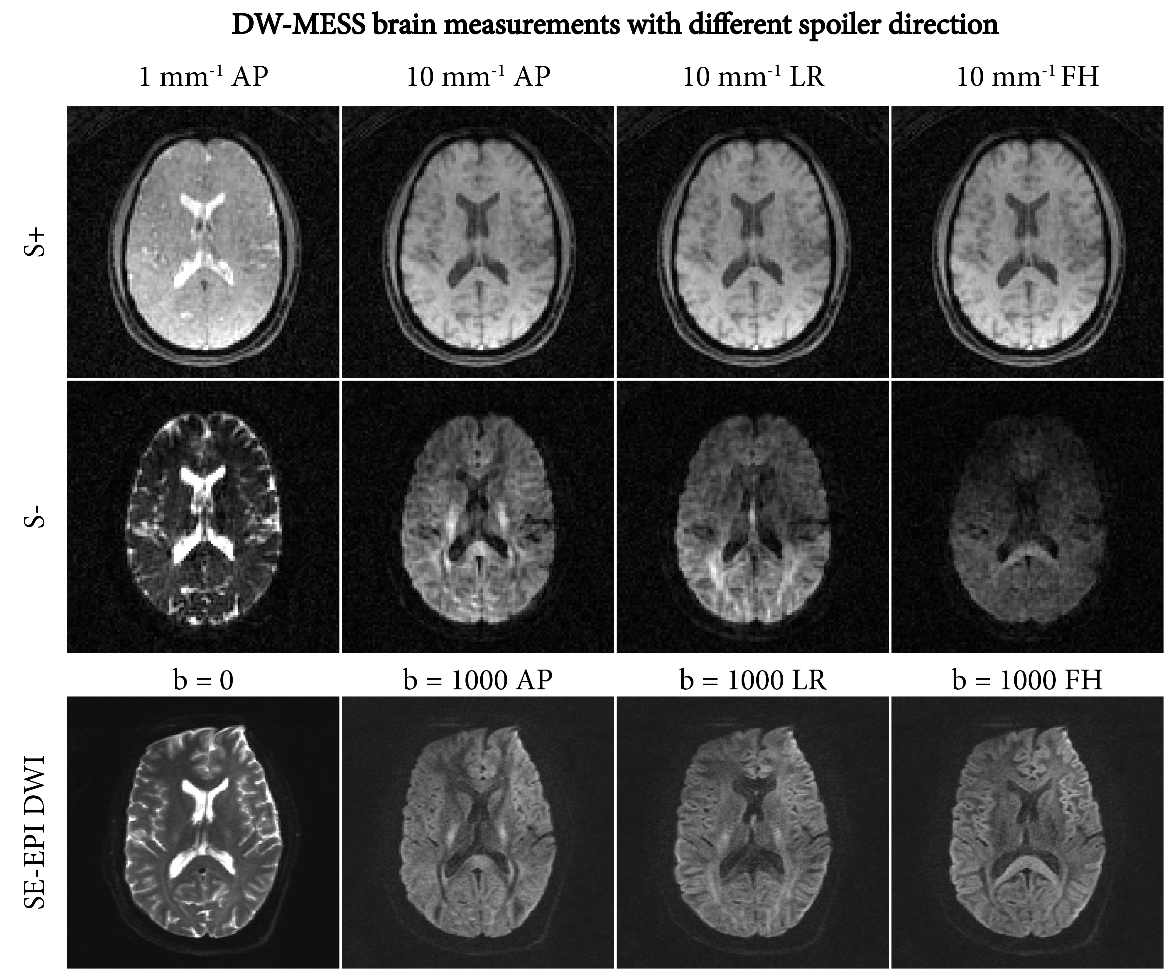
Figure 3 shows DW-MESS images of a diffusion phantom with different spoiler areas. Based on the ratios of the S- signals, we estimated the effective b-values to be ~236 for 5mm-1 spoiler area and ~700 for 10mm-1 spoiler area. Using two DW-MESS acquisitions, diffusion maps can be calculated based on their S- signal ratios (Figure 3C).Figure 4 shows DW-MESS images of the brain with 2mm isotropic resolution with different diffusion encoding directions. Interestingly, the S+ echo is also influenced by the spoiler strength, showing a proton density contrast for low spoiling area and T1-weighted contrast for high spoiling area. The S- with the AP spoiler shows contrast comparable to SE-EPI, whereas the LR and FH directions are deteriorated by artifacts, most likely related to the cardiac cycle16.
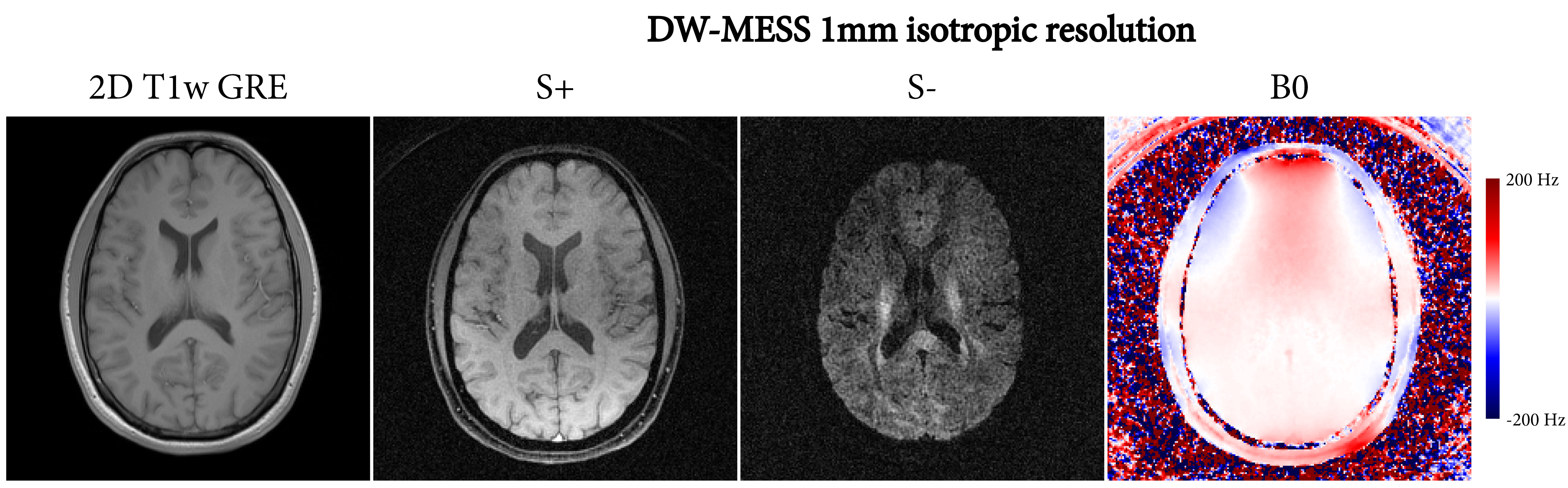
Figure 5 shows the DW-MESS images for a 1mm resolution acquisition. The S+ image is very close to the T1-weighted GRE image, both in resolution and contrast. At this higher resolution, the limited SNR of the S- image becomes visible.
Discussion
DW-MESS enables efficient, distortion-free 3D isotropic T1-, T2- and diffusion-weighted imaging. Our sampling scheme mitigated artifacts originating from the strong diffusion gradients in the AP direction, but additional phase corrections need to be explored for quality improvement in all diffusion directions.DW-MESS enables imaging with longer TRs, which boosts the T2- and diffusion-weighting, though at the cost of SNR, which was especially visible in the high resolution S- image. Imaging with shorter echo trains (e.g.$$$\:3\times3\:$$$beams) could balance TR and SNR. Furthermore, other scan parameters (e.g. flip angle) need to be optimized for optimal SNR.
Reconstruction of the 32 individual echoes could support T2* quantification, quantitative susceptibility mapping, and/or water-fat separation6. As such, the DW-MESS sequence is promising for efficient, multi-parametric imaging.
Acknowledgements
This work was supported by the Research Council of Norway (FRIPRO Researcher Project 302624).References
- Redpath TW, Jones RA. FADE–A new fast imaging sequence. Magn Reson Med. 1988;6(2):224-234
- Bruder H, Fischer H, Graumann R, Deimling M. A new steady-state imaging sequence for simultaneous acquisition of two MR images with clearly different contrasts. Magn Reson Med. 1988;7(1):35-42
- Welsch GH, Scheffler K, Mamisch TC, et al. Rapid estimation of cartilage T2 based on double echo at steady state (DESS) with 3 Tesla. Magn Reson Med. 2009;62(2):544-549
- Bieri O, Ganter C, Scheffler K. Quantitative in vivo diffusion imaging of cartilage using double echo steady-state free precession. Magn Reson Med. 2012;68(3):720-729
- Moran CJ, Cheng JY, Sandino CM, et al. Diffusion-weighted double-echo steady-state with a three-dimensional cones trajectory for non-contrast-enhanced breast MRI. J Magn Reson Imaging. 2021;53(5):1594-1605
- Zijlstra F, Seevinck PR. Multiple-echo steady-state (MESS): Extending DESS for joint T2 mapping and chemical-shift corrected water-fat separation. Magn Reson Med. 2021;86(6):3156-3165
- Granlund KL, Staroswiecki E, Alley MT, Daniel BL, Hargreaves BA. High-resolution, three-dimensional diffusion-weighted breast imaging using DESS. Magn Reson Imaging. 2014;32(4):330-341
- McNab JA, Gallichan D, Miller KL. 3D steady-state diffusion-weighted imaging with trajectory using radially batched internal navigator echoes (TURBINE). Magn Reson Med. 2010;63(1):235-242
- Pipe JG. Motion correction with PROPELLER MRI: Application to head motion and free-breathing cardiac imaging. Magn Reson Med. 1999;42(5):963-969
- Ravi KS, Geethanath S, Vaughan JT. PyPulseq: A Python Package for MRI
Pulse Sequence Design. J Open Source Softw. 2019;4(42):1725
- Buehrer M, Pruessmann KP, Boesiger P, Kozerke S. Array compression for
MRI with large coil arrays. Magn Reson Med. 2007;57(6):1131-1139
- Fessler JA, Sutton BP. Nonuniform fast Fourier transforms using min-max interpolation. IEEE Trans Signal Process. 2003;51(2):560-574
- Pipe JG, Menon P. Sampling density compensation in MRI: Rationale and an iterative numerical solution. Magn Reson Med. 1999;41(1):179-186
- Inati SJ, Hansen MS, Kellman P. A Fast Optimal Method for Coil Sensitivity Estimation and Adaptive Coil Combination for Complex Images. In: Proceedings of the 22nd Annual Meeting of the ISMRM. ; 2014
- Fong DCL, Saunders M. LSMR: An Iterative Algorithm for Sparse Least-Squares Problems. SIAM J Sci Comput. 2011;33(5):2950-2971
- Miller KL, Pauly JM. Nonlinear phase correction for navigated diffusion imaging. Magn Reson Med. 2003;50(2):343-353
Figures