1136
gNET: gSlider Self-Supervised Neural Network for Accelerated Reconstruction of Super-resolution Diffusion MRI1Athinoula A. Martinos Center for Biomedical Imaging, Boston, MA, United States, 2Mechanical Engineering, Escola Politécnica da USP, São Paulo, Brazil, 3Technical University of Munich, Munich, Germany, 4Radiology, Harvard Medical School, Boston, MA, United States, 5Harvard/MIT Health Sciences and Technology, Cambridge, MA, United States, 6Boston Children’s Hospital, Boston, MA, United States, 7Department of Biomedical Engineering, Tsinghua University, Beijin, China
Synopsis
Keywords: Diffusion Reconstruction, Diffusion/other diffusion imaging techniques, gSlider, deep-learning, self-supervised, AI/ML Image Reconstruction
Motivation: gSlider utilizes radio-frequency encoding to acquire high and isotropic resolution brain diffusion-MRI with high SNR. However, this comes at the cost of prolonged acquisition time, which also increases the sensitivity to motion.
Goal(s): This work proposes gSlider Network (gNET) to accelerate gSlider from acquisitions with jointly subsampled RF- and q-space.
Approach: The self-supervised model was trained and tested on a 1mm3 resolution BUDA-gSlider dataset (Tacq = 32 min). FSL and the DIMOND self-supervised were used to estimate the diffusion parameters.
Results: gNET achieved an acceleration factor of R=2 and, when combined with DIMOND, reached a total R=4-fold (Tacq = 8 min).
Impact: gNET facilitates super-resolution dMRI by reducing the acquisition time by 4-fold with high fidelity. Its application may propel new discoveries in the neuroscientific field and the clinical translation of the gSlider framework.
INTRODUCTION
Diffusion MRI yields valuable information on the brain’s structure and connectivity and finds important applications in the diagnosis and monitoring of neurological disorders and in neuroscientific research 1.Unfortunately, high-resolution dMRI is hampered by its inherently low SNR. Super-resolution techniques help boost the SNR and have been deployed with single-shot approaches 2,3. More recently, gSlider-SMS achieved whole-brain in vivo sub-millimeter imaging using a RF-encoding to excite a thick slab, from which thin slice images could be resolved by inverting the RF-encoding basis. The drawback is that gSlider RF-encoding prolongs the acquisition time, which hampers its practical utility 4,5.
gSlider-SR 6 was developed to accelerate the gSlider acquisition by reconstructing RF-encoded data using a basis of spherical ridgelets, exploiting redundancies within RF-encoding basis and q-space. The present work pushes the acceleration further using a deep-learning neural network, which resulted in a final R=4-fold speed-up with high fidelity, thus facilitating the application of this encoding technique.
Code repository: https://anonymous.4open.science/r/gSliderRecon-E592/README.md
METHODS
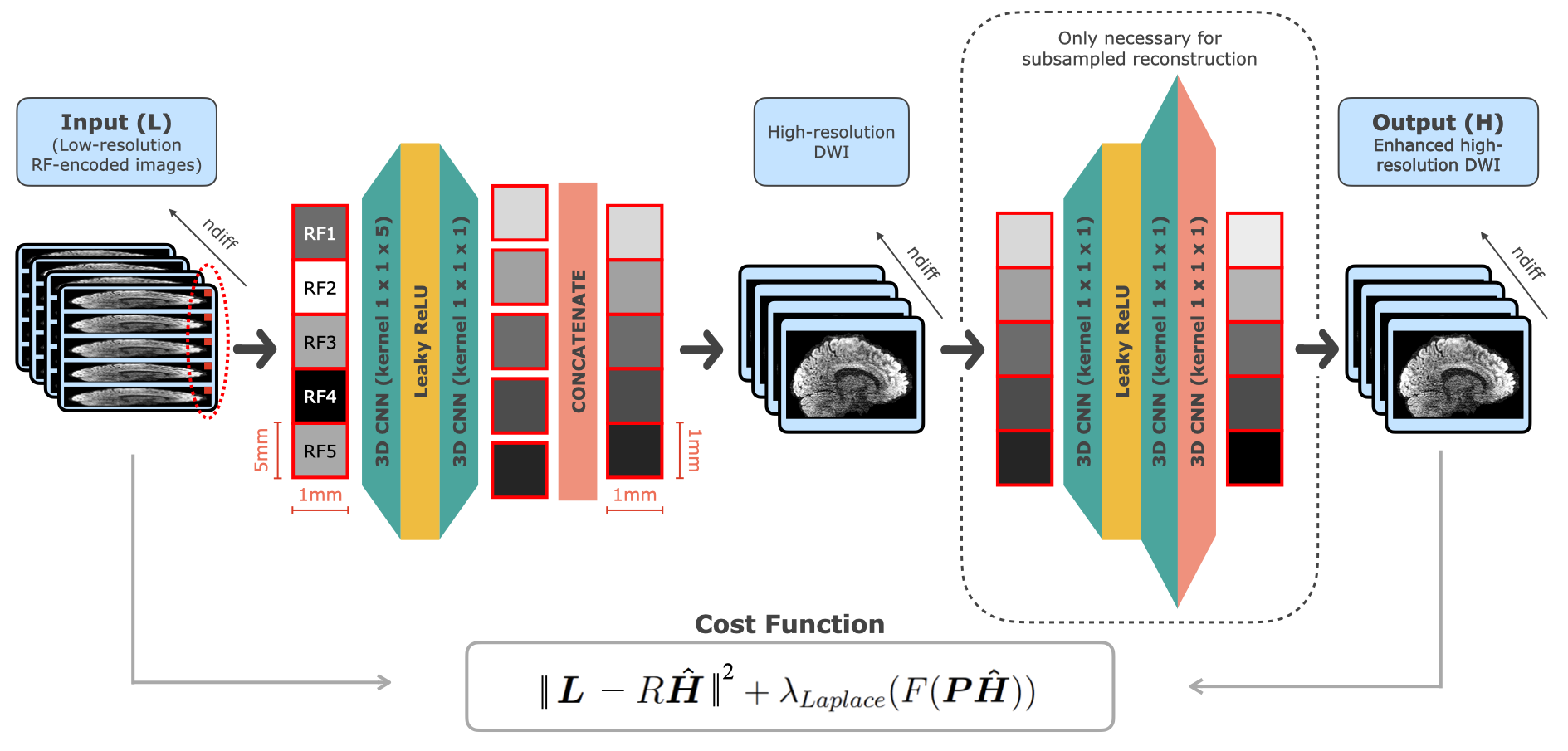
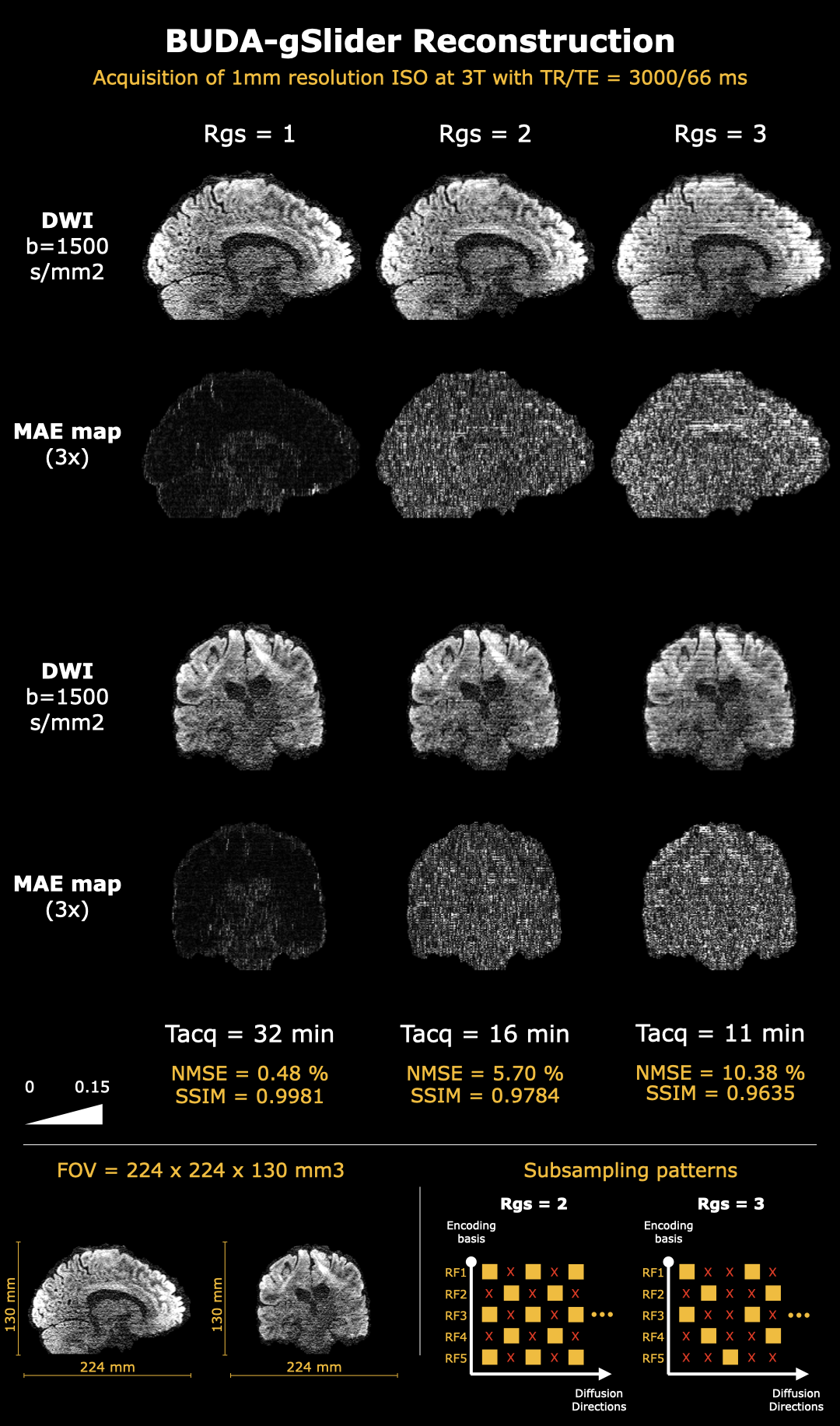
BUDA-gSlider dataset. Acquired 7 on a Siemens 3T MAGNETOM Prisma, it is composed of whole-brain (FOV = 224 x 224 x 130 mm3) data, separated into 26 slabs of 5 mm each, which required a 32 minutes scan. It uses MultiBand (MB) = 1, Rin−plane = 4, partial Fourier = 6/8, TR/TE = 3000/66 ms, and two BUDA shots to yield 60 diffusion directions (b = 1500 s/mm2) and four b=0 images. For accelerated acquisition, the subsampling patterns across RF and q-space at Rgs=2 and Rgs=3 in Figure 2 are employed.gSlider Network (gNET). The proposed NN transforms real-valued RF-encoded low-resolution gSlider data into high-resolution images, by applying two 3D CNN layers and three fully-connected layers (Figure 1). The outputs are then fed to the gSlider forward model to synthesize the input through
$$\boldsymbol{L} = R\boldsymbol{\hat{H}} + \lambda_{Laplace}(F(\boldsymbol{P\hat{H}}))$$
where $$$\boldsymbol{L}$$$ is the low-resolution image, $$$R$$$ is the RF-encoding matrix, $$$\boldsymbol{\hat{H}}$$$ is the high-resolution image, $$$\lambda_{Laplace}$$$ is the penalty parameter, and $$$F(\boldsymbol{P\hat{H}})$$$ is the Laplace regularizer applied to the phase corrected high-resolution image. Constructing the loss function using the difference between the input and synthesized low-resolution volumes allows the model to operate in a self-supervised manner. The NN was implemented using Pytorch and optimized with Rprop 8 and L2 loss. 1/6 of the data was separated for training due to hardware constraints and 5/6 for validation. Learning rate was 0.001, which was reduced to 0.0001 for fine-tuning.
Parameters estimation and validation. Diffusion parameters were firstly estimated using FSL DTIFIT tool 9. Afterwards, Fractional Anisotropy (FA), Mean Diffusivity (MD) and colored FA were obtained using the self-supervised parameter estimator model called DIMOND 10 under fully sampled (Rd = 1) and subsampled q-space (Rd > 1). The final results were compared to a ground truth (fully-sampled conventional gSlider reconstruction) using normalized mean squared error (NMSE) and structural similarity index (SSIM).
RESULTS
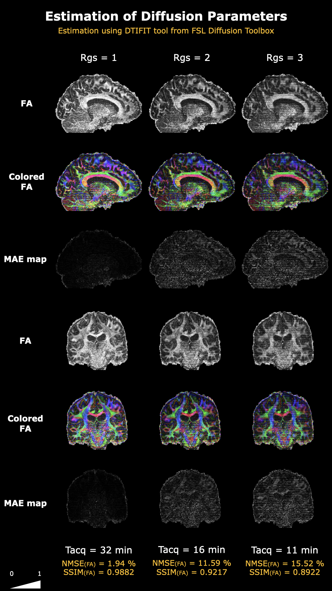
As shown in Figure 2, gNET reconstructed accurate BUDA-gSlider DWIs when fully sampled (Rgs = 1) and when subsampled with Rgs = 2. With NMSE = 5.70% and SSIM = 0.9784, the Rgs = 2 images retain high fidelity to the fully sampled case. At the higher acceleration factor of Rgs = 3, the image quality is still adequate, yet with lower contrast and some stripe artifacts.Figure 3 shows the FSL estimation of the reconstructed DWIs for each case. FA from both subsampled cases appear underestimated compared to the fully sampled case, and the Rgs = 3 contains subtle stripe artifacts. This FA underestimation leads to higher NMSEs (>10%), which signifies a need for better alternatives to FSL’s dtifit.
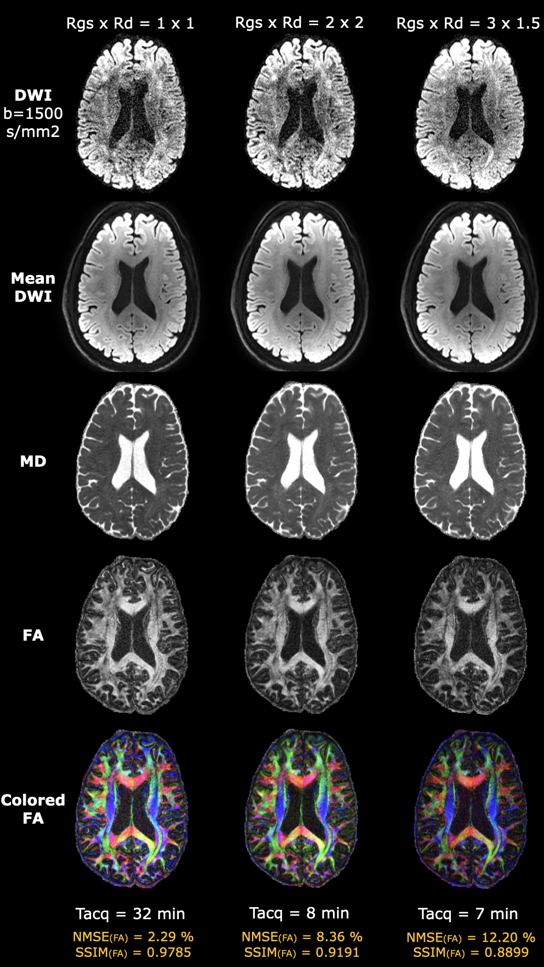
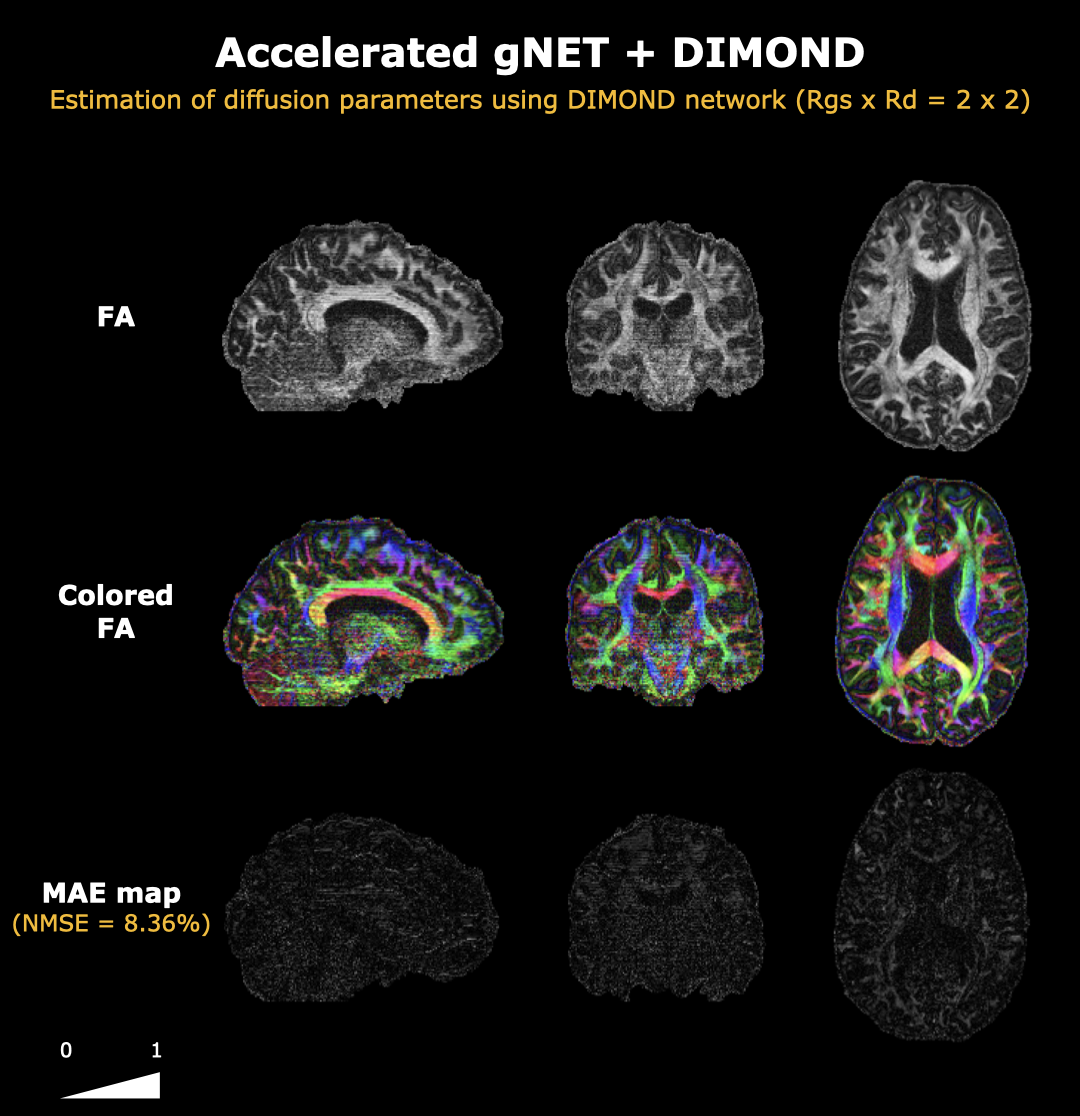
The results from DIMOND NN demonstrate a denoising effect, leading to higher quality images. MD and FA from Rgs x Rd = 2 x 2 are similar to the fully sampled case with NMSE = 8.36% and SSIM = 0.9191. Despite the slight green bias in its colored FA, the regions of the white matter with higher anisotropy are reconstructed adequately.
DISCUSSION AND CONCLUSION
The self-supervised gSlider Network successfully reduced the BUDA-gSlider acquisition time by up to R=4-fold. As shown in Figure 5, gNET achieved Rgs=2-fold acceleration during high-resolution gSlider reconstruction and a further Rd=2 acceleration was provided by DIMOND by directly reducing the number of directions, yielding a total R=4 (Tacq from 32 minutes to 8 minutes). Future work will explore the creation of a single NN capable of directly estimating the diffusion parameters from the low-resolution data.Acknowledgements
This work was supported by research grants NIH R01 EB028797, U01 EB025162, P41 EB030006, U01 EB026996, R03 EB031175, R01 EB032378, UG3 EB034875, NVidia Corporation for computing support.References
1. JONES, D. K. Diffusion MRI. Oxford University Press, 2010.
2. PELED, S.; YESHURUN, Y. Superresolution in MRI: application to human white matter fiber tract visualization by diffusion tensor imaging. Magnetic Resonance in Medicine: An Official Journal of the International Society for Magnetic Resonance in Medicine, Wiley Online Library, v. 45, n. 1, p. 29–35, 2001.
3. GREENSPAN, H. et al. MRI inter-slice reconstruction using super-resolution. Magnetic resonance imaging, Elsevier, v. 20, n. 5, p. 437–446, 2002.
4. SETSOMPOP, K. et al. Slice dithered enhanced resolution simultaneous multislice (slider-SMS) for high resolution (700 μm) diffusion imaging of the human brain. In: 23rd Annual Meeting of International Society for Magnetic Resonance in Medicine (ISMRM), Toronto, Canada, p. 339, 2015.
5. HALDAR, J. P.; FAN, Q.; SETSOMPOP, K. Whole-brain quantitative diffusion MRI at 660 μm resolution in 25 minutes using gSlider-SMS and SNR-enhancing joint reconstruction. In: Proceedings of the 24th Annual Meeting of ISMRM, Singapore, p. 102, 2016.
6. RAMOS-LLORDÉN, G. et al. High-fidelity, accelerated whole-brain submillimeter in vivo diffusion MRI using gSlider-spherical ridgelets (gSlider-SR). Magnetic resonance in medicine, Wiley Online Library, v. 84, n. 4, p. 1781–1795, 2020.
7. RAMOS-LLORDEN, G.; BILGIC, B.; HUANG, S. Y. Rapid, high-spatial resolution in vivo diffusion MRI with joint subsampling and reconstruction in k-, q-and RF-space. ISMRM, London, 2022.
8. RIEDMILLER, Martin; BRAUN, Heinrich. A direct adaptive method for faster backpropagation learning: The RPROP algorithm. In: IEEE international conference on neural networks. IEEE, p. 586-591, 1993.
9. SMITH, Stephen M. et al. Advances in functional and structural MR image analysis and implementation as FSL. Neuroimage, v. 23, p. S208-S219, 2004.
10. ZIHAN, L. et al. Dimond: Diffusion model optimization with deep learning. Magnetic Resonance in Medicine, 2022.
Figures