1135
Hierarchical-µGUIDE: fast and robust Bayesian hierarchical modelling using deep learning simulation-based inference1MIND team - Inria, Palaiseau, France, 2CUBRIC - Cardiff University, Cardiff, United Kingdom
Synopsis
Keywords: Microstructure, Microstructure
Motivation: In-vivo brain microstructure can be estimated using diffusion MRI. However, most approaches do not quantify estimates reliability, although crucial for interpreting the results, and consider every voxel independently, leading to high uncertainties.
Goal(s): Our goal is to develop a new framework to efficiently estimate tissue microstructure and improve data fitting quality.
Approach: We propose Hierarchical-µGUIDE, a Bayesian method that estimates posterior distributions, by combining simulation-based inference with a hierarchical structure.
Results: Hierarchical-µGUIDE bypasses the high computational and time cost of conventional Bayesian approaches. Sharper microstructure parameter maps that preserve tissue heterogeneity are obtained, along with a tissue parcellation that segments an epileptic lesion.
Impact: The proposed Bayesian framework improves single-subject inference for clinical diagnosis, by efficiently estimating posterior distributions, reducing estimates uncertainty, and learning a tissue parcellation. This works unlocks the possibility to apply hierarchical Bayesian methods taylored for microstructure estimation to large datasets.
Introduction
Diffusion-weighted MRI is a promising imaging technique for characterizing brain microstructure in-vivo1,2,3. By fitting a biophysical model to the acquired diffusion signals, one can quantify histologically meaningful microstructural parameters within each voxel1. However, most approaches a) only provide the most likely parameter value without offering insights about their robustness and reliability, although crucial for interpreting the results, and b) fit each voxel independently, without exploiting information redundancy across voxels.A recent approach, µGUIDE4, based on Simulation-Based Inference (SBI)5, estimates full posterior distributions and quantifies the uncertainty of the estimated tissue parameters4,5,6. µGUIDE circumvents the limitations of traditional Bayesian inference approaches (e.g. Markov-Chain-Monte-Carlo) by providing faster and more robust estimates of parameters posteriors in each voxel independently.
However, the estimated voxel-independent posterior distributions can present large variance (i.e. high uncertainty) due to low signal-to-noise ratio (SNR). Resulting parameter maps can be too noisy for diagnosis, since the noise can obscure or be mistaken for pathology.
Here we introduce Hierarchical-µGUIDE, a new method enhancing the fitting quality of microstructure parameters by combining information across voxels presenting a similar microstructure, hypothesising a hierarchical structure7,8,9. Similar voxels are grouped into parcels. The posterior distributions and the parcellation are jointly estimated from the observed data. Voxel parameters are estimated with higher precision, enhancing their clinical utility.
Methods
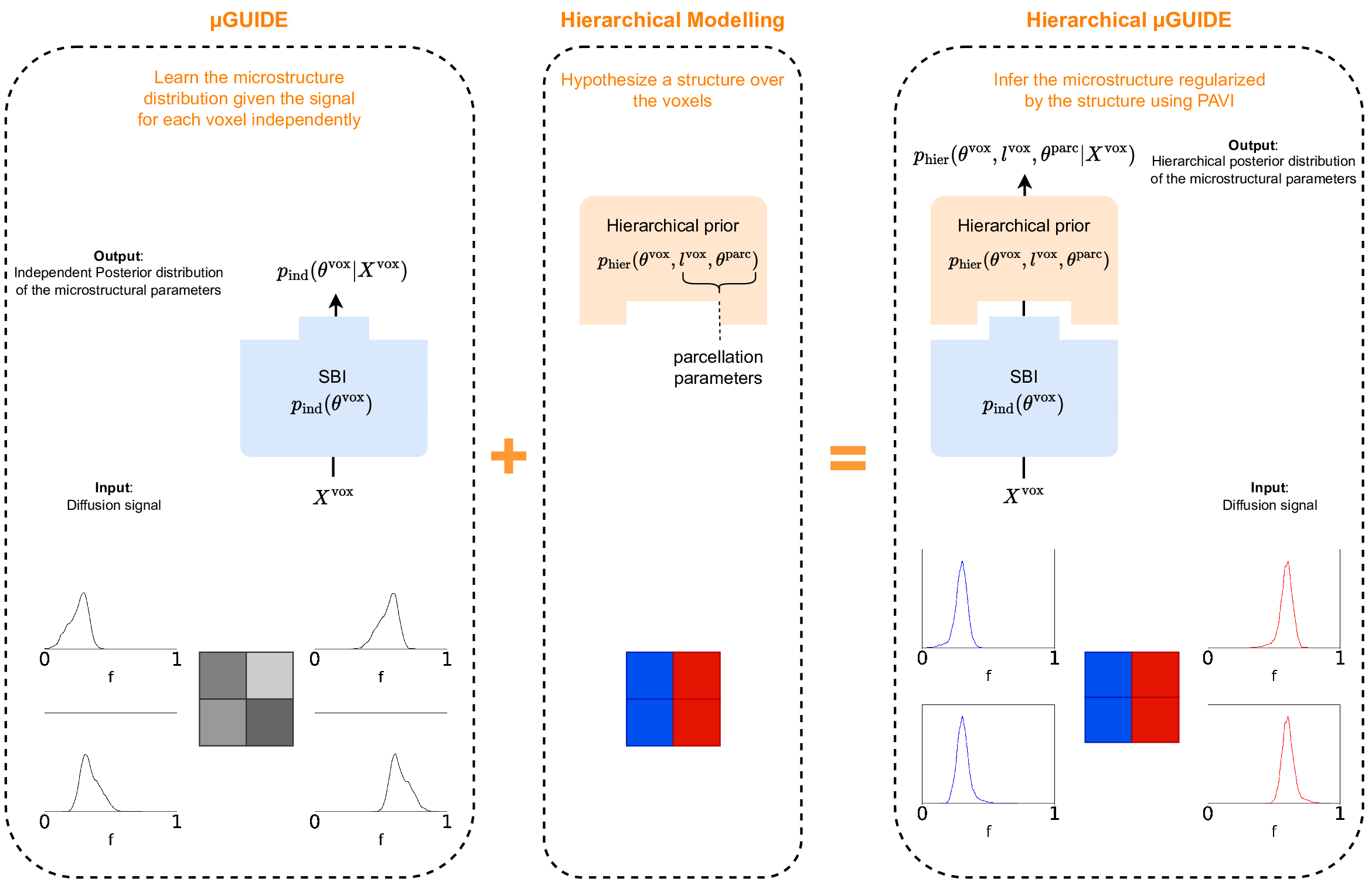
Hierarchical-µGUIDE. Hierarchical-µGUIDE relies on Bayesian inference, which takes as input a multi-shell diffusion-weighted signal for each voxel $$$X^\text{vox}$$$, and outputs the posterior distributions $$$p_\text{hier}(\theta^\text{vox}|X^\text{vox})$$$ of the microstructure parameters $$$\theta^\text{vox}$$$, defined by a given biophysical model of the tissue2,3.Contrary to µGUIDE4, voxels are not considered independently. Instead, we group voxels into $$$L$$$ parcels of similar microstructure. Each parcel is associated with the average parameters $$$\theta^\text{parc}$$$, and each voxel’s microstructure $$$\theta^\text{vox}$$$ is assumed to be a perturbation of the parcel parameters it belongs to, with variability $$$\sigma^\text{parc}$$$. Using Hierarchical-µGUIDE, we 1) learn a parcellation, and 2) reduce the uncertainty in each voxel’s estimation by sharing information across voxels in the same parcel.
Hierarchical-µGUIDE consists of two successive steps (Fig.1). First, similar to μGUIDE4, we learn the independent posteriors $$$p_\text{ind}(\theta^\text{vox}|X^\text{vox})$$$, by training over a large synthetic dataset of microstructure-signal pairs, sampled from a simulator with a uniform prior $$$p_\text{ind}(\theta^\text{vox})$$$. Second, we hypothesise a hierarchical structure over the voxels7,8,9 with prior $$$p_\text{hier}(\theta^\text{vox},l^\text{vox},\theta^\text{parc})$$$, that replaces the independent prior $$$p_\text{ind}(\theta^\text{vox})$$$. Using Plate Amortized Variational Inference (PAVI)7, we start from the posteriors $$$p_\text{ind}(\theta^\text{vox}|X^\text{vox})$$$, and progressively regularise those into $$$p_\text{hier}(\theta^\text{vox}|X^\text{vox})$$$. $$$p_\text{hier}(\theta^\text{vox}|X^\text{vox})$$$ integrates both the SBI-learnt $$$p_\text{ind}(\theta^\text{vox}|X^\text{vox})$$$ and the prior structure $$$p_\text{hier}(\theta^\text{vox})$$$.
Exemplar application. As demonstrator, we applied Hierarchical-µGUIDE to the Standard Model (SM)3, a two-compartment model with five microstructural parameters: neurite signal fraction f, intra-neurite diffusivity Da, orientation dispersion index ODI, and parallel/perpendicular diffusivity within the extra-neurite space De||/De┴.
Training. SBI training was performed on $$$N=10^5$$$ synthetic simulations computed using MISST10 with random combinations of the model parameters, uniformly sampled from biologically plausible ranges. To match experimental data, Rician noise ($$$\text{SNR}=30$$$) was added to the simulated signals.
Experiment I: simulations. We applied Hierarchical-µGUIDE to a synthetic phantom generated similarly to the training set, where the ground-truth values of the model parameters were set to white matter tissue estimates11.
Experiment II: MRI data. We applied Hierarchical-µGUIDE to a healthy participant, and a participant with epilepsy. Data were acquired using a PGSE acquisition with b-value=[200,500,1200,2400,6000]s/mm2, respectively [20,20,30,61,61] diffusion encoding directions uniformly distributed, δ/Δ=7/24ms, and TE/TR=76/3200ms.
Results
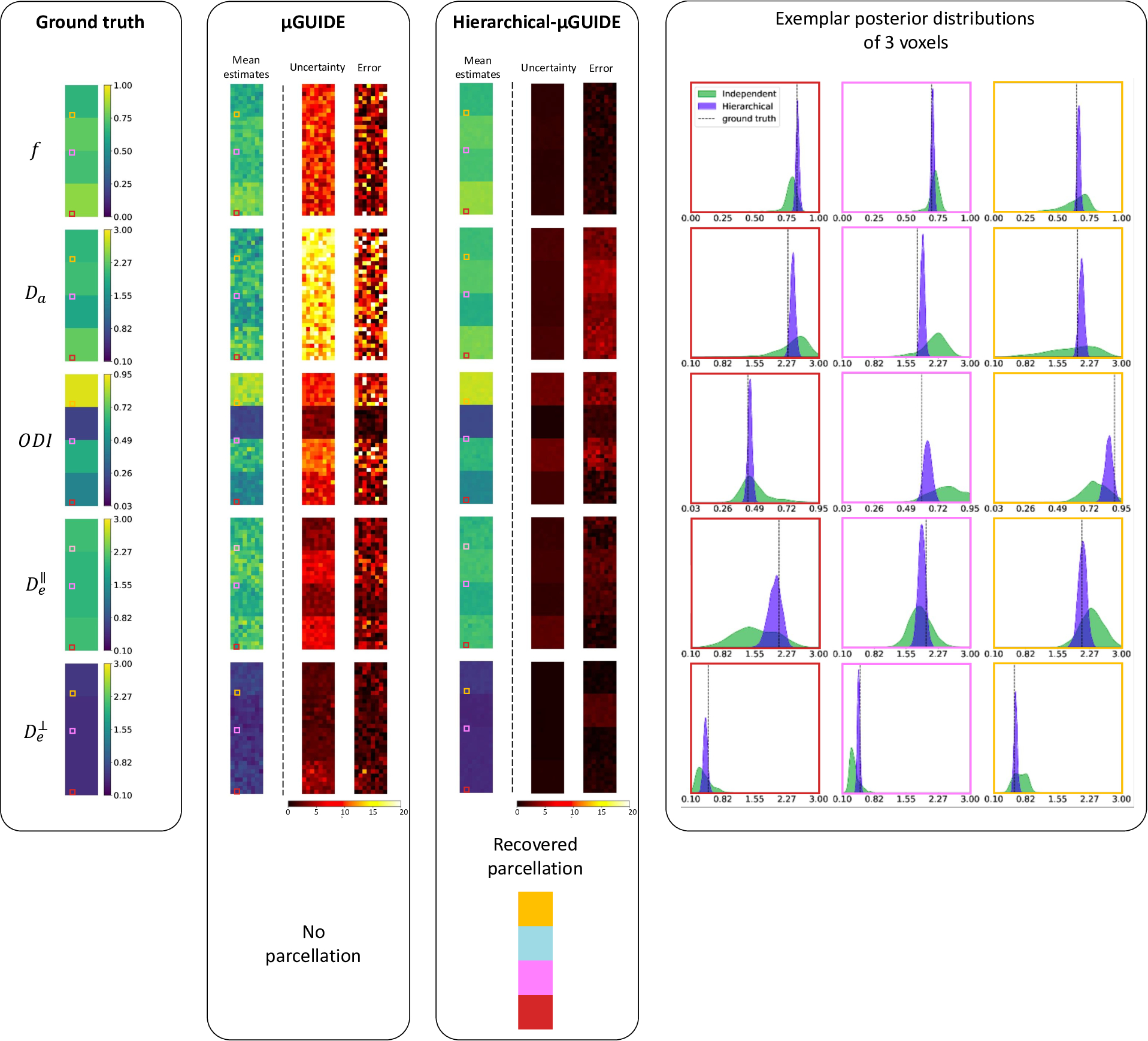
Fig.2 showcases Hierarchical-µGUIDE’s ability to simultaneously learn posterior distributions within each voxel and a parcellation, allowing to reduce uncertainty compared to independent estimates.Fig.3 presents the results over simulated data, fitting the voxels independently using µGUIDE, and hierarchically using Hierarchical-µGUIDE. Hierarchical-µGUIDE provides more accurate and precise parameter estimates. Although full posterior distributions are estimated for each voxel, here we report the mean, relative standard deviation and bias of the estimated parameters as summary statistics.
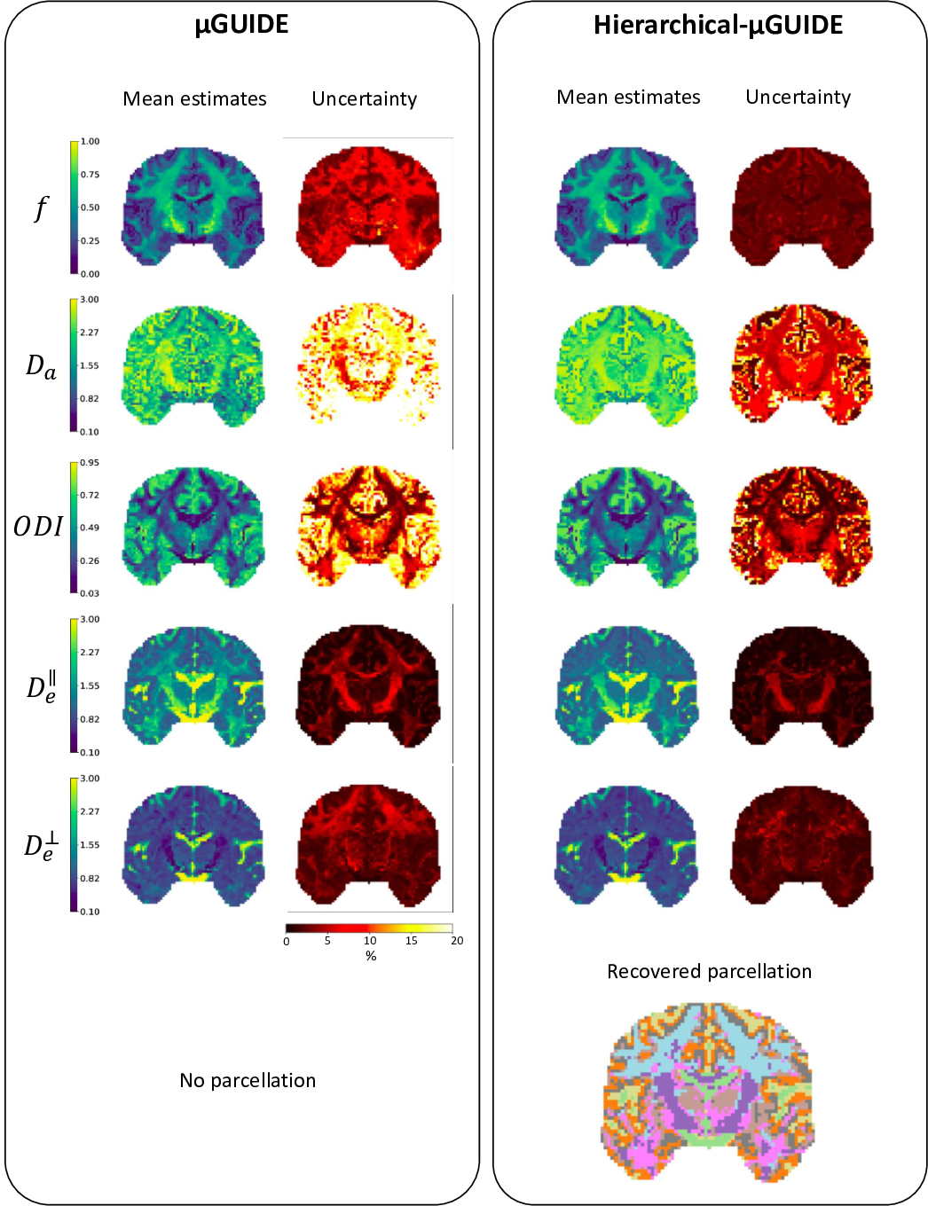
Fig.4 presents the SM parametric maps estimated using µGUIDE and Hierarchical-µGUIDE on a healthy participant, alongside their uncertainty and parcellation.
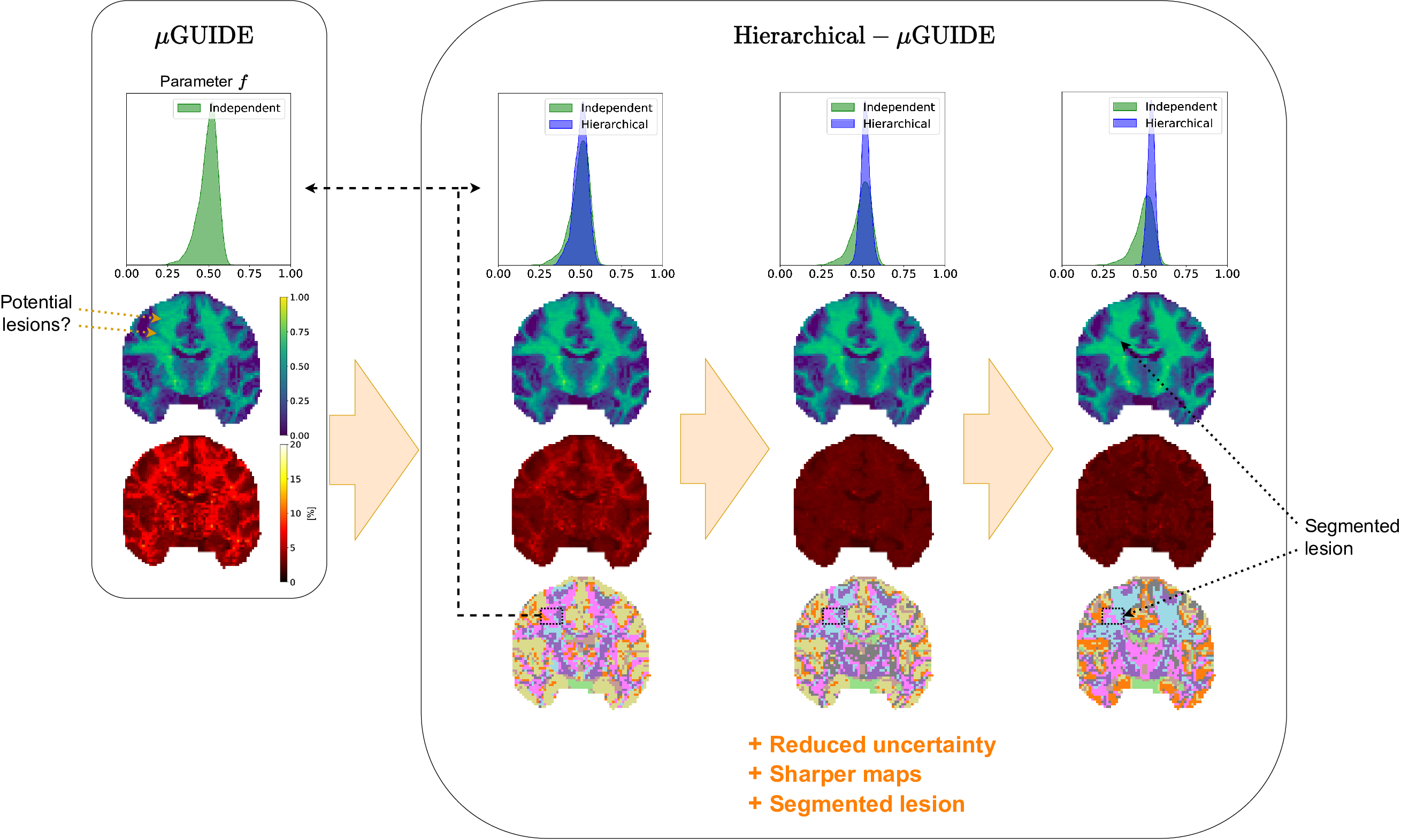
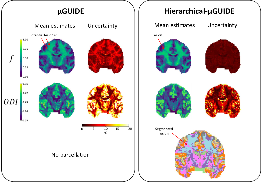
Fig.5 presents Hierarchical-µGUIDE’s application to participant with epilepsy. The sharper parametric maps and the parcellation highlight the lesion.
Discussion
Hierarchical-µGUIDE yields faster posterior distribution estimates compared to existing hierarchical methods relying on MCMC8,9, and returns a parcellation learnt from, and not imposed on, the data.Hierarchical-µGUIDE’s efficiency unlocks applications on large datasets, therefore gaining statistical power.
Hierarchical-µGUIDE can also seamlessly be applied to different biophysical models/representations, and hierarchical structures.
Conclusion
Hierarchical-µGUIDE improves the quality of tissue microstructure estimations by using a hierarchical Bayesian approach to efficiently estimate full posterior distributions along with a tissue parcellation. Hierarchical-µGUIDE reduces estimates uncertainty, while preserving tissue heterogeneity, allowing for pathology detection.Acknowledgements
This work, MJ and MP are supported by UKRI Future Leaders Fellowship (MR/T020296/2). LR and DW are supported by the ERC-StG NeuroLang (ID:757672). We are thankful to Dr. Dmitri Sastin and Dr. Khalid Hamandi for sharing their dataset from a participant with epilepsy.References
- Alexander, DC, Dyrby, TB, Nilsson, M, Zhang, H. Imaging brain microstructure with diffusion MRI: practicality and applications. NMR in Biomedicine. 2019.
- Jelescu IO, Palombo M, Bagnato F, Schilling KG. Challenges for biophysical modeling of microstructure. J Neurosci Methods. 2020.
- Novikov, DS, Fieremans, E, Jespersen, SN, Kiselev, VG. Quantifying brain microstructure with diffusion MRI: Theory and parameter estimation. NMR in Biomedicine. 2019.
- Jallais, M. and Palombo M. µGUIDE: a framework for microstructure imaging via generalized uncertainty-driven inference using deep learning. ISMRM. 2023.
- Cranmer K, Brehmer J, Louppe G. The frontier of simulation-based inference. Proc National Acad Sci. 2020.
- Jallais, M., Rodrigues, P.L.C., Gramfort, A. and Wassermann, D. Inverting brain grey matter models with likelihood-free inference: a tool for trustable cytoarchitecture measurement. Machine Learning for Biomedical Imaging. 2022.
- Rouillard, L., Le Bris, A., Moreau T., and Wassermann, D. PAVI: Plate-Amortized Variational Inference. TMLR 2023 https://openreview.net/forum?id=vlY9GDCCA6.
- Powell, E., Battocchio, M., Parker, C. S., and Slator, P. J. Generalised Hierarchical Bayesian Microstructure Modelling for Diffusion MRI. CDMRI. 2021
- Orton, M.R., Collins, D.J., Koh D.M. and Leach, M.O. Improved intravoxel incoherent motion analysis of diffusion weighted imaging by data driven Bayesian modeling, Magnetic Resonance in Medicine. 2014.
- Ianuş, A., Shemesh, N., Alexander, D.C. and Drobnjak, I. Double oscillating diffusion encoding and sensitivity to microscopic anisotropy. Magn. Reson. Med. 2017.
- Coelho, S., Baete, S.H., Lemberskiy, G., Ades-Aron, B., Barrol, G., Veraart, J., Novikov, D.S., Fieremans, E. Reproducibility of the Standard Model of diffusion in white matter on clinical MRI systems. NeuroImage. 2022.
Figures