1132
Comparison of White Matter Maturation Rates in Young Rhesus Macaques and Humans1Medical Physics, University of Wisconsin-Madison, Madison, WI, United States, 2Pediatrics, University of Wisconsin-Madison, Madison, WI, United States, 3Medicine, University of Wisconsin-Madison, Madison, WI, United States, 4Neuroscience Training Program, University of Wisconsin-Madison, Madison, WI, United States, 5Psychiatry, University of Wisconsin-Madison, Madison, WI, United States, 6Waisman Center, University of Wisconsin-Madison, Madison, WI, United States
Synopsis
Keywords: Large Animals, Nonhuman Primates, White Matter, Non-Human Primates, Modeling, Normal Development
Motivation: Non-human primates are thought to develop 3-4 times faster than humans based on sexual maturity and death; however, there has been a lack of quantitative data to support this ratio to describe brain development.
Goal(s): Our goal was to find a quantitative relationship between the rate of white matter myelination in rhesus macaques and humans.
Approach: We compared rates of change in quantitative relaxometry MRI T1 values in six ROIs for rhesus macaques and human infants.
Results: We found a ratio ranging from 4.7 to 6.2 in the ROIs, corresponding to 4.7-6.2 times faster white matter myelination in rhesus macaques than humans.
Impact: By providing a quantitative approach to comparing early-life rhesus macaques white matter development with human infants, research that relates rhesus macaques and human brain development can make a more informed comparison, assisting researchers in translating results between species.
Introduction
Myelination in white matter during early brain development is crucial, as myelin acts as an insulator for neurons, allowing for new connections between brain regions to form as myelin increases. Myelination rapidly develops in infant brains during the early years of life. Quantitative T1 values from MRI relaxometry is inversely related to myelination (i.e., R1= 1/T1 is linearly related to myelination)1. Non-human primates (NHP) are often used in studies as models for human brain development because of their genetic, neuroanatomical and behavioral similarities. Rhesus macaque NHPs thought to develop 3-4 times faster than humans based on age of sexual maturity, menopause, and death.2 3 Cognitive tests have shown NHPs to develop at different rates depending on the task, indicating that the relative rate of brain development of macaques to humans is both regional and different than the rate based on sexual maturity and death.4 However, there is a lack of quantitative imaging data to support how NHP white matter development rates compare to humans. The goal of this study is to use myelin sensitive measures such as quantitative T1 relaxometry to compare brain development rates in rhesus macaques and humans. This will help inform future research study plans in choosing timepoints that best relate to human development.Methods
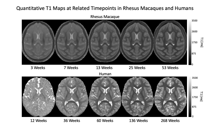
The study included a cross-sectional sample of forty-seven human participants imaged at ages ranging from 2 months to 9.75 years (mean age: 51.4 months)5 and thirty-four rhesus macaque NHPs imaged longitudinally at 5 time points over the first year of life (3, 7, 13, 25, and 53 weeks).6 Both groups were imaged with a 3T GE MR750 scanner using a novel motion-corrected MPnRAGE sequence which provided simultaneous T1-weighted images and quantitative T1 maps.7 Human images were obtained at 1mm isotropic resolution and voxels, while the NHP T1-weighted were acquired at 0.625 mm isotropic resolution and reconstructed to 0.47 mm voxels. We obtained mean qT1 values for six regions of interest, using the JHU white matter region template in humans and an inhouse macaque white matter template.8 For both species, the quantitative T1 values for each ROI were fit to an exponential decay model:$$ T1(t) = a+(b-a)e^{\frac{-t}{c}} $$
where t is age in days. This model was chosen because it estimated the maturational time parameter, c, independent of the asymptote, a, and intercept, b, parameters, that may vary with the acquisition protocol or development state at birth. The ratio of c values between rhesus macaque NHP and humans were calculated to estimate the relative rates of white matter maturation in the NHP.
Results
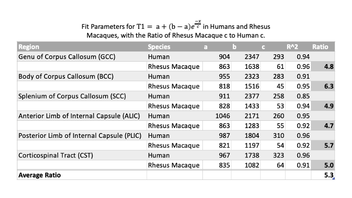
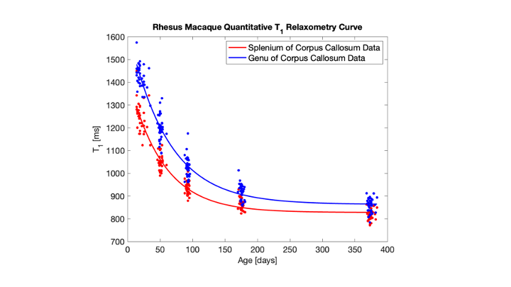
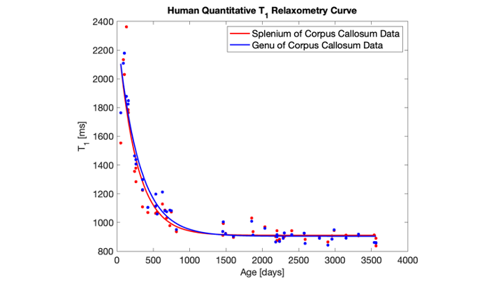
The model parameters and R2 values for each ROI in both humans and rhesus macaques are listed in Figure 2. Figure 3 plots example data and model fits for the genu and splenium of corpus callosum regions of rhesus macaques. Figure 4 plots the data and model fits for the same regions in humans. Figure 2 also lists the ratios of relative NHP maturational rates (cNHP:cHum), which range from 4.7 for the anterior limb of internal capsule, to 6.2 for the body of corpus callosum, with an overall mean across all regions of 5.3.Discussion
In this study, the rhesus macaques’ brain T1 values matured at a rate of roughly 4.7 to 6.2 times faster than humans. As T1 is a marker of white matter myelination,3 this gives insight into how much faster the rhesus macaque brain develops compared to humans. Our results indicate that this rate is slightly higher than the factor of 3-4 previously described, and that these rates are region dependent. These results may assist in informing future studies using non-human primates as a model for human brain development, as it gives insight into how timepoints in non-human primates correspond to human timepoints. Future studies will include comparisons of T1 maturation in other brain regions and other quantitative imaging measurements such as diffusion tensor imaging. Several potential limitations of the study include the relative differences in the voxel scales for NHP and humans, and differences in the cohort design – longitudinal for the NHP and cross-sectional for the human samples.Conclusions
Our results show white matter maturation in rhesus macaques is about 4.7-6.2 times faster than in human infants based on quantitative T1 relaxometry. This allows for comparison of timepoints of similar white matter myelination in humans and rhesus macaques, which will lead to better timepoint correspondence in future studies using rhesus macaques as a model for human development.Acknowledgements
We would like to thank the children and family participated in this study. This project was supported by the following NIH grants P50 HD105353, R01 HD108868, U01 DA055370, R34 DA050258, P50 MH100031, R00 MH11056, MH081884 and MH046729, and the Waisman Center at the University of Wisconsin-Madison.
References
1 Grotheer, M., Rosenke, M., Wu, H., Kular, H., Querdasi, F. R., Natu, V. S., Yeatman, J. D., & Grill-Spector, K. (2022). White matter myelination during early infancy is linked to spatial gradients and myelin content at birth. Nature Communications, 13(1), 1–12. https://doi.org/10.1038/s41467-022-28326-4
2 Simmons, H. A. (2016). Age-associated pathology in rhesus macaques (macaca mulatta). Veterinary Pathology, 53(2), 399–416. https://doi.org/10.1177/0300985815620628
3 Mattison, J. A., & Vaughan, K. L. (2017). An overview of nonhuman primates in aging research. Experimental Gerontology, 94, 41–45. https://doi.org/10.1016/j.exger.2016.12.005
4 Ferrigno, S., Hughes, K. D., & Cantlon, J. F. (2016). Precocious quantitative cognition in Monkeys. Psychonomic Bulletin & Review, 23(1), 141–147. https://doi.org/10.3758/s13423-015-0893-5
5 Dean, D. C., Shah, L. S., DiPiero, M., Pletcher, C., Heinrich, L., Planalp, E., Alexander, A. L., Kecskemeti, S. (2022, 08-12 May). Mapping Brain Development in Infants and Young Children Using MPnRAGE T1 Relaxometry. [Abstract]. The International Society for Magnetic Resonance in Medicine, London, England. Abstract nr 4031.
6 Moody, J. F., Aggarwal, N., Dean, D. C., Tromp, D. P. M., Kecskemeti, S. R., Oler, J. A., Kalin, N. H., & Alexander, A. L. (2022). Longitudinal assessment of early-life white matter development with quantitative relaxometry in nonhuman primates. NeuroImage, 251, 118989. https://doi.org/10.1016/j.neuroimage.2022.118989
7 Kecskemeti, S., Samsonov, A., Hurley, S. A., Dean, D. C., Field, A., & Alexander, A. L. (2016). MPnRAGE: A technique to simultaneously acquire hundreds of differently contrasted MPRAGE images with applications to quantitative T1 mapping. Magnetic Resonance in Medicine, 75(3), 1040–1053. https://doi.org/10.1002/mrm.25674
8 Zakszewski, E., Adluru, N., Tromp, D. P., Kalin, N., & Alexander, A. L. (2014). A diffusion-tensor-based white matter atlas for Rhesus macaques. PLoS ONE, 9(9). https://doi.org/10.1371/journal.pone.0107398
Figures