1128
The resolution dependence of MRI vessel size index varies across brain regions.1Biomedical Engineering, Ulsan National Institute of Science and Technology, Ulsan, Korea, Republic of, 2Korea Basic Science Institute, Cheongju, Korea, Republic of
Synopsis
Keywords: Perfusion, Vessels, vessel size index, VSI, resolution dependence
Motivation: Vessel size index (VSI) MRI of the rodent brain is measured at various in-plane resolutions, but the resolution dependence of VSI has not yet been explored.
Goal(s): Here, we investigated the differences in rat brain VSI at mostly measured in-plane resolutions of 125μm2 and 250μm2.
Approach: Resolution-dependent differences in VSI across brain regions were investigated in in-vivo rat experiments via a steady-state susceptibility contrast method by injection of monocrystalline iron oxide nanoparticles and validated through Monte Carlo simulations.
Results: In the white matter and hippocampus regions, the VSI was measured to be 12% larger as the resolution was lowered from 125μm2 to 250μm2.
Impact: Because the resolution dependence of VSI quantification varies across brain regions depending on the vascular configuration within MRI voxels, caution is required when comparing and analyzing brain VSI MRI obtained at different resolutions.
Introduction
Vessel size index (VSI) MRI provides non-invasive quantification of the in vivo average microvascular radius of the brain1. Although there have been many studies to validate the absolute quantification of VSI MRI for various experimental parameters such as magnetic field strength, contrast agent concentration, echo time, etc.2, to the best of our knowledge, the resolution dependence of VSI in vivo has not been systematically studied. Here, we investigate the resolution dependence of rat brain VSI MRI at mostly measured in-plane resolutions of 125μm2 and 250μm2. A steady-state susceptibility contrast method was performed by injecting monocrystalline iron oxide nanoparticles. In vivo resolution-dependent differences in VSI was calculated by region-by-region basis and verified through Monte-Carlo simulations.Methods
MRI experimentsIn vivo MRI experiments were performed with 13 rats (Wistar, 210‒400g, under 2.0‒1.5% isoflurane anesthesia) on a 7T Bruker scanner. Gradient-echo (GE) and spin-echo (SE) MRIs (in-plane resolutions of 125 and 250µm2 with 1.5mm slice thickness) were performed before and after injection of 300µmol Fe/Kg of monocrystalline iron oxide nanoparticles (MION). ΔR2, ΔR2*, and VSI maps were calculated1 with the correction of declination of ΔR2 and ΔR2* due to washout of the contrast agent based on the general assumption that ΔR2 and ΔR2* is linearly proportional to the tissue contrast agent concentration. The ΔR2, ΔR2*, and VSI maps registered to the Waxholm Space rat brain atlas3,4 via Advanced Normalization Tools registration software5. Brain regional analysis was performed with the co-registered maps.
Monte-Carlo simulations
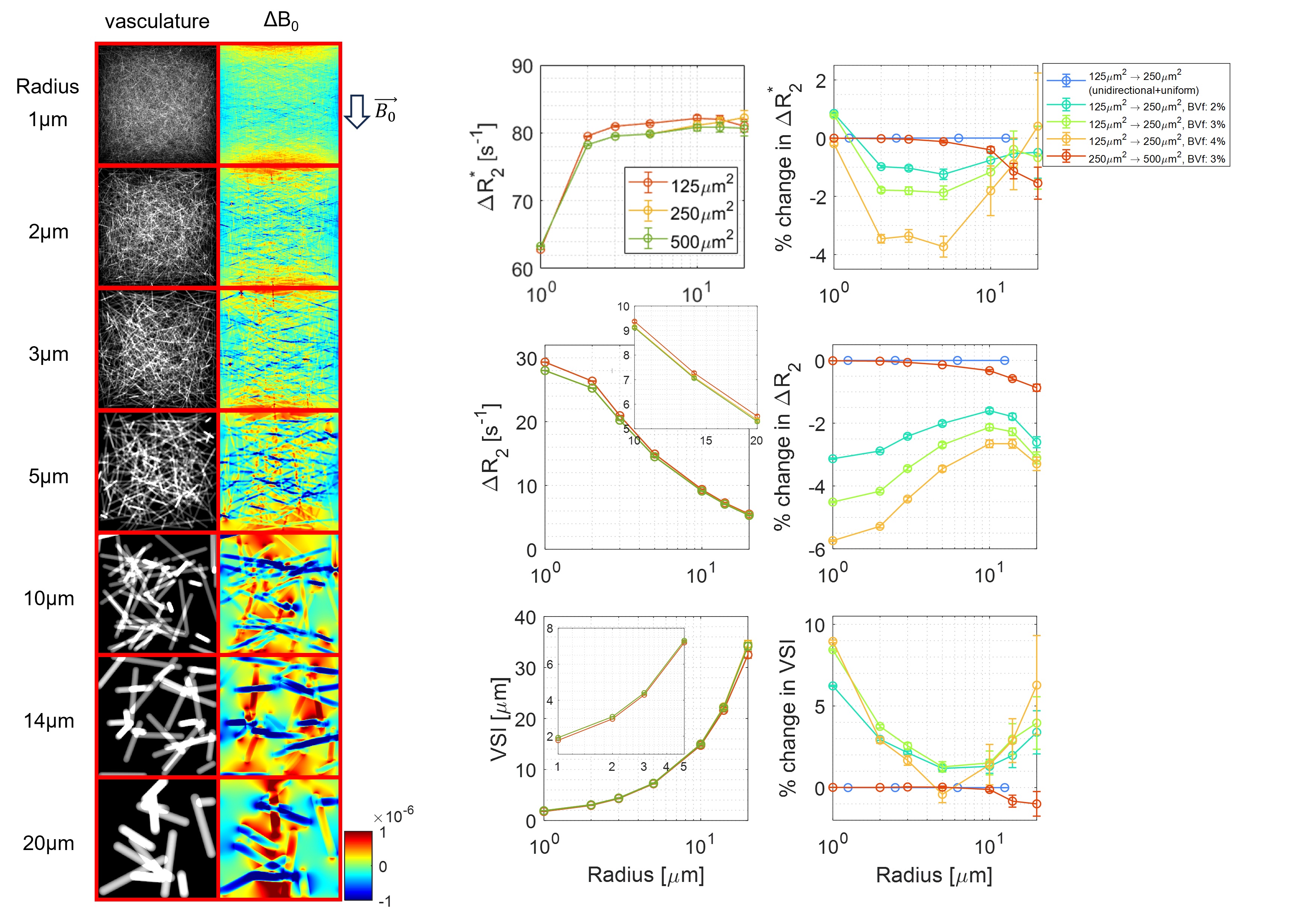
Monte-Carlo proton diffusion simulations were performed to compute ΔR2 and ΔR2* (SE and GE, respectively) based on previously described procedures at 7T (Lee et al.6). The simulation volume of randomly distributed cylindrical models (n=50) was 500×500×500µm3 with various radii (each random cylinder model consisted with a single vessel radius; 1, 2, 3, 5, 10, 14, and 20 (see Figure 1). Each simulation volume was subdivided into 125×125×500 and 250×250×500µm3 sub-volumes, and ΔR2, ΔR2*, and VSI of three different spatial resolutions (125×125×500, 250×250×500, and 500×500×500µm3) was calculated and compared. Blood volume fraction (BVf) effect was also examined for BVf of 2%, 3%, and 4%. Monte-Carlo simulations for unidirectional and uniformly distributed cylinder model was performed to verify that the resolution dependence of VSI originated from accumulated phase distribution within the MRI voxel.
Results
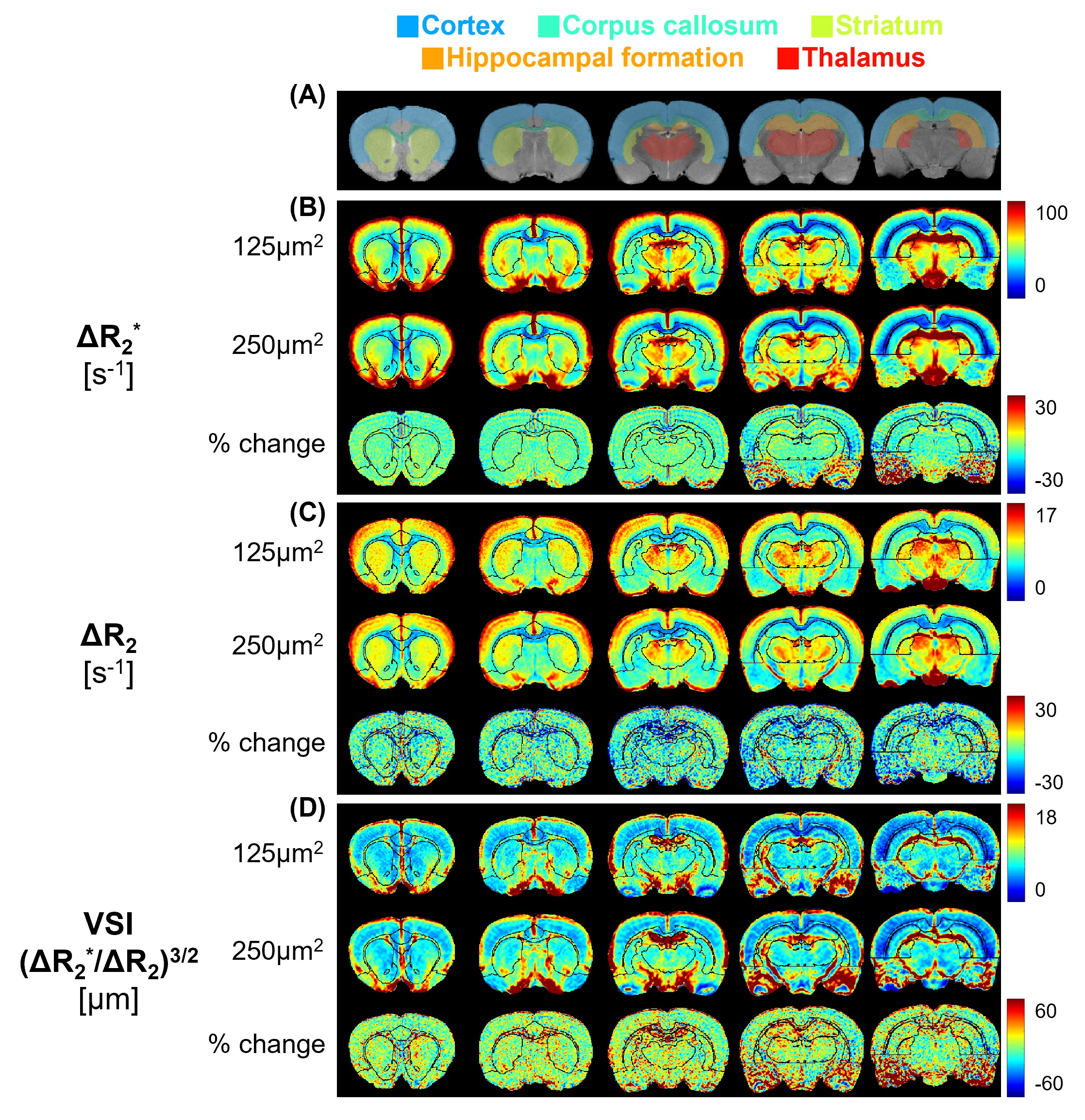
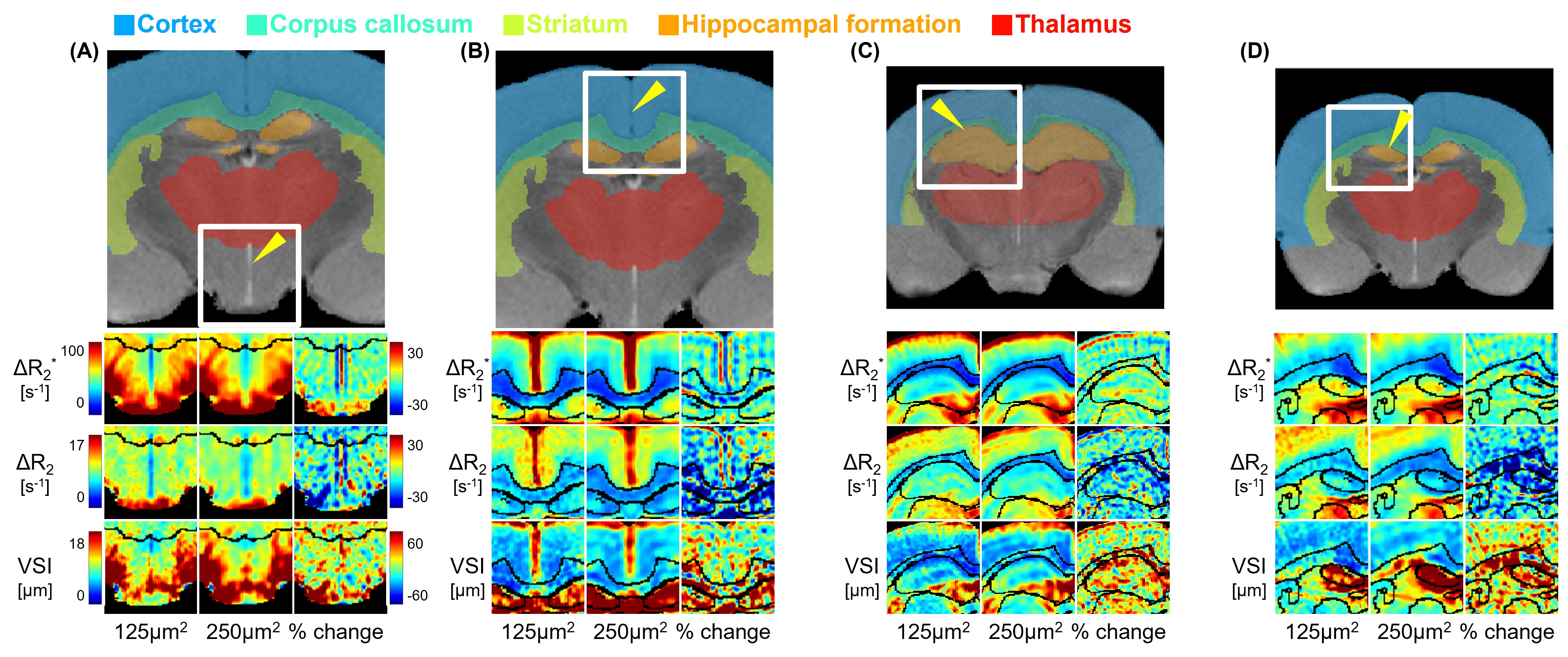
Figure 1 illustrated the Monte-Carlo simulation results for the resolution dependence of VSI MRI. Randomly distributed cylindrical models and corresponding ΔB0 maps were shown. ΔR2 and ΔR2* tend to decrease with lower spatial resolution to varying degrees with respect to vessel radius. As a result, when the in-plane resolution was lowered from 125µm2 to 250µm2, VSI tended to increase in the range of small microvessels with a vessel radius of less than 3um and in large vessels with a radius of 15 or more. When the in-plane resolution was lowered from 250µm2 to 500µm2, the change in ΔR2 and ΔR2* was minimal, and as a result, the change in VSI was also minimal. For the uniform cylinder distribution model, there was no change in ΔR2, ΔR2*, and VSI as the resolution decreases.Figure 2 shows the voxel-wise mean ΔR2, ΔR2*, and VSI maps and corresponding difference maps between 125µm2 and 250µm2 of in-plane resolutions (n=13 rats). Brain regional differences in ΔR2, ΔR2*, and VSI are clearly distinguished. In both ΔR2 and ΔR2* difference maps, large blood vessels and ventricles which have similar size to MRI voxel shows spill in and spill out effects due to partial volume effects, whereas these effects are canceled in VSI maps (Figure 3A,B). ΔR2 difference maps for small vessels (corpus callous and hippocampal formation, Figure 3C) and slightly large vessels (Figure 3D) shows larger reduction than other brain regions. As a result, the VSI difference due to resolution dependence in corpus callous and hippocampal formation regions (~12% increase) was larger than other brain areas (~5% increase).
Conclusion and discussion
The spatial resolution dependence of VSI MRI was investigated at in-plane resolutions of 125 and 250µm2 with 1.5 mm slice-thickness through in vivo rat brain experiments and Monte-Carlo simulations. Partial volume effects due to reduced resolution were present in ΔR2*and ΔR2, but were not significant in VSI. In areas with small VSI (corpus callosum and hippocampal formation) and areas with large VSI, the lower the resolution, the larger the decrease in ΔR2, resulting in a tendency to increase VSI, which was closely correlated with the simulation results. In summary, the resolution dependence of VSI MRI is largely influenced by ΔR2 rather than ΔR2*, and this dependence is strong at in-plane resolutions between 125 and 250µm2 in the rat brain.Acknowledgements
This research was supported by KBRI basic research program through Korea Brain Research Institute funded by Ministry of Science and ICT (23-BR-05-02)References
1. Tropres, Irene, et al. "Vessel size imaging." Magnetic Resonance in Medicine: An Official Journal of the International Society for Magnetic Resonance in Medicine 45.3 (2001): 397-408.
2. Troprès, Irène, et al. "Imaging the microvessel caliber and density: principles and applications of microvascular MRI." Magnetic resonance in medicine 73.1 (2015): 325-341.
3. Waxholm Space Atlas of the Sprague Dawley Rat Brain (RRID:SCR_017124)
4. Papp, Eszter A., et al. "Waxholm Space atlas of the Sprague Dawley rat brain." Neuroimage 97 (2014): 374-386.
5. Sosa, Sebastian, et al. "A multilevel statistical toolkit to study animal social networks: The Animal Network Toolkit Software (ANTs) R package." Scientific reports 10.1 (2020): 1-8.
6. Lee, D. K., M. S. Kang, and H. Cho. "MRI size assessment of cerebral microvasculature using diffusion-time-dependent stimulated-echo acquisition: A feasibility study in rodent." Neuroimage 215 (2020): 116784.
Figures