1127
High resolution pCASL mapping of perfusion in the mouse brain1Champalimaud Research, Champalimaud Foundation, Lisbon, Portugal, 2Institute for Systems and Robotics - Lisboa and Department of Bioengineering, Instituto Superior Técnico, Universidade de Lisboa, Lisbon, Portugal, 3C.J. Gorter Center for High Field MRI, Department of Radiology, Leiden University Medical Center, Leiden, Netherlands, 4Université Grenoble Alpes, Inserm, Grenoble Institut des Neurosciences, Grenoble, France
Synopsis
Keywords: Biology, Models, Methods, Perfusion, Arterial Spin Labelling
Motivation: pCASL perfusion mapping has many potential applications in preclinical imaging, but its use is still challenging particularly in mice and at higher fields due to limited sensitivity and constraints on labelling arising from the mouse’s anatomy.
Goal(s): Here, we set to push the spatial resolution limitations of pCASL in mice by over an order of magnitude.
Approach: For this, we leverage SNR increases provided by cryogenic coils and develop a novel experimental setup optimizing and stabilizing the positioning of the mice.
Results: We then show x11 higher spatial resolution CBF maps compared to the previous state-of-the-art and higher stability and reproducibility of findings.
Impact: We developed a setup optimizing carotid positioning for mice, thereby enabling efficient pCASL labeling. When combined with a cryogenic coil, perfusion images of the mouse brain were enhanced x11 in spatial resolution and were highly reproducible compared to current state-of-the-art.
Introduction
pCASL enables efficient perfusion mapping in-vivo without injection of contrast agents or use of multiple coils1. Despite its great potential, state-of-the-art pCASL in preclinical settings is still characterized by low spatial resolution and high variability in CBF maps1–3. Compared to rats, pCASL measurements are particularly challenging in mice due to the smaller brain, higher susceptibility artifacts, and mouse-specific anatomical constraints that limit labelling efficiency due to the way carotids are placed4. Here, we address this gap and develop a novel experimental setup that optimizes carotid placement and further harnesses dramatic SNR increases from cryogenic coils5 to achieve high-resolution and reliable pCASL imaging in mice at 9.4 T. Our results and the novel paradigm we introduce here augur well for future pCASL applications in mouse models of health and disease.Methods
Animal experiments were conducted according to the European Directive 2010/63 and preapproved by competent authorities.Experimental setup: A custom-built ramp was designed to prevent the angle between the animal’s head and body caused by the ear bars – ramp setup (Figure 1B).
Animal Preparation: Adult female C57BL/g mice (N=16,~12 weeks old, weights 20–25g) were sedated using 1.5–2.5% isoflurane. Respiratory rate was kept at 60-90 bpm.
Stroke induction: In N=3 animals a focal infarct was induced in the somatosensory cortex (S1HL) with a photothrombotic stroke model6. The animals were scanned using the ramp at t=0h, t=24h and t=5days after stroke.
MRI experiments: Experiments were conducted on a 9.4 T Bruker Biospec Scanner with an 86mm volume transmit coil and a 4-element array cryogenic receive coil. An unbalanced pCASL sequence2 was used as described in Hirschler et al.(2018)7. The labelling plane was positioned at the mouse neck (~8mm below the isocenter), labelling duration (LD)=3s and post-labelling delay (PLD)=300ms. N=7 animals were scanned with the labelling plane tilt of 0o (no ramp 0o) and 45o (no ramp 45o) in relation to the imaging region (Fig.1A) and N=6 animals were scanned with the labeling plane angle of 0o and on top of a ramp (Fig.1B). All mice were scanned at low and high-resolution.
Low-resolution pCASL acquisitions consisted of a single-shot EPI: FOV=14x14mm2, slice thickness=1mm, slice gap=0.2mm, matrix=96x96 resulting in a spatial resolution of 146x146μm2, repetition time (TR)/echo time(TE)=4000/17ms, 30 repetitions, Tacq=4min.
For high-resolution pCASL, a single-shot EPI was implemented: FOV=12x12mm2, slice thickness=0.5mm, slice gap=0.35mm, matrix=120x120, resulting in a spatial resolution of 100x100μm2, TR/TE=4000/25ms, 30 repetitions, Tacq=4min. For CBF quantification, the T1 map was obtained from an inversion recovery sequence.7 A pCASL encoded FLASH was employed to estimate the inversion efficiency (IE) 3mm above the labelling plane (PLD of 0ms, LD of 200ms).7
Data Analysis: CBF (ml/100g/min) was calculated pixel-by-pixel from the relative ASL signal difference between control and label images (rASL) as previously described8. In the rASL and CBF high-resolution maps, two ROIs were defined (cortical and thalamic) and used for averaging rASL and CBF. The IE was calculated by manually drawing a ROI for each carotid.
Results
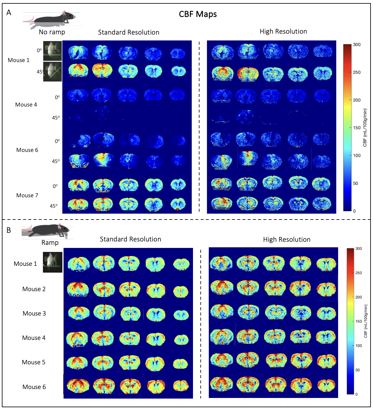
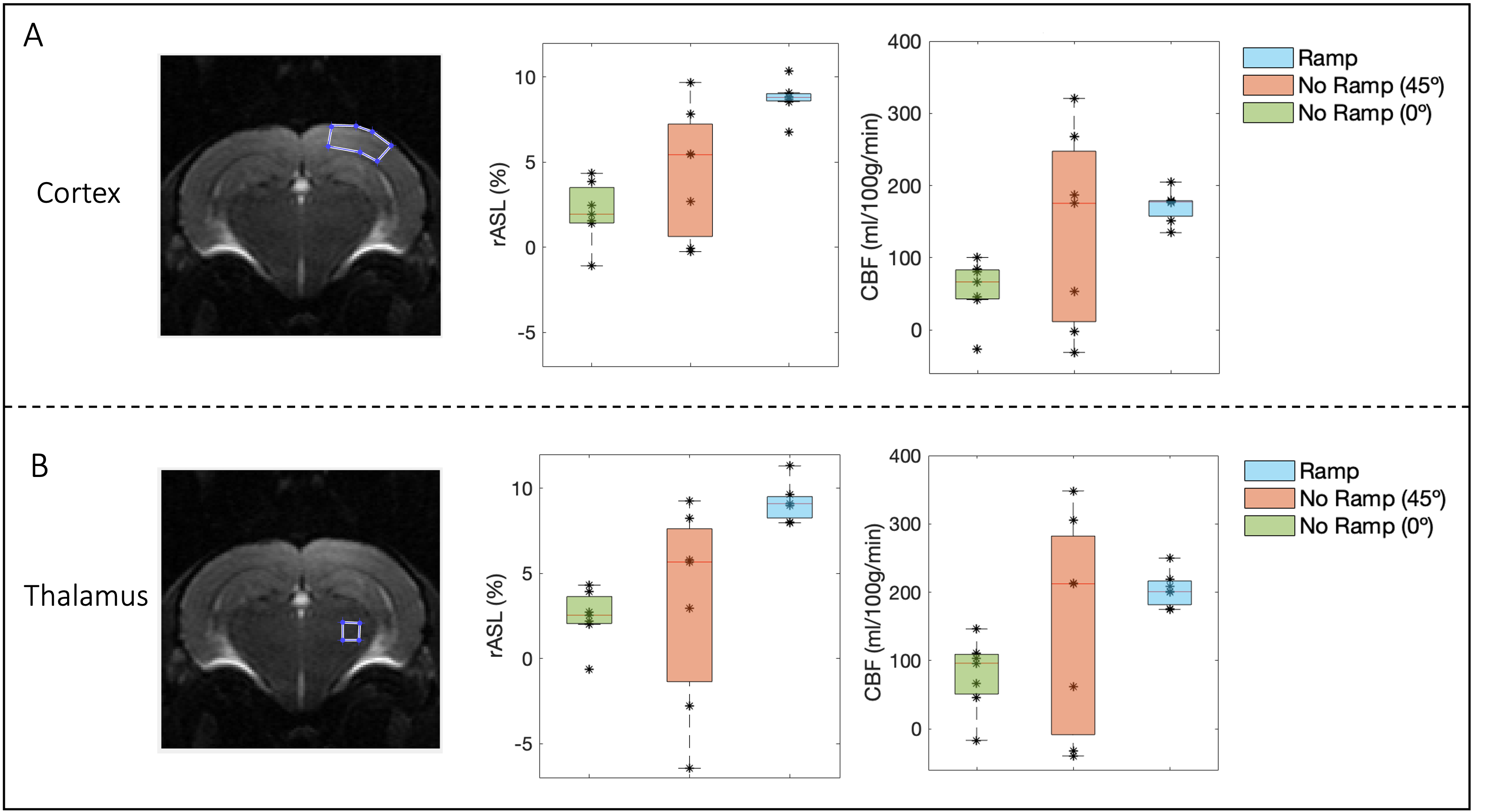
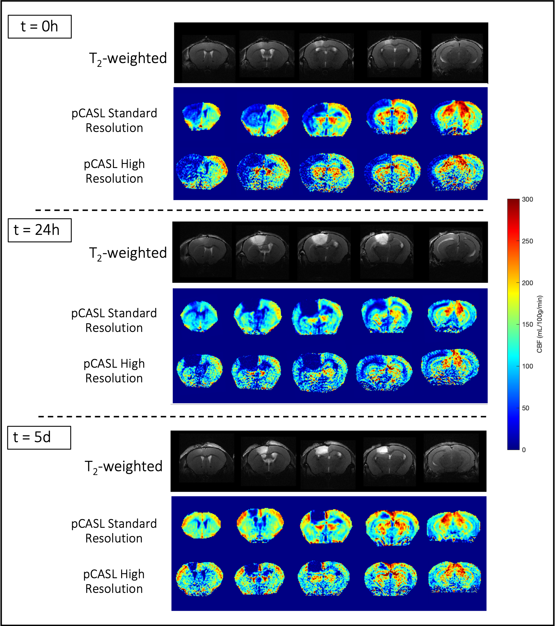
The IE was calculated for each setup as an average of both carotids: no ramp 0o=0.57±0.20, no ramp 45o=0.80±0.63 and ramp=0.78±0.04. Note the dramatically smaller variance in the ramp compared to the other conditions. Additionally, only the ramp setup reveals the expected linear relationship between rASL and IE in the ROIs analysed (Fig.2). Figure 3 shows CBF maps from the mice scanned at the low-resolution and high-resolution for the 3 conditions. In controls, only the acquisitions using the ramp present the expected CBF patterns in the healthy mouse brain, with little variability across animals both at low- and high-resolution. In the high-resolution images, a better delineation of different brain regions can be discerned (e.g., differences in perfusion within different hippocampal layers, descending cortical vessels). Furthermore, the ramp setup shows lower standard deviation and highest average rASL across the two ROIs (Fig.4). In the stroke model, we detected CBF impairments across the brain as well as the lesion volume that were changing over time (Fig.5).Discussion
By using a novel experimental setup, we managed to increase the state-of-the-art resolution of pCASL perfusion images of the mouse brain by a factor of 11 and to obtain highly reliable CBF maps across animals, without prolonging experiment time. Our findings include enhanced stability of the images acquired with the ramp (lower standard deviation in the IE measure) and a better delineation of brain areas including in an animal model of stroke, unveiling pCASL’s potential to detect alterations in CBF in mouse models of disease or healthy longitudinal processes. Our findings bode well for future applications of quantitative CBF mapping in mice.Acknowledgements
The authors acknowledge the vivarium of the Champalimaud Centre for the Unknown, a facility of CONGENTO which is a research infrastructure co-financed by Lisboa Regional Operational Programme (Lisboa 2020), under the PORTUGAL 2020 Partnership Agreement through the European Regional Development Fund (ERDF) and the Champalimaud Communication, Events and Outreach team. We would also like to acknowledge Fundação para a Ciência e Tecnologia (Portugal), under project LISBOA-01-0145-FEDER-022170 and grant 2021.08457.BD.
References
1. Buck J, Larkin JR, Simard MA, et al. Sensitivity of multiphase pseudocontinuous arterial spin labelling (MP pCASL) magnetic resonance imaging for measuring brain and tumour blood flow in mice. Contrast Media Mol Imaging. 2018;2018.
2. Duhamel G, Callot V, Tachrount M, et al. Pseudo-continuous arterial spin labeling at very high magnetic field (11.75 T) for high-resolution mouse brain perfusion imaging. Magn Reson Med. 2012;67(5):1225-1236.
3. Hirschler L, Munting LP, Khmelinskii A, et al. Transit time mapping in the mouse brain using time-encoded pCASL. NMR Biomed. 2018;31(2):1-11.
4. Larkin JR, Simard MA, Khrapitchev AA, et al. Quantitative blood flow measurement in rat brain with multiphase arterial spin labelling magnetic resonance imaging. Journal of Cerebral Blood Flow and Metabolism. 2019;39(8):1557-1569.
5. Rating D, Baltes C, Nordmeyer-Massner J, et al. Performance of a 200-MHz cryogenic RF probe designed for MRI and MRS of the murine brain. Magn Reson Med. 2008;59(6):1440-1447.
6. Watson BD, Dalton Dietrich W, Busto R, et al. Induction of Reproducible Brain Infarction by Photochemically Initiated Thrombosis, Ann. Neurol., vol. 17, no. 5, pp. 497–504, 1985.
7. Hirschler L, Debacker CS, Voiron J, et al. Interpulse phase corrections for unbalanced pseudo-continuous arterial spin labeling at high magnetic field. Magn Reson Med. 2018;79(3):1314-1324.
8. Alsop DC, Detre JA, Golay X, et al. Recommended Implementation of ASL Perfusion MRI for Clinical Applications. Magn Reson Med. 2015;73(1):102-116
Figures
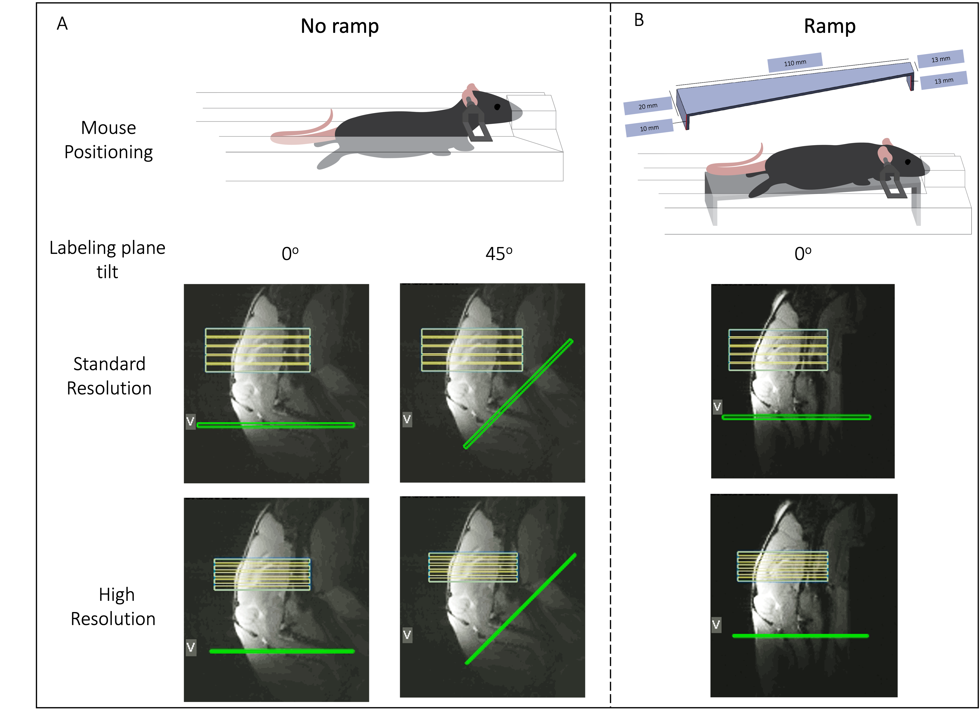
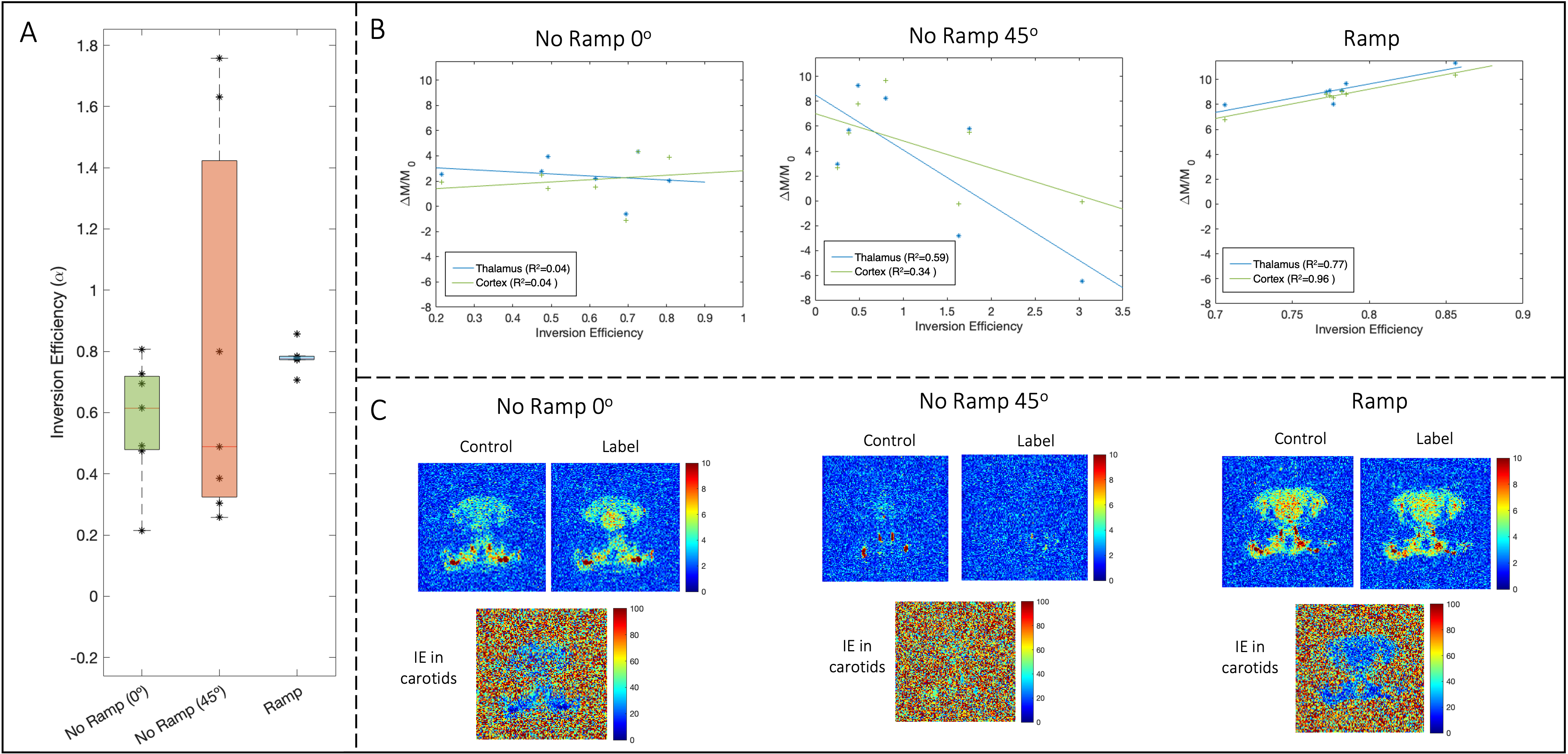
Fig. 2 A) Inversion Efficiency (IE) estimation for the 3 setups at the level of the carotids. Note that there is a dramatically smaller variance in the ramp compared to the other conditions. B) Relationship between ΔM and IE across setups in two specific ROIs (thalamic and cortical). Only in the ramp setup there is the expected linear relationship. C) Magnitude images acquired with the FcFLASH sequence. Note that in the no ramp 45o setup, the carotids can hardly be spotted.