1120
Single-timepoint dynamic functional connectivity patterns in temporal lobe epilepsy1Department of Biomedical Engineering, Vanderbilt University, Nashville, TN, United States, 2Institute of Imaging Science, Vanderbilt University Medical Center, Nashville, TN, United States, 3Department of Electrical and Computer Engineering, Vanderbilt University, Nashville, TN, United States, 4Department of Neurological Surgery, Vanderbilt University Medical Center, Nashville, TN, United States
Synopsis
Keywords: Functional Connectivity, fMRI (resting state), Epilepsy, Functional Connectivity
Motivation: Epileptic tissue generates interictal spikes between seizures, which are used to localize the epileptic focus clinically.
Goal(s): We aimed to detect dynamic functional connectivity (FC) patterns in resting-state fMRI data that may be related to interictal spikes.
Approach: We detected whole-brain dynamic FC patterns at timepoints that had FC characteristics similar to epileptic spikes in both healthy controls and patients with temporal lobe epilepsy (TLE).
Results: We found three dynamic FC patterns, one of which occurred more in TLE than in controls and the occurrence of which was related to clinical measures of epilepsy severity.
Impact: These results suggest the potential clinical utility of fMRI-based dynamic FC to detect interictal spikes. Future studies can evaluate the correspondence of these dynamic FC patterns to interictal spikes using simultaneous electrophysiology and fMRI.
Introduction
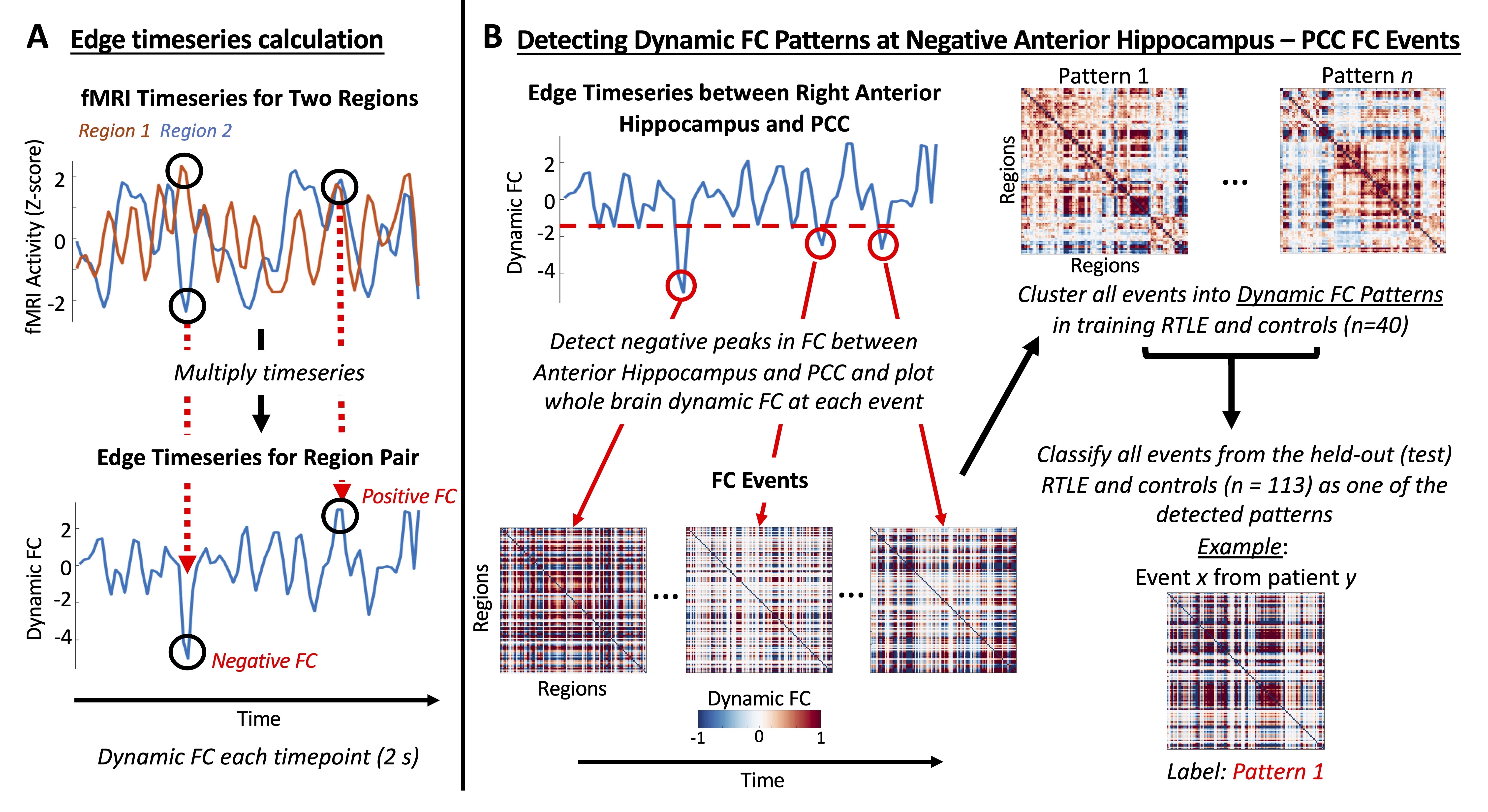
Epileptic foci produce interictal spikes, brief electrical events between seizures that are used to localize epileptic tissue. In patients with temporal lobe epilepsy (TLE), these spikes induce opposing fMRI signal changes in the anterior hippocampus and posterior cingulate cortex (PCC)1,2, producing instances of negative functional connectivity (FC) between these regions. Here we applied edge timeseries, a single-timepoint dynamic FC method3, to resting-state fMRI to detect whole-brain dynamic FC patterns at moments of negative FC between the anterior hippocampus and PCC. We tested whether these dynamic FC patterns occurred more in TLE, if they were related to disease severity, and investigated their spatiotemporal dynamics to assess their potential link to interictal spikes.Methods
This study included 96 healthy controls (37.9 ± 13.4 yrs, 46 female), split into training (n = 20) and testing (n = 76) groups, along with 37 right TLE (RTLE) patients (40.4 ± 11.5 yrs, 19 female) split into training (n = 20) and testing (n = 17) groups. Participants underwent 3T MRI scanning including a T1-weighted scan (1x1x1 mm3) and 20 minutes of resting-state fMRI (TR = 2 s, 3x3x4 mm3). The T1-weighted scan was used to segment 111 cortical and subcortical brain regions4.fMRI data underwent standard preprocessing including RETROICOR5. Normalized timeseries from each pair of regions were multiplied at each timepoint to obtain edge timeseries3 (Fig 1A). Events were detected as negative peaks that exceeded a threshold in the edge timeseries between the right anterior hippocampus and right PCC (Fig 1B). Whole-brain dynamic FC events were extracted as the edge timeseries from all pairs of brain regions at event timepoints. All FC events from the training RTLE and control participants were clustered into dynamic FC patterns with the Louvain algorithm6,7 (Fig 1B). FC events from all participants were classified as one of the detected patterns (Fig 1B).
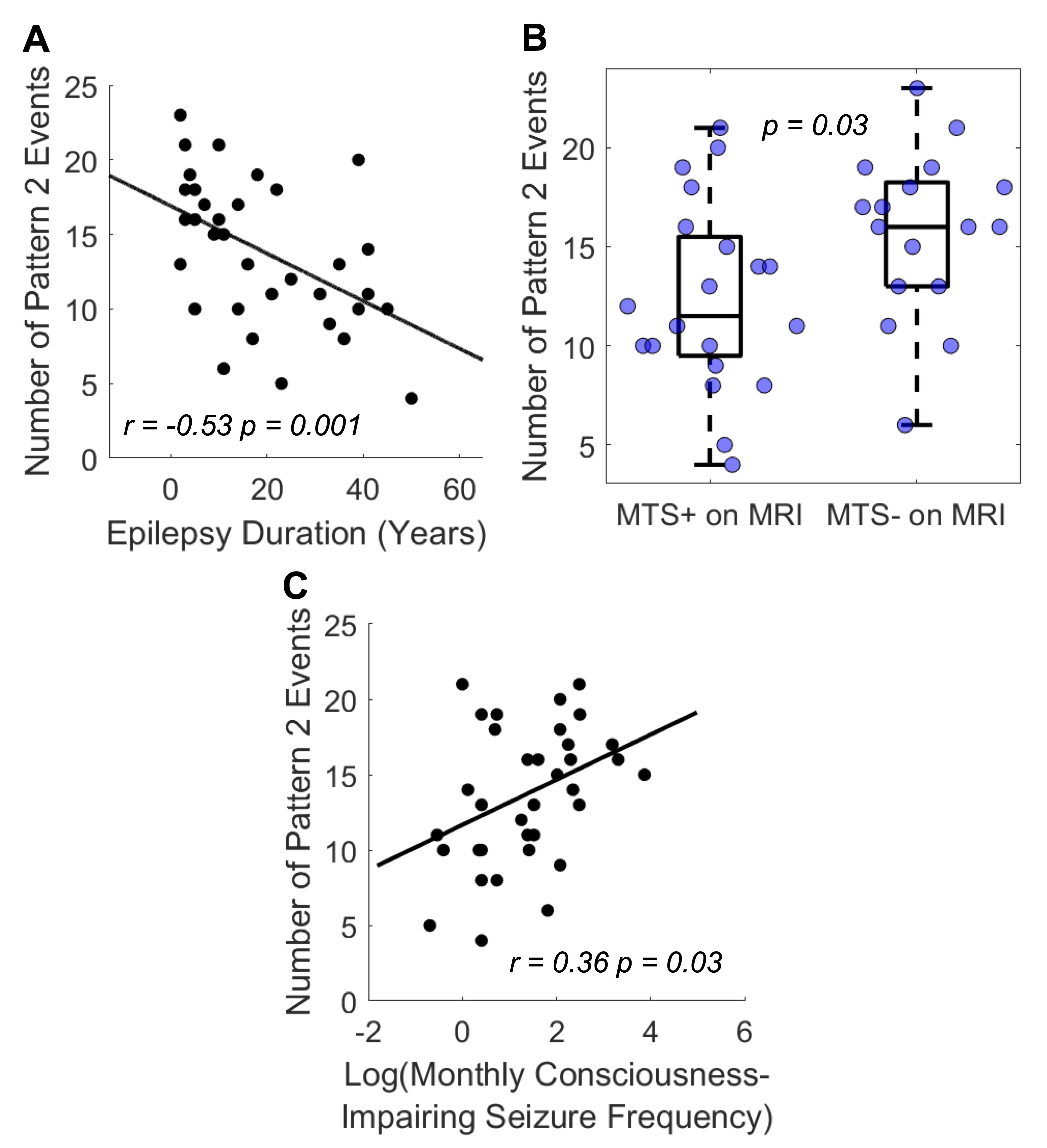
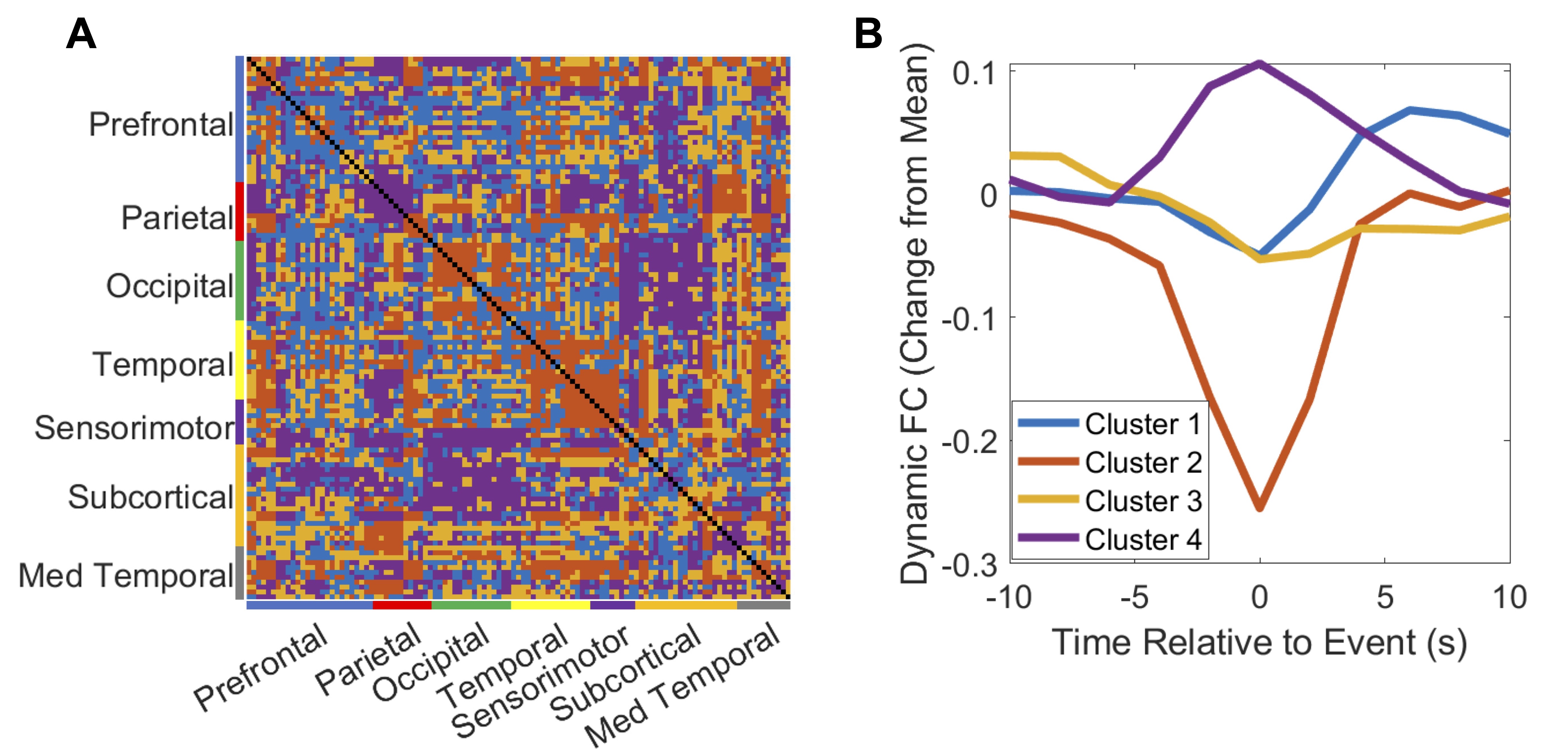
The total number of detected FC events, as well as the occurrence of each dynamic FC pattern, were compared between test RTLE and controls using Wilcoxon rank-sum tests. The occurrence of Pattern 2 in RTLE was compared to epilepsy duration and consciousness-impairing seizure frequency using Spearman correlations, as well as to the presence of mesial temporal sclerosis (MTS) on MRI using a Wilcoxon rank-sum test. To examine the spatiotemporal dynamics of Pattern 2, edges were clustered based on their average dynamic FC surrounding Pattern 2 events with k-means.
Results
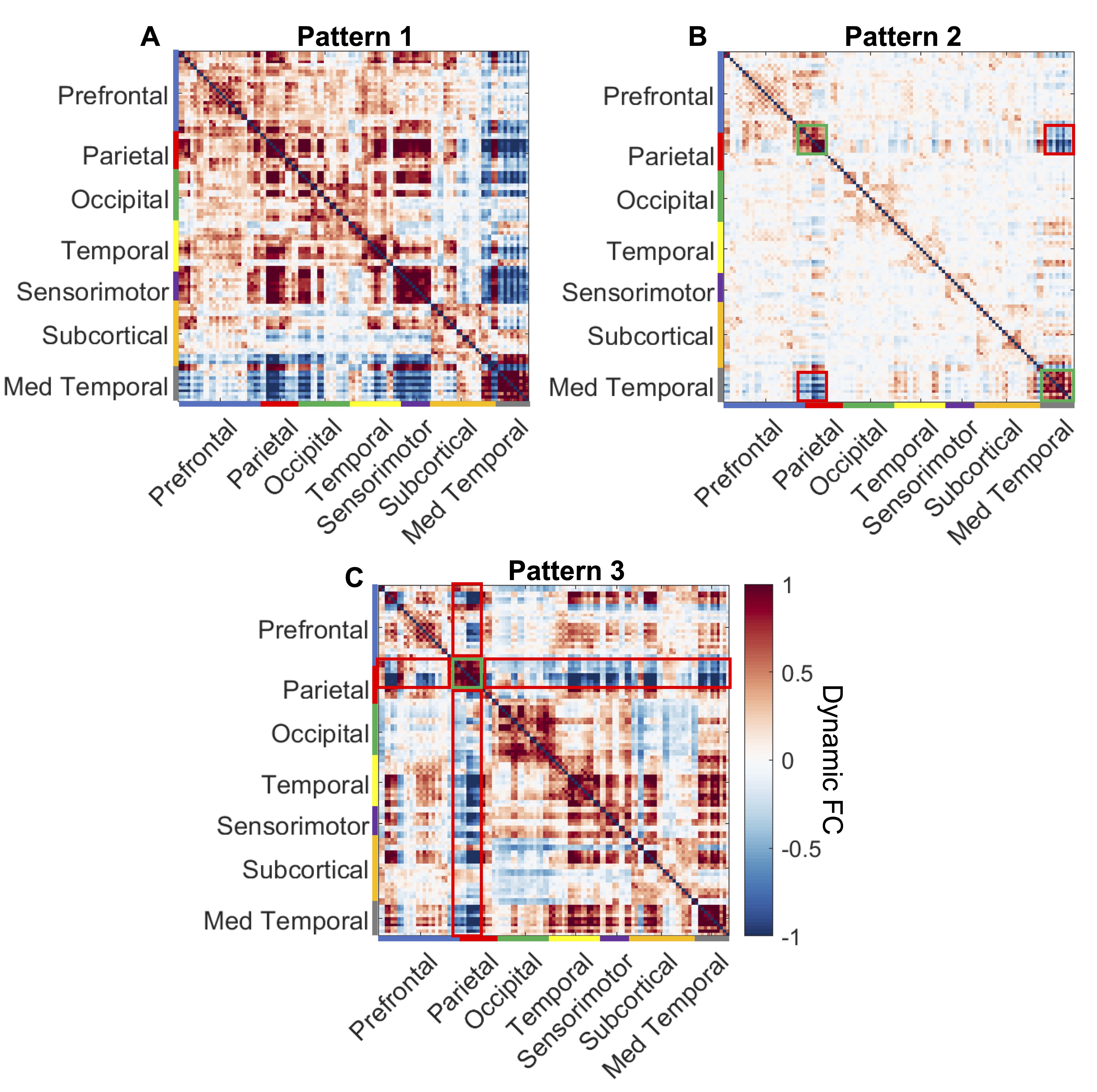
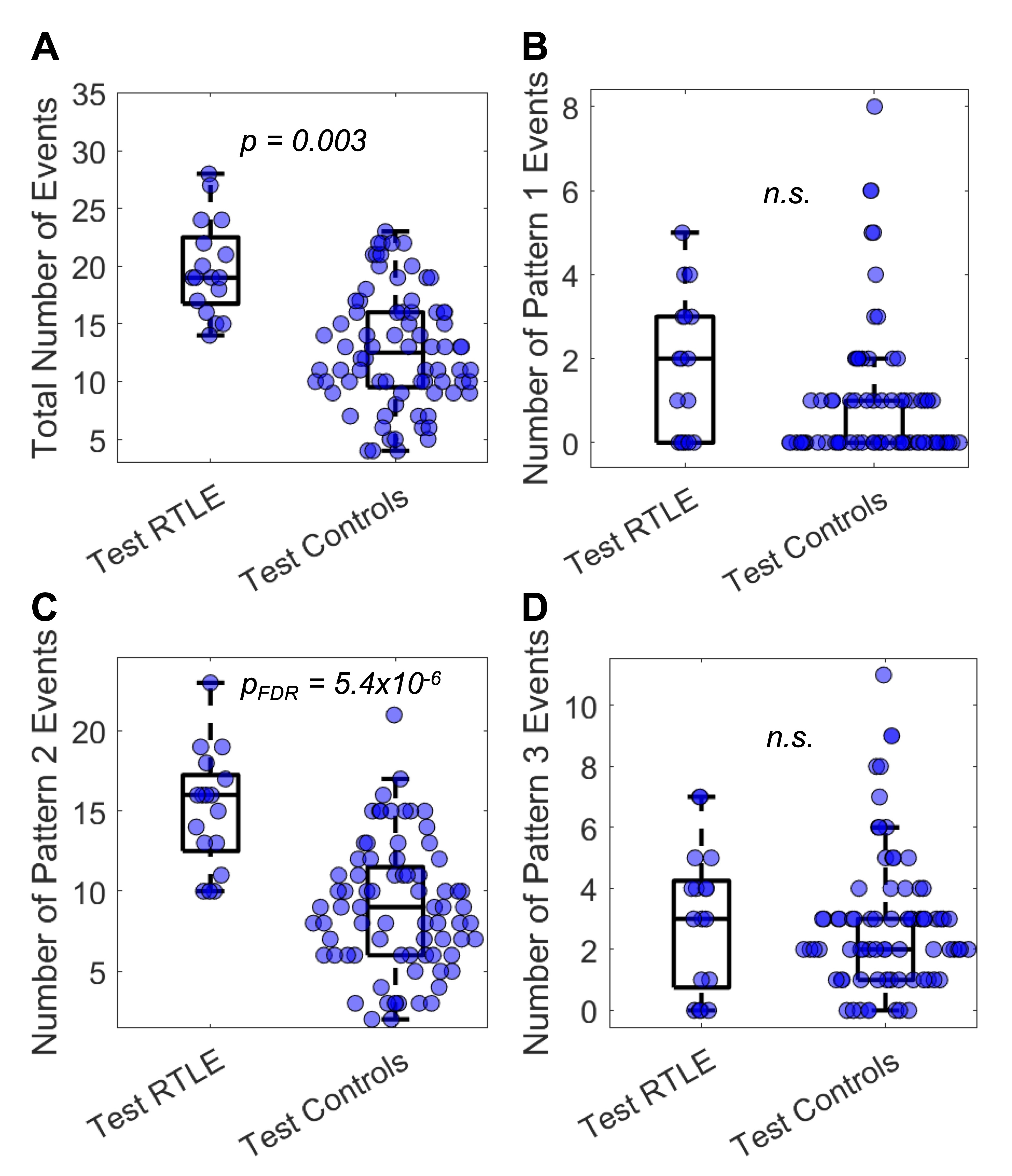
Three dynamic FC patterns were found at timepoints of negative FC between the right anterior hippocampus and right PCC. Pattern 1 displayed positive FC within the cortex and within the subcortex, but negative FC between the cortex and subcortex (Fig 2A). Pattern 2 had low amplitude FC overall, but strong positive FC within the default-mode network (DMN) and within medial temporal regions (green boxes, Fig 2B) along with negative FC between DMN and medial temporal regions (red boxes, Fig 2B). Pattern 3 showed positive FC within the DMN (green box, Fig 2C) and within much of the rest of the cortex, along with negative FC between the DMN and the rest of the cortex (red boxes, Fig 2C).Patients with RTLE had more frequent negative anterior hippocampus-PCC FC events, regardless of pattern, (p = 0.003, Fig 3A) as well as more Pattern 2 events (pFDR = 5.4x10⁻⁶, Fig 3C) than controls. The number of Pattern 2 events was correlated with epilepsy duration (r = -0.53, p = 0.001, Fig 4A) and consciousness-impairing seizure frequency (r = 0.36, p =0.03, Fig 4C). Patients with RTLE who were MTS positive on MRI had fewer Pattern 2 events than those who were MTS negative on MRI (p = 0.03, Fig 4B). The spatiotemporal dynamics surrounding Pattern 2 was found to be described by four clusters of edges (Fig 5).
Discussion
We detected three whole-brain dynamic FC patterns that occur at timepoints of negative FC between the anterior hippocampus and PCC. Two of the dynamic FC patterns were global events, while Pattern 2 was focal to DMN and medial temporal regions. Both RTLE and controls had each kind of pattern occur, however, patients with RTLE had more FC events than controls overall, which was largely due to a higher occurrence of Pattern 2. Moreover, the number of Pattern 2 events was correlated with multiple disease measures across patients with RTLE.Conclusion
This study found an increased occurrence of a specific dynamic FC pattern in RTLE compared to controls with negative hippocampus-PCC FC, an attribute of interictal spikes. Future studies can determine whether these dynamic FC patterns are directly related to interictal spikes using simultaneous electrophysiology and fMRI.Acknowledgements
Funded by NIH T32 EB021937, R01 NS075270, R01 NS108445, R01 NS110130, and R00 NS097618.References
1. Kobayashi E, Grova C, Tyvaert L, Dubeau F, Gotman J. Structures involved at the time of temporal lobe spikes revealed by interindividual group analysis of EEG/fMRI data. Epilepsia. 2009;50(12):2549-2556. doi:https://doi.org/10.1111/j.1528-1167.2009.02180.x2.
2. Laufs H, Hamandi K, Salek‐Haddadi A, Kleinschmidt AK, Duncan JS, Lemieux L. Temporal lobe interictal epileptic discharges affect cerebral activity in “default mode” brain regions. Human Brain Mapping. 2007;28(10):1023-1032. doi:https://doi.org/10.1002/hbm.203233.
3. Esfahlani FZ, Jo Y, Faskowitz J, et al. High-amplitude cofluctuations in cortical activity drive functional connectivity. PNAS. 2020;117(45):28393-28401. doi:10.1073/pnas.20055311174.
4. Sainburg LE, Little AA, Johnson GW, et al. Characterization of resting functional MRI activity alterations across epileptic foci and networks. Cerebral Cortex. 2022;32(24):5555-5568. doi:10.1093/cercor/bhac0355.
5. Glover GH, Li TQ, Ress D. Image-based method for retrospective correction of physiological motion effects in fMRI: RETROICOR. Magnetic Resonance in Medicine. 2000;44(1):162-167. doi:10.1002/1522-2594(200007)44:1<162::AID-MRM23>3.0.CO;2-E6.
6. Lancichinetti A, Fortunato S. Consensus clustering in complex networks. Sci Rep. 2012;2(1):336. doi:10.1038/srep003367.
7. Betzel RF, Cutts SA, Greenwell S, Faskowitz J, Sporns O. Individualized event structure drives individual differences in whole-brain functional connectivity. NeuroImage. 2022;252:118993. doi:10.1016/j.neuroimage.2022.118993
Figures