1118
Longitudinal resting-state network changes in treatment-resistant OCD patients following MR-guided Focused Ultrasound Capsulotomy1Hotchkiss Brain Institute, Cumming School of Medicine, University of Calgary, Calgary, AB, Canada, 2Radiology and Clinical Neurosciences, Cumming School of Medicine, University of Calgary, Calgary, AB, Canada, 3Dept of Surgery, University of Calgary, Calgary, AB, Canada, 4Dept of Psychiatry, Cumming School of Medicine, University of Calgary, Calgary, AB, Canada
Synopsis
Keywords: Task/Intervention Based fMRI, fMRI (resting state), Obsessive compulsive disorder, Focused Ultrasound, Surgery
Motivation: MR-guided focused ultrasound capsulotomy is a surgical procedure to treat intractable OCD. While demonstrating clinical success, the mechanisms of symptom decrease are poorly understood.
Goal(s): This study sought to explore how intrinsic brain networks change following surgery.
Approach: Seed-based resting state fMRI was used to analyze intrinsic networks in a group of 6 OCD patients prior to and following surgery, along with a matched control group.
Results: Prior to surgery, OCD patients showed greater connectivity within internally-focused networks, and less connectivity in those involved in external cognition. One year following surgery, these connectivity differences were substantially reduced relative to controls.
Impact: This study will appeal to neuroscientists interested in resting-state networks involved with OCD, as well as in how these networks change following a MRgFUS surgical procedure which produced substantial clinical improvement.
INTRODUCTION
Obsessive-compulsive disorder (OCD) is defined by obsessions (unwanted repetitive thoughts) and compulsions (ritualistic behaviour or situational avoidance). Surgical options exist for OCD patients with severe symptoms who are unresponsive to medication or behavioural therapy. Magnetic resonance-guided focused ultrasound (MRgFUS) lesioning of the anterior limbs of the internal capsule is an appealing surgical procedure for intractable OCD, due to its precision and minimal invasiveness.Dysfunction between various brain networks, including Default Mode Network (DMN), Central Executive Network (CEN), and Salience Network (SAL) may contribute to OCD pathology1,2. DMN is considered task-negative and associated with internal cognition, while CEN is associated with external goal-oriented behaviour. The SAL network serves as a "switch" between DMN and CEN, identifying novel stimuli and prompting activation of CEN and other attentional networks. The dorsal anterior cingulate cortex (dACC) is a key node of the SAL network3, and dysfunction in this region has been associated with OCD3.
Purpose: Investigate dACC-seeded resting-state fMRI for exploration of network changes in OCD patients presurgically and 1-year post-MRgFUS capsulotomy.
METHODS
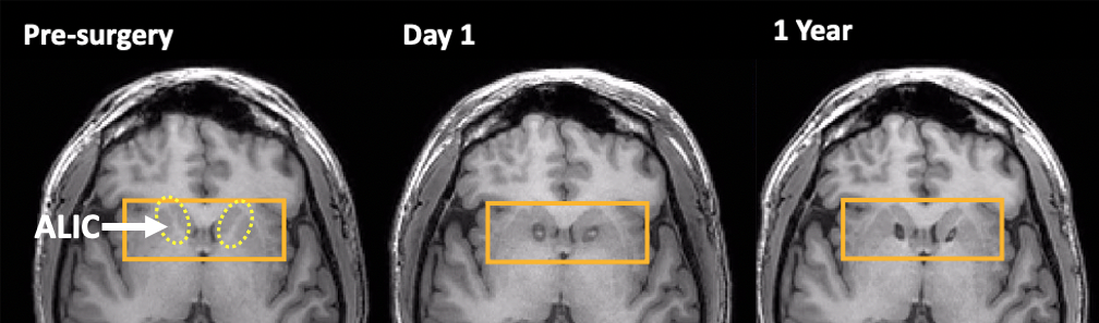
Participants: 6 patients with extreme4 treatment-resistant OCD (35.5±11.4y, 3M/3F, Y-BOCS 32.3±1.2) who underwent MRgFUS bilateral capsulotomy (Fig. 1) in our centre June 2019-September 2022, and a matched n=6 healthy control group (HC/NC; 35.8±11.6y, 3M/3F). The Yale-Brown Obsessive Compulsive Score5 (Y-BOCS; max 40) was used to assess OCD symptom severity, with pre-surgical and 12-month timepoints considered in this analysis.MRI: MRI data were acquired using a 3T Discovery 750 (GE Healthcare), and included T1-weighted anatomical [T1w; 3D FSPGR (BRAVO)] and resting-state functional [gradient-echo EPI [TR/TE: 3500/30ms, FA=90o, 128x128matrix, 224x224mm FOV, 50 slices, 200 volumes, eyes open, 11:40 scantime]]. MRI data were obtained prior to (PRE) and 12-months following surgery (1Yr).
Resting-state preprocessing (using FSL6 unless noted): registration between fMRI, T1w, and MNI; motion correction; slice-timing correction; 4mm spatial filtering, bandpass 0.01-0.1Hz, and ICA-based cleaning (FIX7).
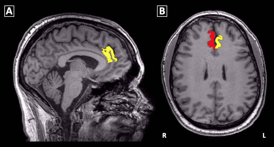
ROI analysis: Left and right dACC were defined on T1w (Fig. 2) for each participant using the Destrieux atlas within Freesurfer8, and transformed to rs-fMRI space. Seed-based analysis using dACC ROIs was then performed for OCD patients (PRE, 1Yr) and HC.
Statistics: OCD_PRE vs HC and OCD_1Yr vs HC were separately analyzed using between-group analyses. Resting-state fMRI analysis used cluster-based thresholds of Z=2.7 and p=0.01 in group analyses, conducted using FEAT9.
RESULTS
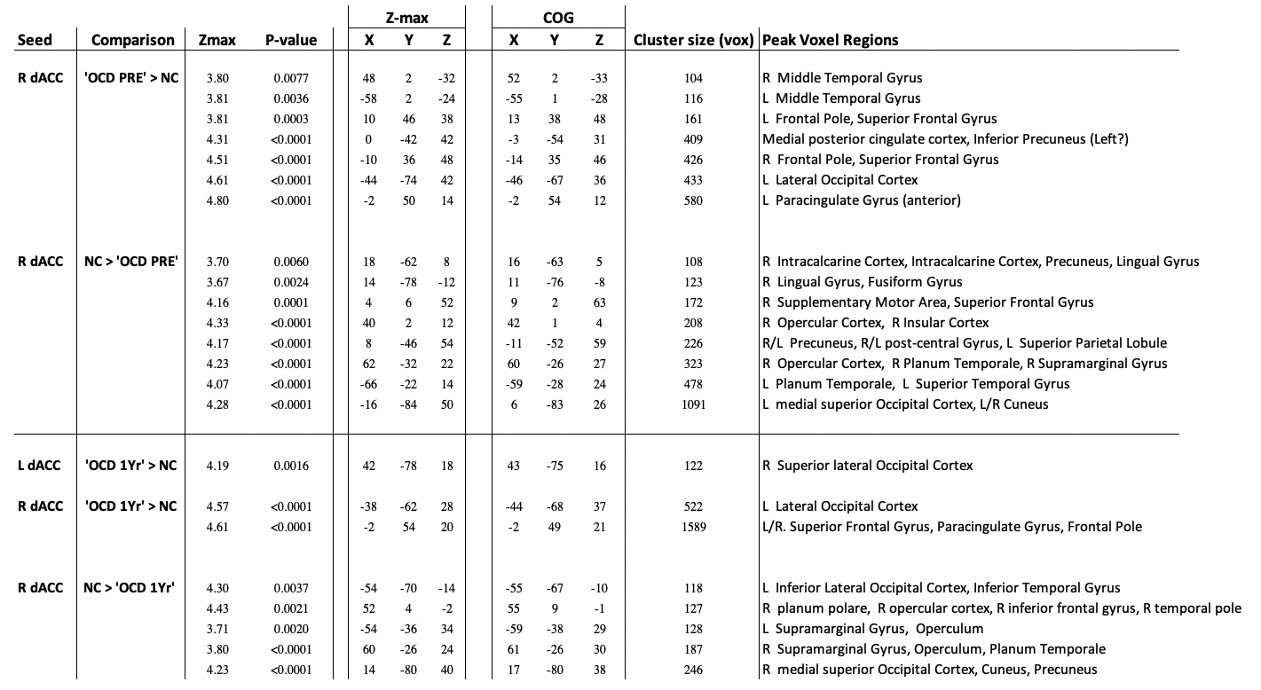
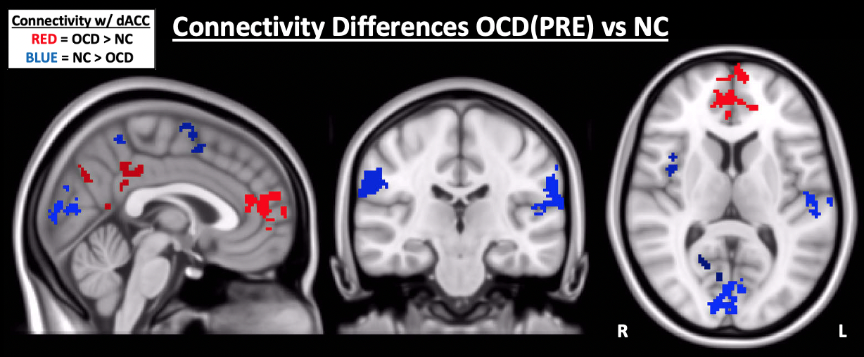
Clinical Measures: Pre-surgical Y-BOCS scores were 32.3±1.2 (extreme OCD4), and 19.2±2.7 1-year post-surgery (moderate OCD). Y-BOCS reduction was 41±7% (p<0.005), regarded as full response to treatment10.rs-fMRI: Between Groups ('OCD_PRE' vs HC): Only right dACC produced significant clusters (Fig. 3 Table). Notable regions of greater dACC-connectivity within presurgical OCD patients included bilateral middle temporal gyrus, bilateral frontal pole and superior frontal gyrus, medial posterior cingulate cortex, and left superior lateral occipital cortex (Fig. 4).
Conversely, HC showed greater dACC-connectivity in posterior medial regions such as lingual and fusiform gyri, cuneus, dorsal precuneus, bilateral medial superior occipital lobe, as well as lateral regions such as inferior parietal lobule and right operculum/insula.
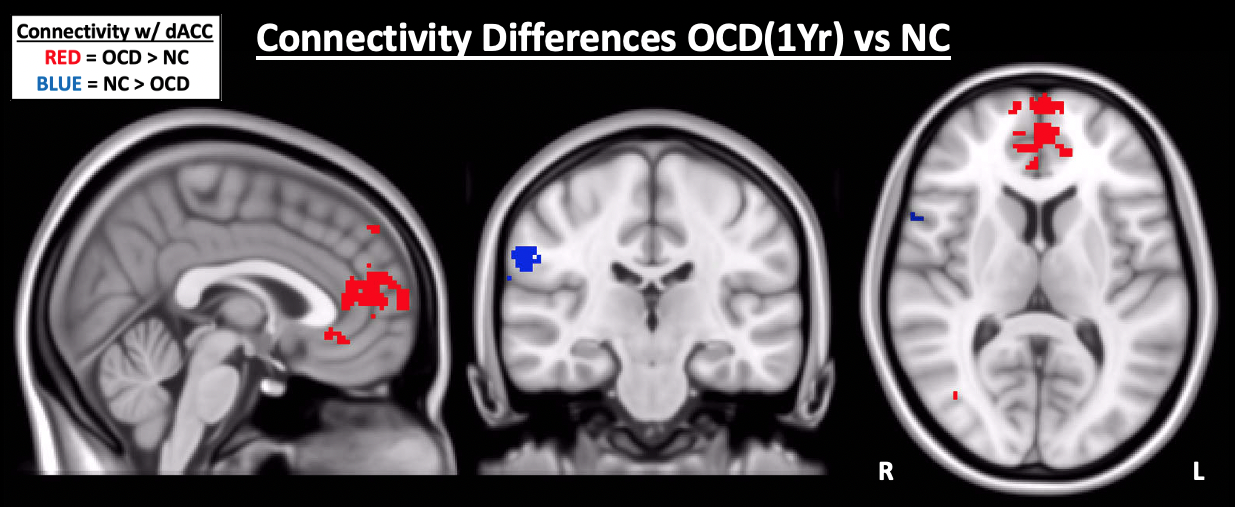
rs-fMRI: Between Groups ('OCD_1Yr' vs HC): The number of significant clusters between OCD_1Yr and HC was drastically reduced in comparison to the OCD_PRE vs HC comparison (Fig. 3 Table), notable absences including medial posterior cingulate cortex, the right insula, dorsal precuneus, and medial occipital lobe regions (Fig. 5).
However, in the OCD group, significant prefrontal cortex clusters remain, along with left superior lateral occipital lobe. Furthermore, a left-dACC-seeded cluster is present in right superior lateral occipital lobe. In the HC group, the right frontal operculum and inferior frontal gyrus clusters remain, as well as those in the bilateral inferior parietal lobule.
DISCUSSION
Our results demonstrate differences in resting state dACC-connectivity between treatment-resistanct OCD patients and controls, as well as a marked reduction in these differences one year following MRgFUS-capsulotomy.Pre-surgically, OCD patients showed elevated dACC-connectivity in medial prefrontal regions, posterior cingulate cortex, and superior lateral cingulate cortex. These regions are key nodes in the DMN11, associated with inwardly-directed cognition11. Conversely, the HC group showed greater dACC-connectivity than pre-surgical OCD patients in regions associated with Salience and Central Executive networks, such as insula and operculum (SAL)12, as well as dorsal precuneus and inferior parietal lobule (CEN)13.
Differences of dACC-connectivity between OCD patients and controls were drastically reduced one year following surgery, particularly within posterior cingulate cortex (DMN)11, right insula (SAL)12, and dorsal precuneus (CEN13), suggesting increased salience of external versus internally-driven stimuli in OCD patients following MRgFUS-capsulotomy.
CONCLUSION
The therapeutic effect of MRgFUS-capsulotomy may relate to a shift from DMN-dominance to externally-related networks in OCD patients.Acknowledgements
No acknowledgement found.References
1. Stern ER, Fitzgerald KD, Welsh RC. Resting-state functional connectivity between fronto-parietal and default mode networks in obsessive-compulsive disorder. PLoS One 2012;7:e36356.
2. Gursel DA, Avram M, Sorg C, Brandl F, Koch K. Frontoparietal areas link impairments of large-scale intrinsic brain networkds with aberrant fronto-striatal interactions in OCD: a meta-analysis of resting-state functional connectivity. Neurosci Biobehav Rev. 2018;87:151-160.
3. McGovern RA, Sheth SA. Role of the dorsal anterior cingulate cortex in obsessive-compulsive: converging evidence from cognitive neuroscience and psychiatric neurosurgery. J Neurosurg. 2017;126:132-147.
4. http://med.stanford.edu/ocd/about/diagnosis.html
5. Goodman WK, Price LH, Rasmussen SA, et al. The Yale-Brown Obsessive Compulsive Scale. Arch Gen Psychiatry. 1989;46(11):1006-1011.
6. Jenkinson M, Beckmann CF, Behrens TE, Woolrich MW, Smith SM. FSL. NeuroImage 2012;62:782-790.
7. Salimi-Khorshidi G, Douaud G, Beckmann CF et al. Automatic denoising of functional MRI data: Combining independent component analysis and hierarchical fusion of classifiers. NeuroImage 2014;90:449-468.
8. Fischl B, van der Kouwe A, Destrieux C, et al. Automatically parcellating the human cerebral cortex. Cereb Cortex. 2004;14:11-22.
9. Woolrich MW, Behrens TEJ, Beckmann CF, Jenkinson M, Smith SM. Multilevel linear modelling for FMRI group analysis using Bayesian inference. NeuroImage 2004;21(4):1748-1781.
10. van Westen M, Rietveld E, Figee M, Denys D. Clinical Outcome and Mechanisms of Deep Brain Stimulation for Obsessive-Compulsive Disorder. Curr Behav Neurosci Rep. 2015;2(2):41-48.
11. Andrews-Hanna JR, Smallwood J, Spreng RN. The default network and self-generated thought: component processes, dynamic control, and clinical relevance. Ann N Y Acad Sci 2014;1316(1): 29-52.
12. Seeley WW. The Salience Network: A Neural System for Perceiving and Responding to Homeostatic Demands. J Neurosci. 2019;39(50):9878-9882.
13. Uddin LQ, Yeo BTT, Spreng RN. Towards a Universal Taxonomy of Macro-scale Functional Human Brain Networks. Brain Topography 2019;32:926-942.
Figures