1117
Probabilistic Template Matching for Detection of Language Network with resting-state fMRI in Patients with Brain Tumors1Department of Imaging Physics, The University of Texas MD Anderson Cancer Center, Houston, TX, United States, 2Medical Physics, The University of Texas MD Anderson Cancer Center UTHealth Graduate School of Biomedical Sciences, Houston, TX, United States, 3Deparment of Neuroradiology, The University of Texas MD Anderson Cancer Center, Houston, TX, United States, 4Neuro-Oncology, The University of Texas MD Anderson Cancer Center, Houston, TX, United States, 5Department of Neurosurgery, The University of Texas MD Anderson Cancer Center, Houston, TX, United States
Synopsis
Keywords: fMRI Analysis, fMRI (resting state), Language Function
Motivation: Automated detection of resting-state language network with independent components analysis (ICA) of brain tumor patients is challenging.
Goal(s): Develop an algorithm to detect the language network with ICA guided by a probabilistic overlap map (POM).
Approach: POM was generated from sentence completion presurgical fMRI of 283 patients. Probabilistic template matching performs a direct search over probability thresholds and component numbers. Independent dataset of 28 patients was used for testing in comparison to an existing method.
Results: Recommended ICA components from our algorithm agreed better with tb-fMRI language localizations, demonstrating significantly higher Dice coefficients and Pearson correlation scores in left hemisphere primary language areas.
Impact: The proposed method can improve the accuracy of automated detection of rs-fMRI language network. This may benefit presurgical evaluation for patients whose tumors are adjacent to language areas but have limited tb-fMRI.
Introduction
Resting-state (rs) functional MRI (fMRI) is a promising alternative to task-based (tb) fMRI for detection of language networks for presurgical planning of brain tumor patients. Independent component analysis (ICA) is a blind source separation technique to detect rs-networks, however, automated identification of the language network component(s) remains challenging. Goodness-of-fit template matching leverages on functional templates developed primarily from healthy subjects and is commonly applied to recommend the component resembling the language network1-3. However, significant inter-subject variations of functional anatomy in brain tumor patients may adversely affect the recommendations. This study aims to develop a template matching algorithm using a probabilistic overlap map of brain tumor patients for recommending the language network from ICA.Methods
Sentence completion (SENT) tb-fMRI was acquired on clinical 3T scanners using T2*-weighted gradient-echo echo-planar-imaging (EPI) (TR/TE=2000 ms/25 ms, 32 slices, voxel size = 3.4 x 3.4 x 4 mm3, 120 dynamics) and rs-fMRI were acquired with the same sequence on clinical 3T scanners (TR/TE=2000 ms/25 ms, 32 slices, voxel size = 3.75 x 3.75 x 4 mm3, 180 dynamics). SENT paradigm consists of a 20-s control block, followed by six 20-s task blocks interleaving with 20-s contrast blocks. Developing the probability overlap map (POM) involved 283 SENT presurgical tb-fMRI scans from brain tumor patients (53 males and 49 Females, mean age 50 ± 15 y; age range 16-80 y). Independent testing dataset consisted of 28 patients (13 males and 15 females, mean age 47 ± 14 y; age range 25-73 y). Two board-certified neuroradiologists outlined activated clusters within anterior primary language area (PLA) (pars triangularis and pars opercularis) and posterior PLA (posterior superior and middle temporal gyri).Single-template matching method1 inclusive of preprocessing of rs-fMRI with slice timing correction, motion correction, nuisance regression and ArtRepair4 was implemented with Nipype5. Inclusion criteria were less than 2mm translation and 2⁰ rotation for head motion (SENT and rs-fMRI) and less than 20% bad volumes (rs-fMRI). Preprocessing for SENT tb-fMRI included motion correction, slice timing correction, co-registration with 3D T1-weighted images, normalization to MNI space and isotropic 6.0mm FWHM smoothing. General linear modelling returned t-value maps of activation that were thresholded at FWE-corrected p<0.05.
SENT POM was obtained by overlaying binary masks of activations for each patient followed by inverse normalization to patient space. Automated Anatomical Labelling 3 (AAL3) atlas6 was used as an anatomical guide. Watershed segmentation was used to generate parcels from the POM with lower 5% threshold. For each parcel, the highest sum of voxel values of the POM intersecting each AAL3 region was used to assign AAL3 anatomical label. Varying the threshold on the POM from 5-10% using 0.1% steps, left hemisphere PLA templates were generated from parcels assigned AAL3 labels inferior frontal gyrus opercular part and triangular part, superior temporal gyrus and middle temporal gyrus. Bi-hemisphere templates were obtained by performing left-right hemisphere flip.
Probabilistic template matching calculates goodness-of-fit ($$$\bar{z}_{in}-\bar{z}_{out}$$$) across different thresholds on the bi-hemisphere masked POM, n=10-50 and component number determined by FSL MELODIC ICA. The highest goodness-of-fit component is recommended as the language network. Within the 5% left hemisphere PLA template, Dice coefficients and Pearson correlation were calculated for recommended components of single-template matching and probabilistic template matching at Z=3.29 threshold, with respect to radiologist outlined significant activations.
Results
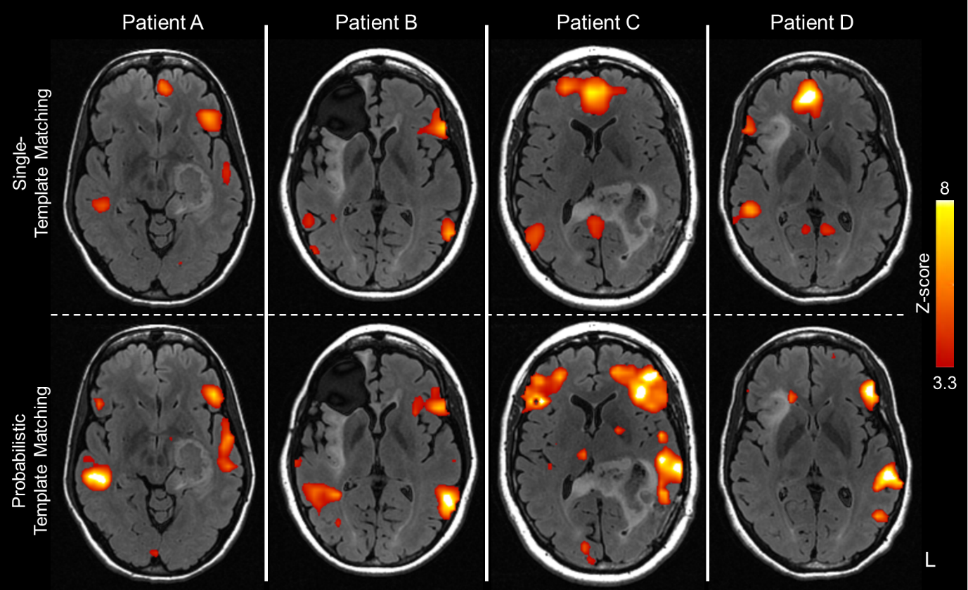
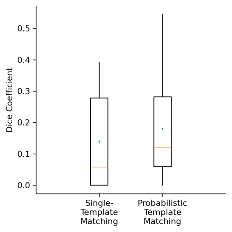
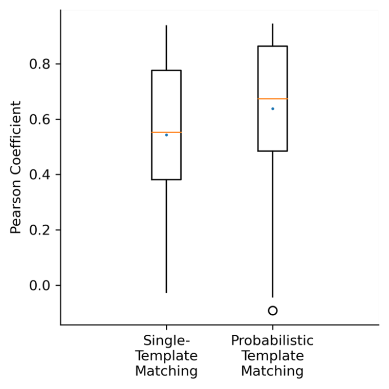
Figure 1 presents the ICA component recommended as the language network for four representative patients by single-template matching in comparison to probabilistic template matching. Recommended components for Patient A and B are similar between the two methods. Recommended components by probabilistic template matching for Patient C and D resemble the language network, while single-template matching recommends components representing parts of the default mode network.Figure 2 and 3 presents boxplots comparing the left hemisphere PLA Dice coefficients and Pearson correlation respectively for probabilistic template matching vs single-template matching. Wilcoxon signed-rank test shows that probabilistic template matching had significantly higher Dice coefficients and Pearson correlation with respect to the tb-fMRI activations (p < 0.05).
Discussion
Dice coefficient indicate spatial similarity while Pearson correlation indicate the temporal correlation of recommended component and language areas with tb-fMRI activations. Probabilistic template matching uses template variation of POM probabilities and ICA components to recommend the language network for each patient. Significantly higher Dice and Pearson correlations suggest probabilistic template matching performs better than single template matching in recommending the language network.Conclusion
Probabilistic template matching uses template variation of a POM developed from brain tumor population to recommend ICA component as the language network and demonstrates better recommendations as compared to a single-template matching method.Acknowledgements
This study was supported by NIH/NCI under award number R01 CA258788.References
- Zaca, D., et al. ReStNeuMap: a tool for automatic extraction of resting-state functional MRI networks in neurosurgical practice. J Neurosurg 131, 764-771 (2018).
- Greicius, M.D., Srivastava, G., Reiss, A.L. & Menon, V. Default-mode network activity distinguishes Alzheimer's disease from healthy aging: evidence from functional MRI. Proc Natl Acad Sci U S A 101, 4637-4642 (2004).
- Tie, Y., et al. Defining language networks from resting-state fMRI for surgical planning--a feasibility study. Hum Brain Mapp 35, 1018-1030 (2014).
- Mazaika, P.H., F. Glover, GH. Reiss, AL. . Methods and software for fMRI analysis of clinical subjects. Neuroimage 47(2009).
- Gorgolewski, K., et al. Nipype: a flexible, lightweight and extensible neuroimaging data processing framework in python. Front Neuroinform 5, 13 (2011).
- Rolls, E.T., Huang, C.C., Lin, C.P., Feng, J. & Joliot, M. Automated anatomical labelling atlas 3. Neuroimage 206, 116189 (2020).
Figures