1115
Two functional connectivity based subtypes of MDD and related biological mechanisms1Department of Radiology and Huaxi MR Research Center (HMRRC), Functional and Molecular lmaging Key Laboratory of Sichuan Province, West China Hospital, Sichuan University, Chengdu 610041, Sichuan Province, China
Synopsis
Keywords: fMRI Analysis, fMRI (resting state), MR value; major depressive disorder; subtypying; genetic mechanisms; neurotransmitter; cognition
Motivation: There’s a large clinical heterogeneity presented in MDD and inconsistent MRI evidence on abnormal functional connectivity (FC) in MDD, let alone the unclear biological mechanisms underlying the neuroimaging alterations.
Goal(s): To identify FC based subtypes of MDD and their genetic mechanisms and neurotransmission patterns.
Approach: Consensus clustering of FC was applied to subtyping MDD. Correlation analyses were used to explore the underlying biological mechanisms of FC alterations in each subtype.
Results: Two stable neurophysiological MDD subtypes were found. While the two subtypes were indistinguishable by clinical symptoms, FC alterations of each subtype had distinct spatial correlations with cognition, gene, and neurotransmission profiles.
Impact: Our findings suggested the presence of two neuroimaging subtypes in MDD and the two subtypes can be characterized by different genetic mechanisms, neurotransmitter receptor/transporter profiles, and cognition types, providing new clues to understand the pathophysiology of MDD.
Introduction
Major depressive disorder (MDD) had a larger heterogeneity in clinical and treatment outcomes 1. The melancholic, atypical, and anxious subtype of MDD have been suggested by DSM-5 2. Individual response to antidepressant treatment is also inconsistent 3. On the other hand, the heterogeneity of MDD also exists in neuroimaging alterations including brain structure 4 and function 5. Subtyping is one promising solution to characterize the heterogeneity 6. Furthermore, the Allen Human Brain Atlas (AHBA) 7 with brain gene expression data from six donors provides us an opportunity to excavate the genetic mechanisms underlying the MDD neurophysiological subtypes. Neurotransmitter has a close relationship with brain neural activity 8. Investigating the association between biological and neuroimage features 9 can help reveal the molecular substrates of MDD subtypes. Thus, in this study, we aimed to identify specific neurophysiological subtypes of MDD based on brain functional connectivity (FC) derived from resting-state functional magnetic resonance imaging data and investigate its related biological mechanisms.Methods
Consensus clustering of FC patterns was applied to a population of 829 MDD patients from REST-Meta-MDD database to find FC based subtypes of MDD. The differences in clinical symptoms were compared between the subtypes. Cognition data extracted from neuromap toolbox and lasso regression were applied to investigate the differences of FC-related cognition between subtypes. Gene transcriptomic data derived from AHBA and neurotransmitter receptor/transporter density data derived from PET/SPECT studies using neuromap toolbox. These biological data with partial least squares regression analysis and spatial autocorrelation corrected method were used to characterize the molecular mechanism underlying each FC-based subtype by identifying the gene set and neurotransmitters/transporters showing high spatial similarity with the profiles of FC alterations between each subtype of MDD and 770 healthy controls (HCs).Results
Two stable neurophysiological MDD subtypes were found and labeled as hypoconnectivity (n=527) and hyperconnectivity (n=299) characterized by the FC differences in each subtype relative to controls, respectively. The two subtypes did not differ in age, sex, and symptom severity. However, the FC in hypoconnectivity subtype group correlated with response inhibition, selective attention, face recognition, sleep, empathy, expertise, uncertainty, and anticipation, while the FC in hyperconnectivity subtype group was specifically related to inference, speech perception, and reward anticipation.The genes related to FC alterations were enriched in ion transmembrane transport, synaptic transmission/organization, axon development, and regulation of neurotransmitter level for both subtypes of MDD, but specifically enriched in glial cell differentiation for hypoconnectivity subtype, while enriched in regulation of presynaptic membrane and regulation of neuron differentiation for hyperconnectivity subtype.
FC alterations were spatially associated with the density of 5-HT2a receptor and 5-HTT in both subtypes. For hyperconnectivity subtype, FC alterations were also correlated with the density of norepinephrine transporter, glutamate receptor, GABA receptor, 5-HT1b receptor, and cannabinoid receptor.
Discussion
Our findings revealed the neuroimaging heterogeneity of MDD in FC alterations, providing new evidence of FC’s contribution to MDD subtyping. Previous works have shown the value of FC in classifying MDD and HCs 10, and some studies started to focus on MDD subtyping with FC deviation from HCs5. Altogether, accumulated evidence emphasizes the FC alterations’ vital role in the neuropathology of MDD. The identified two FC subtypes in MDD had no differences in age, sex, and symptom severity, but had distinguishable cognition characteristics, highlighting the different cognitive problems in MDD with distinct neural basis, consistent with prior results 11.Transcriptomic signatures of FC alteration in MDD suggested the neuronal development, synaptic function, and neurotransmitter release contribute to the pathophysiology of MDD. Hypoconnectivity subtype specifically associated with glial cell differentiation. Glial deficits that contribute to neuronal pathology in MDD 12 damaged neural activity 13, leading to connectivity impairments. While, dysregulation of neurogenesis and neuronal differentiation were the specific characteristics for hyperconnectivity subtype, and excessive activation of such regulation may cause excessive neuronal regeneration 14.
Aberrant neurotransmitter release also contributes to the pathogenesis of MDD 15, in agreement with our findings regarding correlations of FC alterations with neurotransmitter density profile in MDD. Many types of neurotransmitters were related to FC alterations in hyperconnectivity subtype, but only serotonin was related to that in hypoconnectivity subtype. One possible explanation is that neuronal activity regulates neurotransmitter release 16 and hypoconnectivity subtype was correlated to excessive activation of regulation of neuronal function which may promote the neurotransmitter release.
Conclusion
Our findings suggested the presence of two neuroimaging subtypes of MDD characterized by hypo- or hyper-FC which were related to different genetic expressions, neurotransmitter profiles, and cognition, providing a deep insight into the varied biological mechanisms for different neuroimaging-based subtypes of MDD.Acknowledgements
None.References
1. Musil R, Seemüller F, Meyer S, et al. Subtypes of depression and their overlap in a naturalistic inpatient sample of major depressive disorder. Int J Methods Psychiatr Res 2018;27(1).2. Diagnostic and statistical manual of mental disorders : DSM-5. 5th ed. ed. Arlington, VA :: American Psychiatric Association; 2013.
3. Rost N, Binder EB, Brückl TM. Predicting treatment outcome in depression: an introduction into current concepts and challenges. Eur Arch Psychiatry Clin Neurosci 2023;273(1):113-127.
4. Shao J, Qin J, Wang H, et al. Capturing the Individual Deviations From Normative Models of Brain Structure for Depression Diagnosis and Treatment. Biol Psychiatry 2023.
5. Sun X, Sun J, Lu X, et al. Mapping Neurophysiological Subtypes of Major Depressive Disorder Using Normative Models of the Functional Connectome. Biol Psychiatry 2023.
6. Rasero J, Diez I, Cortes JM, Marinazzo D, Stramaglia S. Connectome sorting by consensus clustering increases separability in group neuroimaging studies. Netw Neurosci 2019;3(2):325-343.
7. Hawrylycz MJ, Lein ES, Guillozet-Bongaarts AL, et al. An anatomically comprehensive atlas of the adult human brain transcriptome. Nature 2012;489(7416):391-399.
8. Dukart J, Holiga Š, Chatham C, et al. Cerebral blood flow predicts differential neurotransmitter activity. Sci Rep 2018;8(1):4074.
9. Xiao Y, Zhao L, Zang X, Xue SW. Compressed primary-to-transmodal gradient is accompanied with subcortical alterations and linked to neurotransmitters and cellular signatures in major depressive disorder. Hum Brain Mapp 2023;44(17):5919-5935.
10. Chen Y, Zhao W, Yi S, Liu J. The diagnostic performance of machine learning based on resting-state functional magnetic resonance imaging data for major depressive disorders: a systematic review and meta-analysis. Front Neurosci 2023;17:1174080.
11. Hack LM, Tozzi L, Zenteno S, et al. A Cognitive Biotype of Depression and Symptoms, Behavior Measures, Neural Circuits, and Differential Treatment Outcomes: A Prespecified Secondary Analysis of a Randomized Clinical Trial. JAMA Netw Open 2023;6(6):e2318411.
12. Öngür D, Bechtholt AJ, Carlezon WA, Jr., Cohen BM. Glial abnormalities in mood disorders. Harv Rev Psychiatry 2014;22(6):334-337.
13. Volterra A, Meldolesi J. Astrocytes, from brain glue to communication elements: the revolution continues. Nat Rev Neurosci 2005;6(8):626-640.
14. An J, Chen B, Tian D, Guo Y, Yan Y, Yang H. Regulation of Neurogenesis and Neuronal Differentiation by Natural Compounds. Curr Stem Cell Res Ther 2022;17(8):756-771.
15. Borroto-Escuela DO, Ambrogini P, Chruścicka B, et al. The Role of Central Serotonin Neurons and 5-HT Heteroreceptor Complexes in the Pathophysiology of Depression: A Historical Perspective and Future Prospects. Int J Mol Sci 2021;22(4).
16. Meng D, Li HQ, Deisseroth K, Leutgeb S, Spitzer NC. Neuronal activity regulates neurotransmitter switching in the adult brain following light-induced stress. Proc Natl Acad Sci U S A 2018;115(20):5064-5071.
Figures
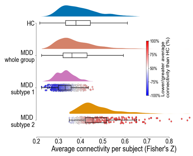
Figure 1. Two stable neurophysiology MDD subtypes were found based on consensus clustering of brain functional connectivity patterns. Subtype 1 is characterized by hypoconnectivity and subtype 2 is characterized by hyperconnectivity. Additionally, for subtypes 1 and 2, two colors were used to show within-group connectivity differences: blue for hypoconnectivity, and red for hyperconnectivity. Abbreviations: MDD, major depressive disorder; HC, healthy controls.