1107
Aberrant brain structural–functional connectivity coupling related to cognitive impairment in different cerebral small vessel disease burden1Department of Radiology, Shandong Provincial Hospital Affiliated to Shandong First Medical University, Jinan, China, 2Department of Radiology and Nuclear medicine, Xuanwu Hospital, Capital Medical University, Beijing, China, 3School of Psychology, Southwest University, Chongqing, China
Synopsis
Keywords: Functional Connectivity, Aging
Motivation: The impact of different cerebral small vessel disease (CSVD) burden on brain structural and functional connectivity coupling and their correlation with neurocognitive outcomes remain largely unknown.
Goal(s): To explore the alterations of structural and functional connection network (SC-FC) coupling in the whole brain and different functional modules of patients with different CSVD burden compared with healthy controls.
Approach: Diffusion tensor imaging (DTI) and Resting-state blood-oxygen-level-dependent (BOLD) fMRI techniques were used to analyze structural and functional brain connections.
Results: Severe CSVD burden patients exhibited significantly decreased whole-brain SC-FC coupling, reduced modular SC-FC coupling and associated with impairment of cognitive outcomes.
Impact: SC-FC coupling might provide a more sensitive neuroimaging biomarker of CSVD burden as well as new insights into the pathophysiologic mechanisms of the clinical development of CSVD.
Introduction
Cerebral small vessel disease (CSVD) is a primary cause of cognitive dysfunction and vascular dementia [1] and considered to be an important pathological basis for vascular cognitive impairment [2]. Emerging evidence suggests that cerebral small vessel disease (CSVD) pathology changes brain structural connectivity and functional connectivity (FC) networks [3-4]. Despite network-level SC and FC are closely coupled in healthy population [5], how SC-FC coupling and functional network changes correlated with neurocognitive outcomes in patients with different CSVD burdens remains largely unknown. In this study, we aimed to construct whole brain SC and FC networks in patients with different CSVD burden and healthy controls using multimodal MRI. We hypothesized that the SC-FC coupling and functional efficiency would decrease with the increased burden of CSVD, and are associated with more severe cognitive decline.Methods
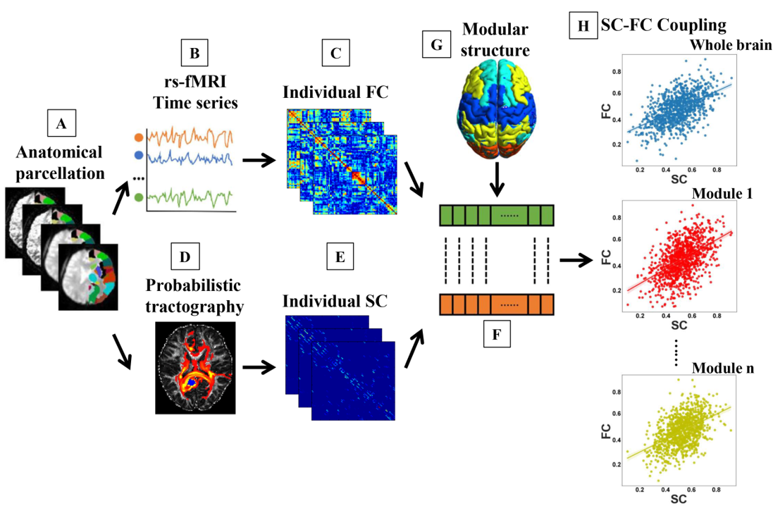
We reconstruct whole-brain SC and FC networks for 54 patients with severe CSVD burden (CSVD-s), 106 patients with mild CSVD burden (CSVD-m) and 79 healthy controls. We then investigated the aberrant SC-FC coupling and functional network topology in CSVD and their correlations with cognitive dysfunction. MRI scans were obtained using a 3.0-Tesla MR system (Siemens Healthcare, Erlangen, Germany). Diffusion tensor imaging (DTI) data were acquired using a simultaneous multislice (SMS) accelerated single-shot echo planar imaging (EPI) sequence. Resting-state blood-oxygen-level-dependent (BOLD) fMRI data were acquired using a gradient-echo echo-planar imaging (GE-EPI) sequence. For each individual, Pearson’s correlations were calculated between the time series of all regions to calculate FC and result in a 90×90 symmetric FC network/matrix (Figure 1A-C). For SC-FC coupling and functional network topological metrics, one-way analysis of covariance (ANCOVA) was performed to investigate differences among three groups while controlling age, sex, education and head motion as covariates, with LSD tests performed for pairwise comparisons. We further calculated the partial correlation coefficients between the network metrics and cognitive parameters for all groups using SPSS v24.0 software.Results
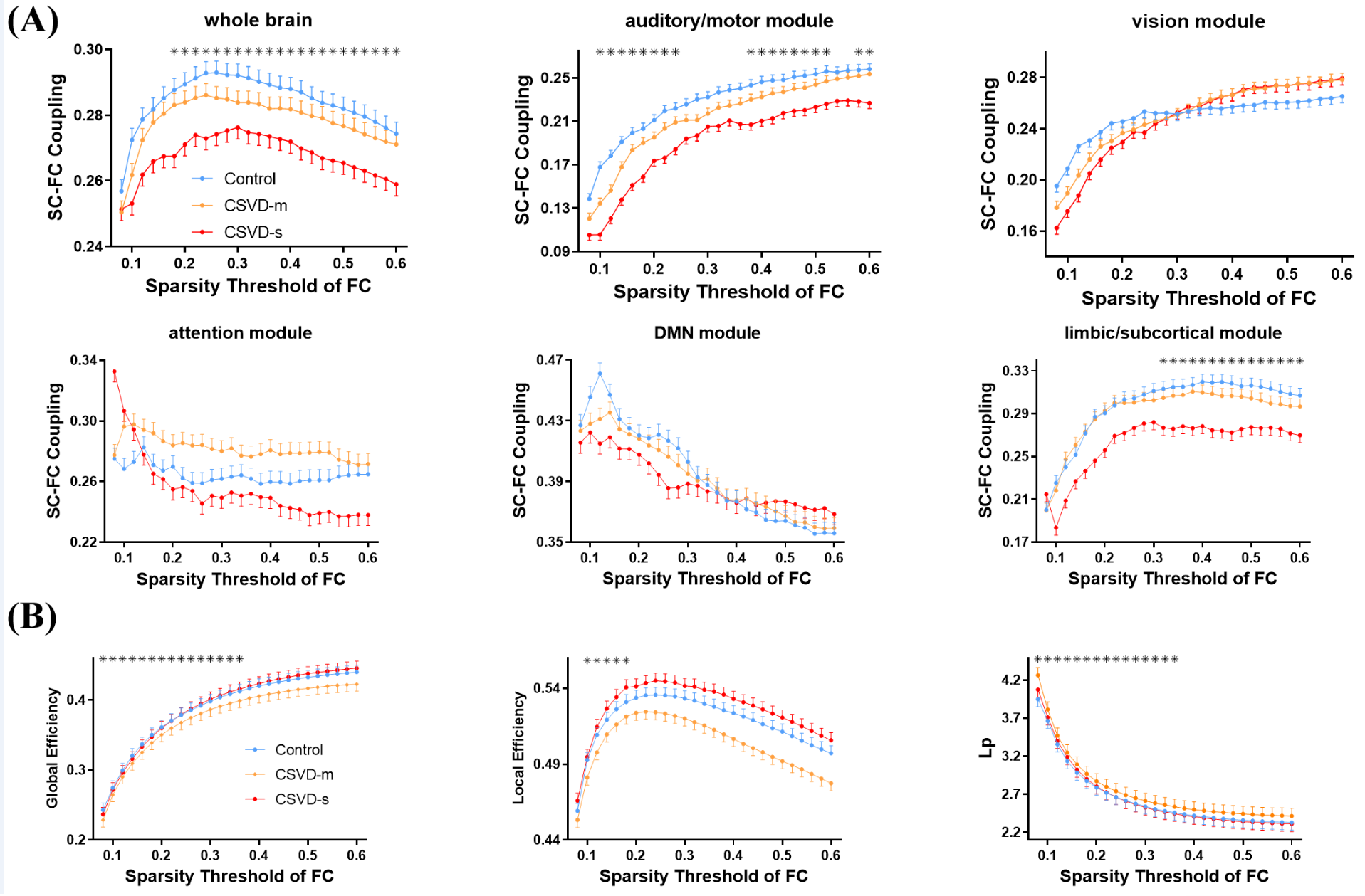
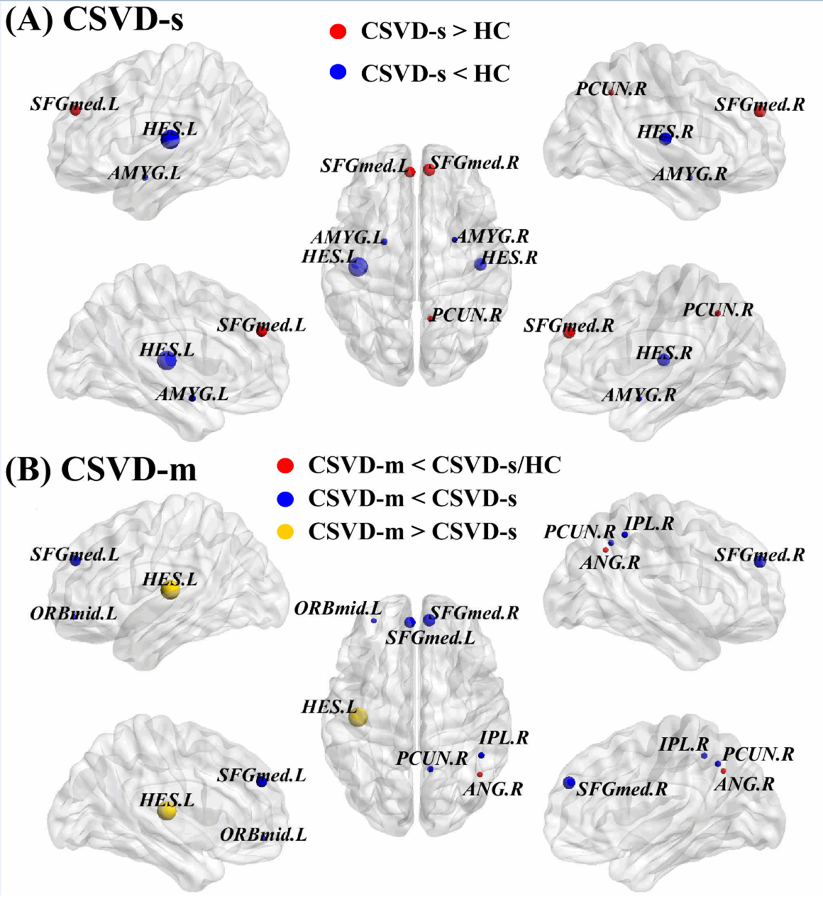
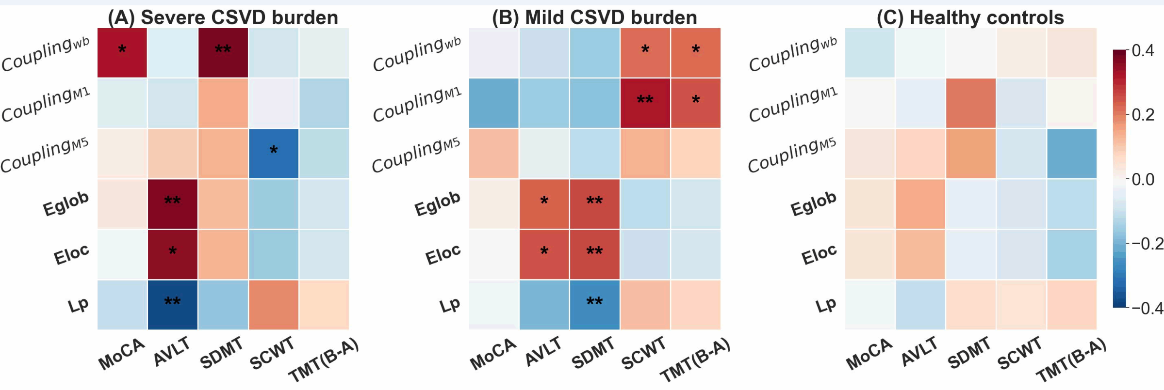
Compared with control group, the CSVD-s group showed significantly decreased SC-FC coupling within the whole brain, sensory/motor and limbic/subcortical functional modules over a wide range of sparsity thresholds of FC (Figure 2). Meanwhile, the CSVD-m patients also showed significantly decreased nodal efficiency in the right angular gyrus and left heschl gyrus (Figure 3). For significantly altered coupling and efficiency metrics among groups, their correlations to cognitive performance were calculated. Intriguingly, we observed significant correlations (p<0.05, FDR corrected) in both CSVD-s and CSVD-m groups (Figure 4A-B), while there was no significant correlation in control (Figure 4C). Briefly, for CSVD-s group, whole-brain SC–FC coupling was positively correlated with MoCA and SDMT scores, and limbic/subcortical modular SC–FC coupling was negatively correlated with SCWT score, as well as global and local efficiencies were positively correlated with AVLT score (Figure 5A). For CSVD-m group, whole-brain and auditory/motor modular SC–FC couplings were positively correlated with SCWT and TMT scores, as well as global and local efficiencies were positively correlated with AVLT and SDMT scores (Figure 5B).Conclusion
Our findings demonstrated that decreased whole-brain and module-dependent SC-FC couplings associated with reduced functional efficiency might underlie more severe burden and worse cognitive decline in CSVD patients. SC-FC coupling might provide a more sensitive neuroimaging biomarker of CSVD burden as well as new insights into the pathophysiologic mechanisms of the clinical development of CSVD. Early detection of cognitive decline in patients with CSVD is crucial for improving and protecting their function and promoting the reversal of their cognitive deficits. The emergence of new biomarkers opens new avenues for better understanding and early intervention of this disorder.Acknowledgements
The informed consent forms were signed by all the volunteers and patients participating in the study. This work was supported by grants from the National Natural Science Foundation of China (32100902), the Natural Science Foundation of Shandong Province (ZR2020MH288) and the Technology Development Plan of Jinan (201301049, 201602206, 201907052).References
1.WARDLAW, J. M., SMITH, E. E., BIESSELS, G. J., CORDONNIER, C., FAZEKAS, F., FRAYNE, R., LINDLEY, R. I., O'BRIEN, J. T., BARKHOF, F., BENAVENTE, O. R., BLACK, S. E., BRAYNE, C., BRETELER, M., CHABRIAT, H., DECARLI, C., DE LEEUW, F. E., DOUBAL, F., DUERING, M., FOX, N. C., GREENBERG, S., HACHINSKI, V., KILIMANN, I., MOK, V., OOSTENBRUGGE, R., PANTONI, L., SPECK, O., STEPHAN, B. C., TEIPEL, S., VISWANATHAN, A., WERRING, D., CHEN, C., SMITH, C., VAN BUCHEM, M., NORRVING, B., GORELICK, P. B. & DICHGANS, M. 2013. Neuroimaging standards for research into small vessel disease and its contribution to ageing and neurodegeneration. Lancet Neurol, 12, 822-38.
2. MARKUS, H. S. & DE LEEUW, F. E. 2023. Cerebral small vessel disease: Recent advances and future directions. Int J Stroke, 18, 4-14
3. FENG, M., WEN, H., XIN, H., WANG, S., GAO, Y., SUI, C., LIANG, C. & GUO, L. 2023. Decreased Local Specialization of Brain Structural Networks Associated with Cognitive Dysfuntion Revealed by Probabilistic Diffusion Tractography for Different Cerebral Small Vessel Disease Burdens. Mol Neurobiol.
4. XIN, H., WEN, H., FENG, M., GAO, Y., SUI, C., ZHANG, N., LIANG, C. & GUO, L. 2022. Disrupted topological organization of resting-state functional brain networks in cerebral small vessel disease. Hum Brain Mapp, 43, 2607-2620.
5. OSMANLıOĞLU, Y., TUNç, B., PARKER, D., ELLIOTT, M. A., BAUM, G. L., CIRIC, R., SATTERTHWAITE, T. D., GUR, R. E., GUR, R. C. & VERMA, R. 2019. System-level matching of structural and functional connectomes in the human brain. Neuroimage, 199, 93-104.
Figures