1102
Exploration of a low-SAR ihMT-RAGE approach for human whole brain imaging at 7T1Aix Marseille Univ, CNRS, CRMBM, Marseille, France, 2APHM, Hôpital Universitaire Timone, CEMEREM, Marseille, France, 3Siemens Healthcare SAS, Courbevoie, France, 4Division of MR Research, Radiology, Beth Israel Deaconess Medical Center, Harvard Medical School, Boston, MA, United States
Synopsis
Keywords: Magnetization Transfer, Pulse Sequence Design, qMRI ihMT Neuro Brain
Motivation: Inhomogeneous Magnetization Transfer (ihMT) is a recent MRI technique that has raised great interest for myelin imaging. Several ihMT protocols have been proposed for whole brain imaging at clinical field strengths. However, ultra-high field (UHF, 7T) translation remains challenging.
Goal(s): ln this work we explore ways to perform ihMT at UHF for clinical applications.
Approach: A low-SAR ihMT-RAGE sequence is proposed by shortening the ihMT preparation and enabling partial Fourier MT saturation.
Results: This original sequence addresses SAR limitations within relatively short scan times, allowing for whole brain 1.2mm isotropic resolution (resp. 1mm) in 12 minutes (resp. 16 min) at 7T.
Impact: Ultra-high field ihMT enables high resolution (1mm iso) myelin specific imaging, opening new perspectives for neuroscience and clinical research. Future developments, such as reduced FOV and compressed sensing sequences could bring scan times further down to 5-10 minutes.
Introduction
Inhomogeneous Magnetization Transfer (ihMT) is a recent MRI technique that has raised great interest for myelin imaging1. Several ihMT protocols have been proposed for whole brain imaging at clinical field strengths (1.5T and 3T) 2–6. However, ultra-high field (UHF, 7T) translation remains challenging7. Aspirations for higher spatial resolution combined with Specific Absorption Rate (SAR) constraints result in long scan times, and strong RF inhomogeneities lead to signal artifacts. Hereafter, we explore an original ihMT-RAGE sequence design targeted to UHF applications that addresses SAR limitations within relatively short scan times, allowing for whole brain 1.2mm isotropic resolution (resp. 1mm) in 12 minutes (resp. 16 min).ihMT-RAGE sequence design and in vivo experiments
At 3T, a previously optimized ihMT-RAGE sequence 3,4 consisted of relatively long MT preparations (5 ms saturation pulses repeated 10 times every 100 ms) followed by RAGE readouts and recovery delays (Fig 1a). The low saturation duty cycle (DC) of 5% was chosen to enhance the ihMT signal2,8.Challenges for UHF translation include dealing with strong $$$B_1^+$$$ inhomogeneities which can drop to 40% of its nominal value in certain brain areas. As the ihMT signal starts proportional to $$$(B_1^{saturation})^4$$$, maximizing $$$B_1^{saturation}$$$ is essential in order to reveal signal in $$$B_1$$$ hypointense regions. However, this has a high SAR payload at UHF, typically leading to long recovery delays.
Preliminary simulations of ihMTR signal buildup through the saturation preparation suggest that a reduced number of MT pulses delivers most of the ihMT signal (Fig. 2). This opens perspectives for SAR reduction, and therefore scan time reduction. An additional way to mitigate SAR consists in using partial Fourier saturation, for which only the k-space central part is subject to ihMT preparation (Fig. 1b). This technique has been used in steady-state ihMT-GRE sequences2,9, and was generalized here to more prepared ihMT-RAGE sequences.
We combined all presented features to propose a prototype low SAR ihMT-RAGE sequence suitable for UHF applications. The efficiency of the sequence was evaluated on a clinically approved 7T (MAGNETOM Terra, Siemens Healthineers) on healthy volunteers ($$$N=3$$$). The protocol included two acquisitions with varying number of readout segments per repetition, as well as a non-optimized (‘classical’) ihMT-RAGE sequence for comparison. $$$T_1$$$ MP2RAGE and $$$B_1^+$$$-mapping sequences were also acquired as in [10]. All sequence parameters are indicated in Table 1.
Processing
Preprocessing of acquired volumes involved proprietary gradients distortion corrections. Quantitative $$$T_1$$$ mapping was performed from MP2RAGE data and included $$$B_1^+$$$ corrections10 using MATLAB 9.14.0. The Postprocessing of ihMT volumes involved motion-correction and denoising11,12. Brain segmentations were obtained from the MP2RAGE UNI image using FreeSurfer 7.3.213. Coregistration was performed using ANTs 2.4.214.Results
All ihMT sequences were run at ~90% SAR level to accommodate varying volunteer coil-loads, under first level supervision mode. The whole brain 1.2mm isotropic ‘classical’ ihMT-RAGE sequence was acquired in 16’14”. In contrast, whole brain 1.2 mm iso low-SAR ihMT-RAGE benefited from a much-reduced TR, and hence shorter scan time of 11’46”. This, even though it was run at a 60% higher $$$B_1^{peak\,saturation}$$$ compared to the ‘classical’ protocol. Moreover, running the whole brain 1.2mm iso ‘classical’ ihMT-RAGE sequence at maximal $$$B_1^{peak\,saturation}$$$ (~23µT) would have led to a scan time of ~30min to stay within SAR limitations, and was not acquired. Finally, whole brain 1 mm iso low-SAR ihMT-RAGE acquisitions only took 15’38”.Acquired ihMTR images are shown on Figures 3 and 4, along with reference anatomical $$$T_1$$$ scan and $$$B_1^+$$$ field. The proposed low-SAR protocol demonstrated capacity in generating ihMT signal over the whole brain with more efficient use of MT RF power. Importantly, running the protocol at maximum allowed $$$B_1^{peak\,saturation}$$$ seems beneficial to reveal ihMT signal in hypointense-$$$B_1$$$ regions (red overlays on Figure 3), for better visualization of these hypointense regions. Finally, high resolution 1mm isotropic acquisitions show improved cortex boundaries, and cerebellum structures delineation.
Discussions and Conclusion
The proposed low-SAR ihMT-RAGE sequence benefits from shorter scan times while keeping a strong ihMT signal by applying saturation pulses at maximal peak $$$B_1$$$. This was made possible by: 1/ shortening the ihMT preparation, and 2/ enabling partial Fourier MT saturation. This reduced SAR constraints, allowing for subsequent scan time reduction by shortening the RAGE recovery delay. For localized brain applications, e.g., thalamus or brain stem, the proposed sequence could run even faster. Future studies will address the impact of k-space sampling scheme on the point-spread function and effective spatial resolution3,6, as well as the validation of $$$B_1^+$$$ correction strategies. Ultimately, this work demonstrates the feasibility of UHF ihMT at millimetric resolution in the clinical setting.Acknowledgements
This work received support from the french government under the France 2030 investment plan, as part of the Initiative d’Excellence d’Aix-Marseille Université – A*MIDEX: AMX-21-HAN-01, and by the National Research Agency (ANR; NormaBRAIN ; ANR-22-CE17-0060). This work was performed by a laboratory member of France Life Imaging network (grant ANR-11-INBS-0006).
References
1. Alsop DC, Ercan E, Girard OM, et al. Inhomogeneous magnetization transfer imaging: Concepts and directions for further development. NMR Biomed. August 2022:e4808. doi:10.1002/nbm.4808 SMASH
SMASH
2. Mchinda S, Varma G, Prevost VH, et al. Whole brain inhomogeneous magnetization transfer (ihMT) imaging: Sensitivity enhancement within a steady-state gradient echo sequence. Magn Reson Med. 2018;79(5):2607-2619. doi:10.1002/mrm.26907 SMASH
3. Varma G, Munsch F, Burns B, et al. Three-dimensional inhomogeneous magnetization transfer with rapid gradient-echo (3D ihMTRAGE) imaging. Magn Reson Med. 2020;84(6):2964-2980. doi:10.1002/mrm.28324 SMASH
4. Munsch F, Varma G, Taso M, et al. Characterization of the cortical myeloarchitecture with inhomogeneous magnetization transfer imaging (ihMT). NeuroImage. 2021;225:117442. doi:10.1016/j.neuroimage.2020.117442 SMASH
5. Malik SJ, Teixeira RPAG, West DJ, Wood TC, Hajnal JV. Steady-state imaging with inhomogeneous magnetization transfer contrast using multiband radiofrequency pulses. Magn Reson Med. 2020;83(3):935-949. doi:10.1002/mrm.27984 SMASH
6. Rowley CD, Campbell JSW, Leppert IR, Nelson MC, Pike GB, Tardif CL. Optimization of acquisition parameters for cortical inhomogeneous magnetization transfer (ihMT) imaging using a rapid gradient echo readout. Magn Reson Med. 2023;90(5):1762-1775. doi:10.1002/mrm.29754 SMASH
7. Girard OM, Soustelle L, Troalen T, et al. Feasibility of inhomogeneous Magnetization Transfer (ihMT) imaging of the human brain at 7T. In: Proceedings of the ISMRM. London, UK; 2022:375.
8. Varma G, Girard OM, Mchinda S, et al. Low duty-cycle pulsed irradiation reduces magnetization transfer and increases the inhomogeneous magnetization transfer effect. J Magn Reson. 2018;296:60-71. doi:10.1016/j.jmr.2018.08.004 SMASH
9. Soustelle L, Troalen T, Hertanu A, et al. A strategy to reduce the sensitivity of inhomogeneous Magnetization Transfer (ihMT) imaging to radiofrequency transmit field variations at 3 T. Magn Reson Med. November 2021. doi:10.1002/mrm.29055 SMASH
10. Massire A, Seiler C, Troalen T, et al. T1-Based Synthetic Magnetic Resonance Contrasts Improve Multiple Sclerosis and Focal Epilepsy Imaging at 7 T. Invest Radiol. August 2020. doi:10.1097/RLI.0000000000000718 SMASH
11. Soustelle L, Lamy J, Troter AL, et al. A Motion Correction Strategy for Multi-Contrast based 3D parametric imaging: Application to Inhomogeneous Magnetization Transfer (ihMT). September 2020:2020.09.11.292649. doi:10.1101/2020.09.11.292649 SMASH
12. ihMT Proc. https://github.com/lsoustelle/ihmt_proc. Accessed November 8, 2023.
13. Sled JG, Zijdenbos AP, Evans AC. A nonparametric method for automatic correction of intensity nonuniformity in MRI data. IEEE Trans Med Imaging. 1998;17(1):87-97. doi:10.1109/42.668698 SMASH
14. Avants BB, Tustison NJ, Song G, Cook PA, Klein A, Gee JC. A reproducible evaluation of ANTs similarity metric performance in brain image registration. NeuroImage. 2011;54(3):2033-2044. doi:10.1016/j.neuroimage.2010.09.025 SMASH
Figures
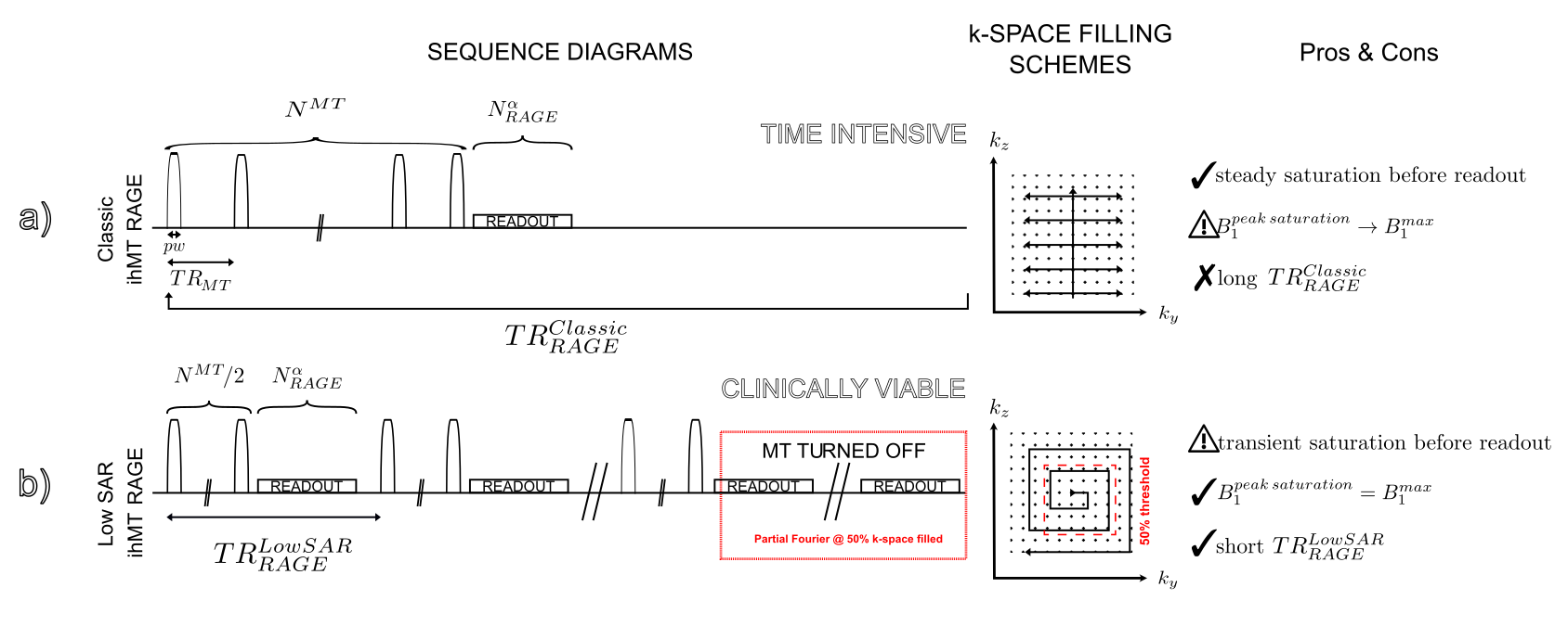
Fig.1: a/ Classical ihMT-RAGE sequence diagram: long MT preparation followed by RAGE readout and a relatively long recovery delay, sampled in linear + centric-out fashion providing steady saturation and high $$$B_1^{saturation}$$$ at the cost of a long TR, b/ Low SAR ihMT-RAGE sequence diagram: half MT preparation followed by RAGE readout and limited recovery delay, sampled in a 2D centric-out fashion, providing maximal $$$B_1^{saturation}$$$ and short TR at the cost of a transient saturation.
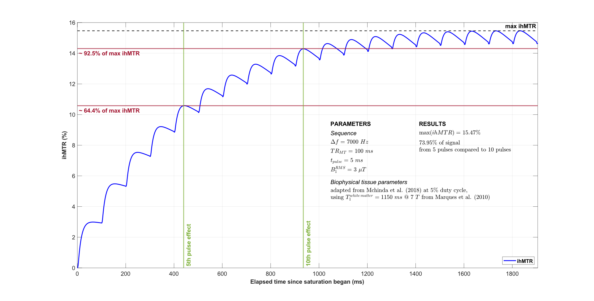
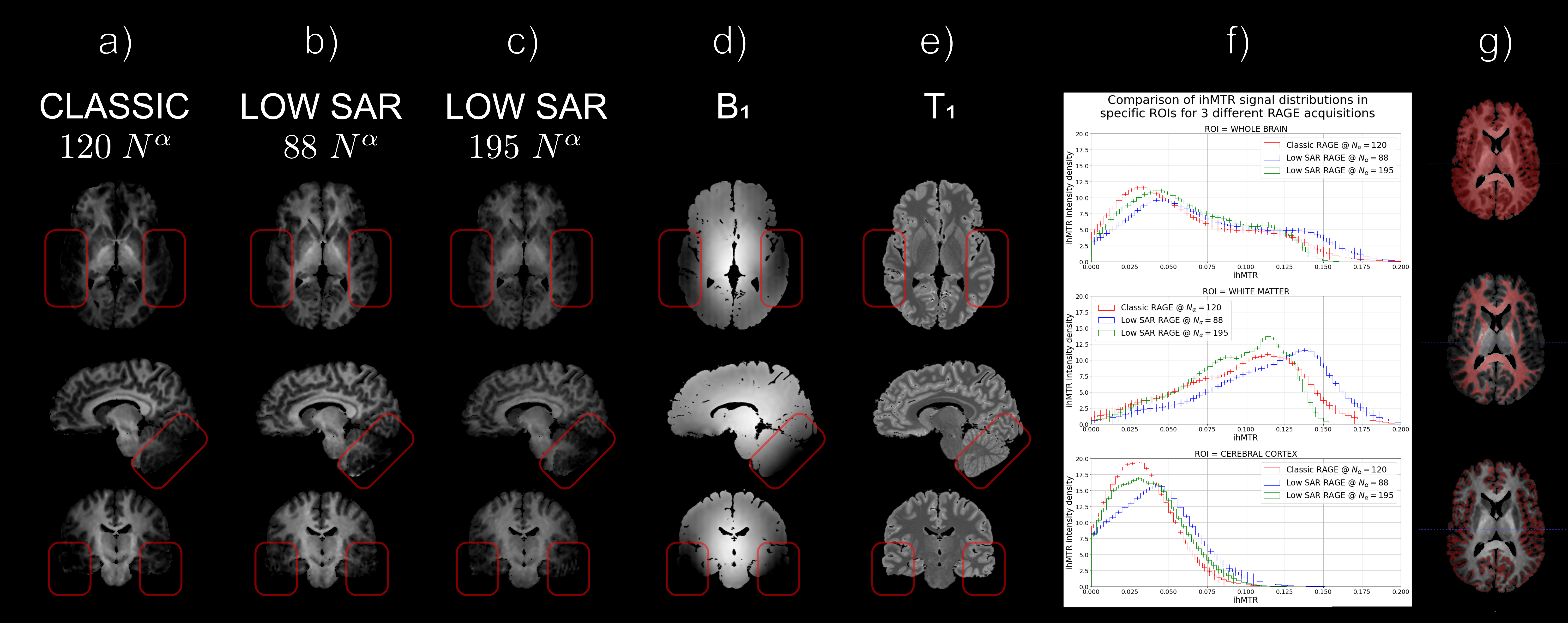
Fig.3: a-e/ Axial (top), Sagittal (middle), and Coronal (bottom) views of a/ Classic, low SAR ihMT-RAGE with b/ 88 readout segments, c/ 195 readout segments, d/ $$$B_1$$$ field, and e/ $$$T_1$$$ maps. Red overlays emphasize hypointense $$$B_1$$$ regions. f/ Signal distributions of displayed ihMT maps are compatible in f-g/ whole brain (top), white matter (middle), and cerebral cortex (bottom). g/ Single slice of associated 3D ROI masks (red), overlaid upon the $$$N^\alpha=88$$$ ihMT-RAGE map.
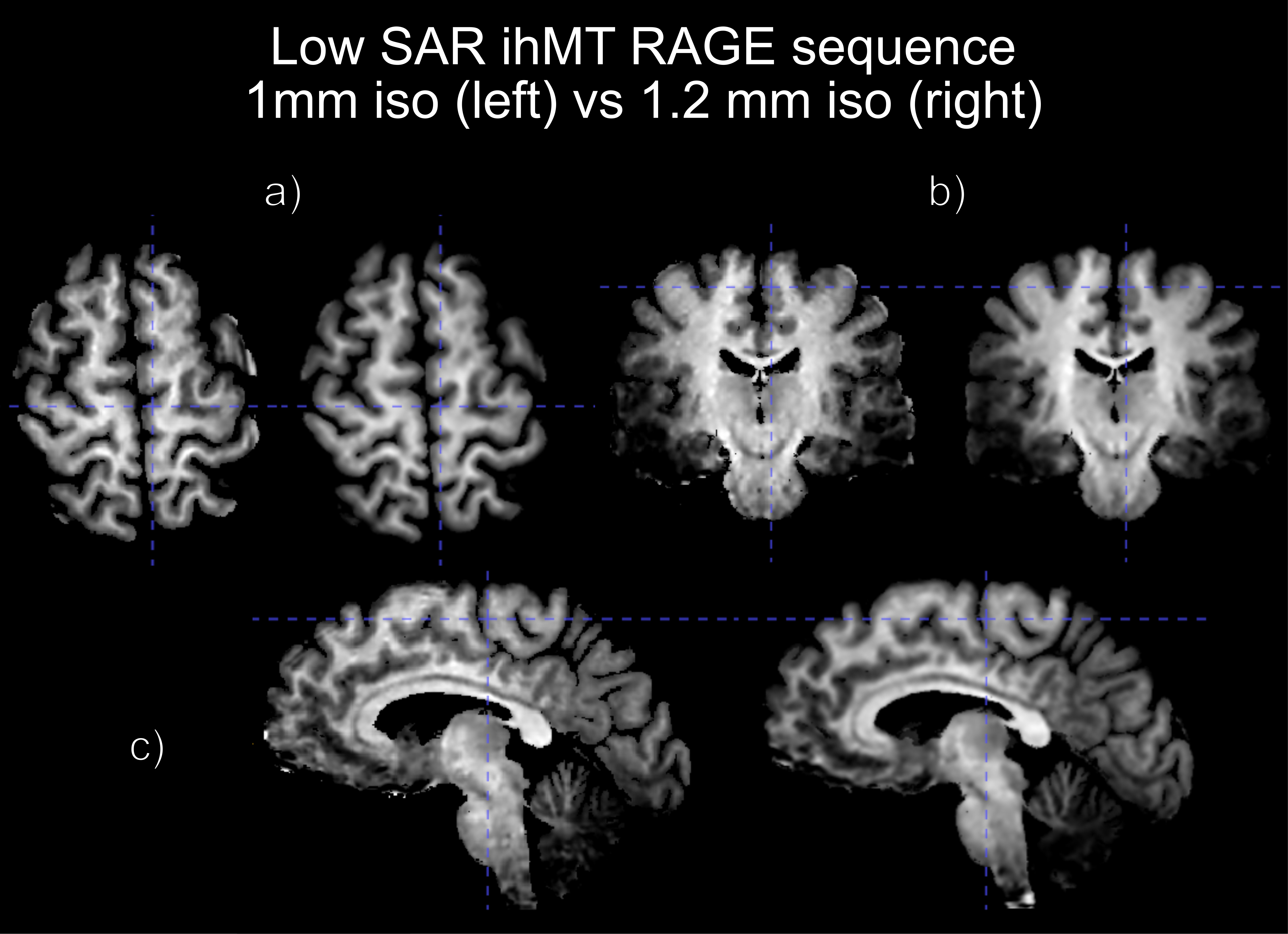
Fig.4: Comparison of a/ axial, b/ coronal, and c/ sagittal views of the ihMTR signal from a low SAR ihMT-RAGE sequence at 1mm iso (left) to a low SAR ihMT-RAGE sequence at 1.2mm iso (right). Both acquisitions were done at 5% duty cycle. The 1mm iso shows clearer cortex boundaries and cerebellum structures delineations.
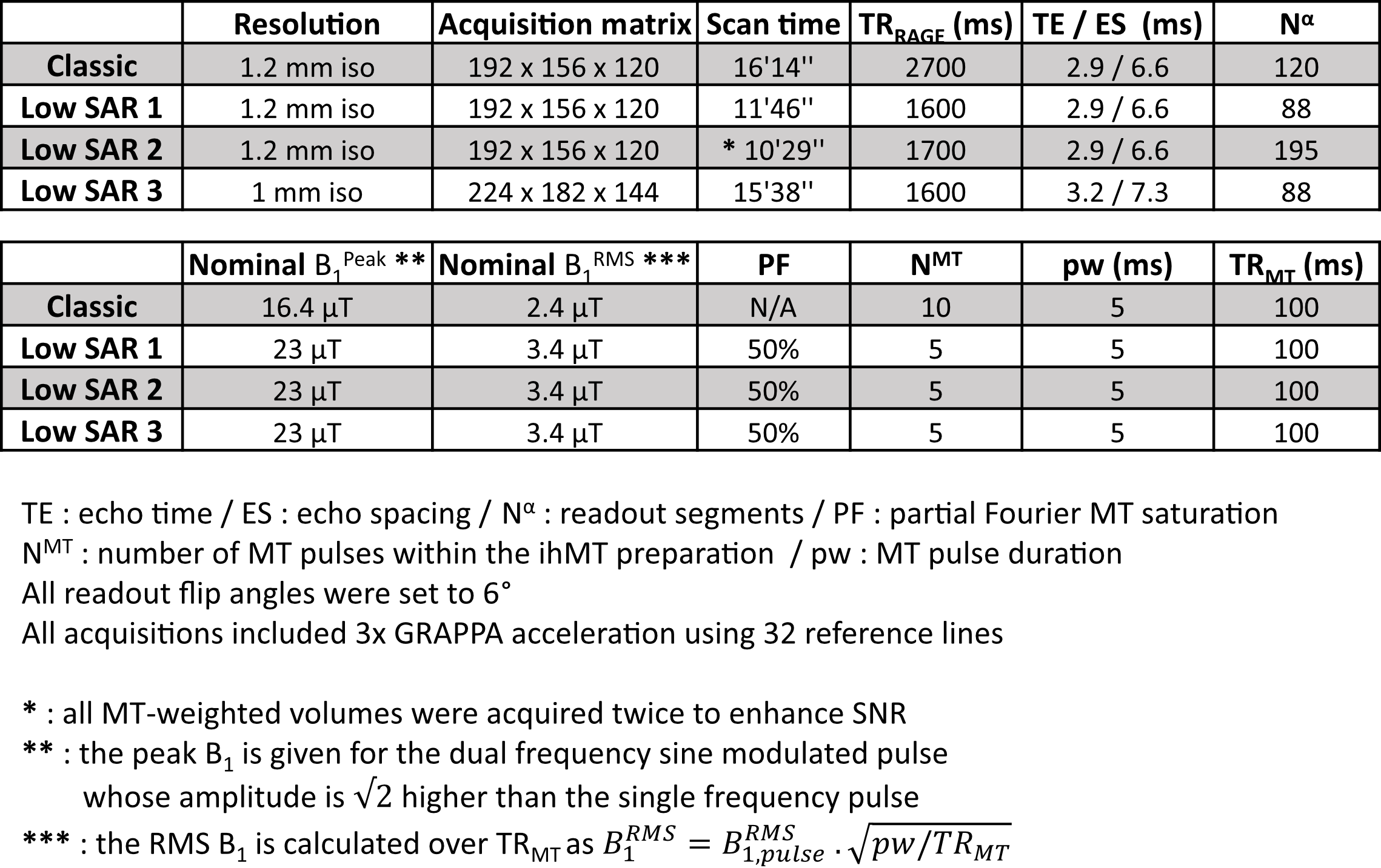
Table.1: Parameters used for (top to bottom): a/ classic ihMT-RAGE, b/ low SAR #1 ihMT-RAGE 1.2mm iso 88 segments, c/ low SAR #2 hMT-RAGE 1.2mm iso 195 segments, d/ low SAR #3 ihMT-RAGE 1mm iso. ihMT saturation pulses followed a Tukey waveform using $$$r=0.2$$$10 with sine modulation to achieve dual frequency saturation. $$$\max(B_1)$$$ using a Nova 1Tx/32Rx head coil was $$$\approx 23$$$ $$$\mu T$$$.