1095
Optimization of sparse saturation transfer MR fingerprinting (ST-MRF) by ranking the importance of saturation transfer contrast features1Russell H. Morgan Department of Radiology and Radiological Science, Johns Hopkins University, Baltimore, MD, United States
Synopsis
Keywords: CEST / APT / NOE, CEST & MT
Motivation: Optimizing ST-MRF sequence design is critical to accelerate image acquisition and improve reconstruction accuracy.
Goal(s): To develop a deep-learning framework that can optimize MRF acquisition for tissue parameter determination with a minimal number of scan parameter settings.
Approach: An interpretable neural network was designed to optimize MRF sequences by ranking the importance of saturation contrast features and evaluated using numerical phantoms and in vivo experiments at 3T.
Results: Importance-ranking network-based sequence optimization demonstrated its ability to improve the choice of scan parameter values for quantification of tissue parameters. Sequence optimization achieved 1.7-fold acquisition acceleration without compromising the fidelity of the tissue parameter quantification.
Impact: Ranking the importance of saturation transfer contrast features facilitates choosing the best combination of sequence parameters for tissue quantification with fingerprinting (ST-MRF). An interpretable network based on importance ranking can significantly accelerate data acquisition for ST-MRF and conventional Z-spectral acquisition.
Introduction
Saturation transfer MRF (ST-MRF) enables simultaneous acquisition of free water, semisolid magnetization transfer (MT), and CEST parameter maps1,2. A pseudo-randomized MRF schedule can be used to create unique saturation signal profiles for different tissue properties. Previous studies utilized deep-learning networks to optimize the acquisition schedule to improve scan efficiency and reconstruction accuracy3,4. However, the complexity of the networks could not allow us to gain insight into how specific acquisition parameters influence the performance of tissue parameter quantification. Recently, the linear projection-based L1-regularization (LASSO) method was successfully applied to reduce the number of frequency offsets in the Z-spectra5, which relies on linear modeling at the expense of lower prediction accuracy in comparison to non-linear deep-learning models. Herein, we developed an interpretable importance-ranking network (IRnet) to optimize MRF acquisition with a minimal number of sequence parameter settings for tissue parameter determination.Methods
IRnet was trained with ST-MRF signals simulated by Bloch-McConnell equations with a three-pool exchange model, free bulk water (w), semisolid macromolecule (m), and amide protons (s). A pre-defined reference MRF schedule (N =103) and tissue parameters, such as relaxation times (T1i and T2i), pool size ratios (Fi) for each pool (i), exchange rate from i- to j- pool (kij), and B0 inhomogeneity were used for the simulation. IRnet consisted of (i) an encoder network for feature extraction and (ii) a replicator network with a single middle layer for interpretation (Fig. 1). The IRnet is defined as follows:$$\widetilde{\theta}_1=arg \min_{\theta_1}(E_{S_{MRF}\sim P(S_{MRF})}[\parallel reconN(S_{MRF}\mid\theta_1)-P_t\parallel_2])\quad{[1]}$$
$$\widetilde{\theta}_2=arg \min_{\theta_2}(E_{S_{MRF}\sim P(S_{MRF})}[\parallel repN(S_{MRF}\mid\theta_2)-encN(S_{MRF}\mid\widetilde{\theta}_1)\parallel_2])\quad{[2]}$$
where $$$reconN(S_{MRF}\mid\theta_1):S_{MRF}\rightarrow\widehat{P}_t$$$ is a reconstruction network with trainable parameters θ1 and trained parameter $$$\widetilde{\theta}_1$$$ is used in the encoder network (encN) in Eq. [2], Pt denotes ground-truth tissue parameters, SMRF =[s1,s2,…sk,…sN] denotes input ST-MRF signals, and $$$E_{S_{MRF}\sim P(S_{MRF})}[.]$$$ denotes the expectation operator, given that SMRF belongs to P(SMRF) distribution. $$$repN(S_{MRF}\mid\theta_2):S_{MRF}\rightarrow{\widehat{P}_{en}}$$$ is a replicator network estimating encoded tissue parameters $$$\widehat{P}_{en}$$$ which is used to calculate a loss function with encoded tissue parameters Pen from an encoder network, $$$encN(S_{MRF}\mid\widetilde{\theta}_1):S_{MRF}\rightarrow{{P}_{en}}$$$. As the replicator network has a single middle layer, weights (wnp, n = the number of dynamic scans for ST-MRF and p = the number of encoded tissue parameters or nodes in the single middle layer) assigned to each input node (SMRF) represent the importance of the corresponding sequence parameters for tissue parameter quantification. The importance can be ranked as follows:
$${Importance}\ {ranking} = arg sort (\sum_{P=1}^L{W^{np}})\quad{[3]}$$
where weight matrix $$$W^{np}\in\widetilde{\theta}_2$$$, L denotes the number of encoded tissue parameters; weight matrix Wnp=[w11,w12,…wnp]. For in-vivo studies, nine healthy subjects participated after informed consent was obtained in accordance with IRB requirements. ST-MRF images were obtained from a multi-shot 3D TSE pulse sequence with varied frequency offsets (Ω), RF saturation powers (B1), RF saturation times (Ts), and relaxation delay times (Td).
Results and Discussion
The IRnet ranked the importance of each dynamic scan and then important scans were selected. In the simulation study (Fig. 2A), the reference schedule (Ref, N = 103) had the lowest normalized root mean square error (nRMSE) against ground-truth tissue parameters. Reducing the number of scans from #103 to #60 resulted in a marginal increase in the nRMSE. To ensure high reconstruction accuracy, the dynamic scan number of #60 was selected for in vivo studies. Individual tissue parameter accuracies obtained with the optimal schedule (IRnet) were comparable with the reference schedule (Ref), as shown in Fig. 2B. The MRF schedules for the four scan parameters optimized by IRnet are shown in Fig. 3, which reduced a total scan time by ~42%. In vivo brain tissue parameter maps were reconstructed from the reference schedule with 103 dynamic scans, as well as pseudo-random, LASSO, and IRnet-based optimized schedules with 60 dynamic scans (Fig. 4). All tissue parameters from IRnet were in good agreement with reference, despite the ~2-fold scan time reduction. Their SSIM outperformed the tissue parameters obtained from pseudo-random and LASSO, particularly for the exchange rates (kmw and ksw), In Fig. 5A, compared with the average tissue parameters obtained from the reference schedule (#103), tissue parameter quantification with #60 dynamic scans was accurate, which is in line with the simulation result. The tissue quantification accuracy was evaluated by calculating nRMSE between the reference and tissue parameters estimated with the different MRF schedules (Fig. 5B). The lowest nRMSE values for all tissue parameters were obtained with the IRnet method.Conclusions
The importance-ranking network could optimize MRF acquisition with a minimal number of scan parameters for tissue parameter determination. The interpretable IRnet framework could significantly reduce data acquisition time for ST-MRF without compromising the fidelity of the tissue parameter quantification.Acknowledgements
This work was supported in part by grants from the National Institutes of Health.References
[1] O. Cohen, S. Huang, M. T. McMahon, M. S. Rosen, and C. T. Farrar, "Rapid and quantitative chemical exchange saturation transfer (CEST) imaging with magnetic resonance fingerprinting (MRF)," Magnetic resonance in medicine, vol. 80, no. 6, pp. 2449-2463, 2018.
[2] H.-Y. Heo, Z. Han, S. Jiang, M. Schär, P. C. van Zijl, and J. Zhou, "Quantifying amide proton exchange rate and concentration in chemical exchange saturation transfer imaging of the human brain," Neuroimage, vol. 189, pp. 202-213, 2019.
[3] B. Kang, B. Kim, H. Park, and H. Y. Heo, "Learning‐based optimization of acquisition schedule for magnetization transfer contrast MR fingerprinting," NMR in Biomedicine, vol. 35, no. 5, p. e4662, 2022.
[4] O. Cohen and R. Otazo, "Global deep learning optimization of chemical exchange saturation transfer magnetic resonance fingerprinting acquisition schedule," NMR Biomed, vol. 36, no. 10, p. e4954, Oct 2023, doi: 10.1002/nbm.4954.
[5] F. Glang et al., "Linear projection-based chemical exchange saturation transfer parameter estimation," NMR Biomed, vol. 36, no. 6, p. e4697, Jun 2023, doi: 10.1002/nbm.4697.
Figures
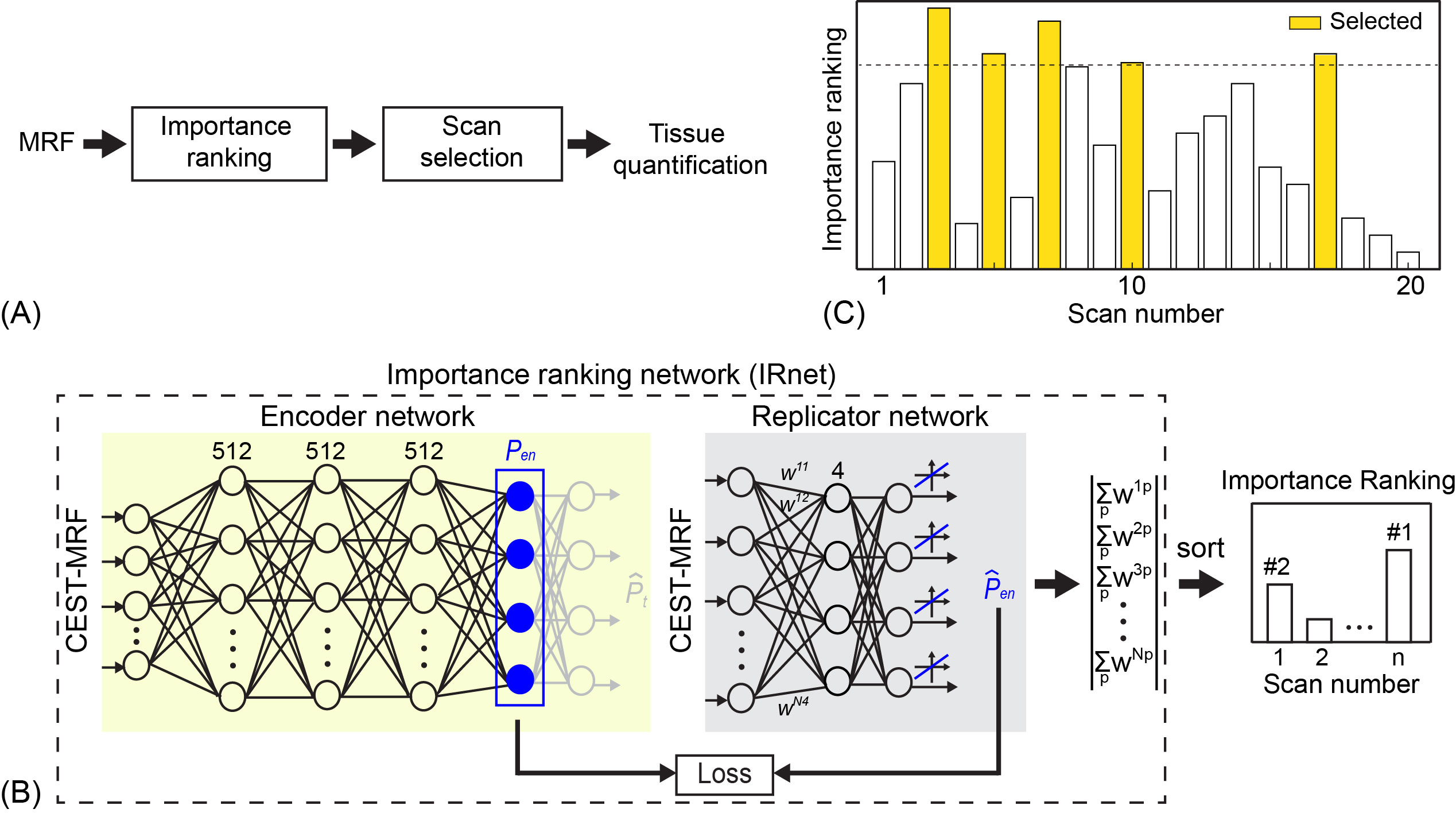
Fig. 1. (A) A framework of the optimization approach includes ranking of the importance of saturation transfer contrast and selecting corresponding dynamic scans. (B) Architecture of the importance ranking network. The output of the replicator network is a weight matrix representing the importance of the sequence parameters for tissue parameter quantification. (C) An example of the importance-ranking score for each dynamic scan. Yellow-colored bars indicate the top 5 highest importance scores.
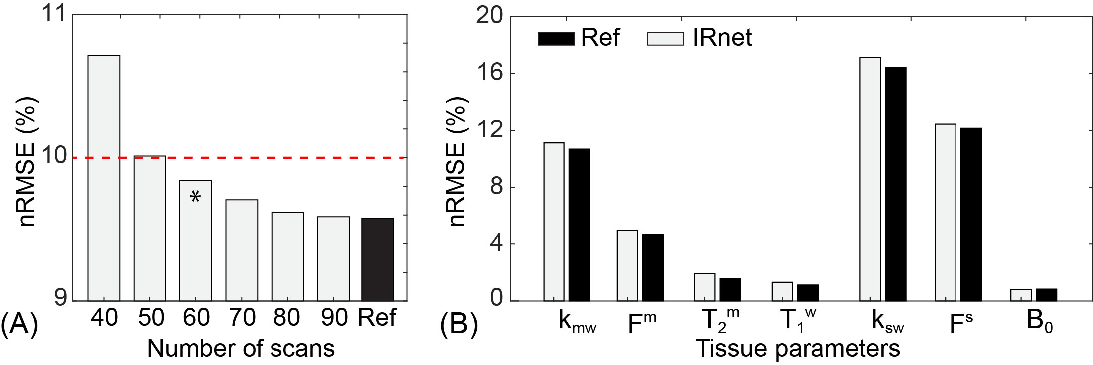
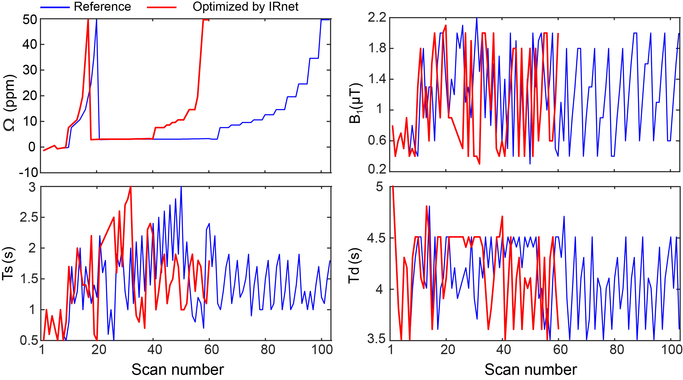
Fig. 3. ST-MRF schedules for in vivo imaging. The IRnet-optimized schedule with #60 dynamic scans (red) and the reference schedule with #103 dynamic scans (blue) are shown. In the ST-MRF sequence, RF frequency offsets (Ω), saturation times (Ts), saturation strengths (B1), and relaxation delay times (Td) are dynamically changed.
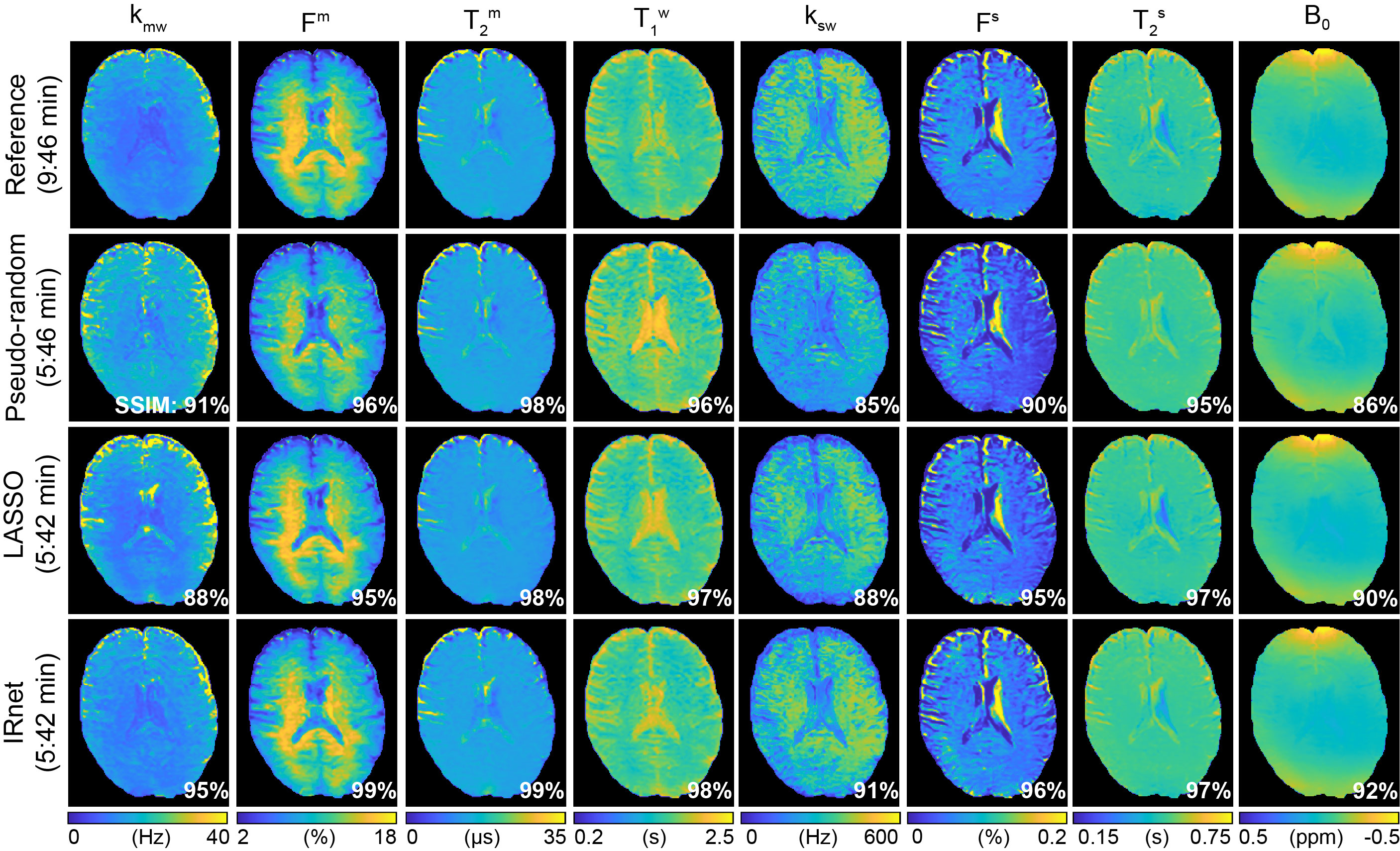
Fig. 4. In vivo tissue parameter maps reconstructed using different MRF schedules. The tissue parameter maps acquired from both the reference schedule and the optimized schedules were compared using the structural similarity index measure (SSIM). The SSIM values are displayed at the right bottom of each image. The IRnet, Random, and LASSO reduced the acquisition time to 5:42 mins. (vs. 9:46 mins for the reference schedule).
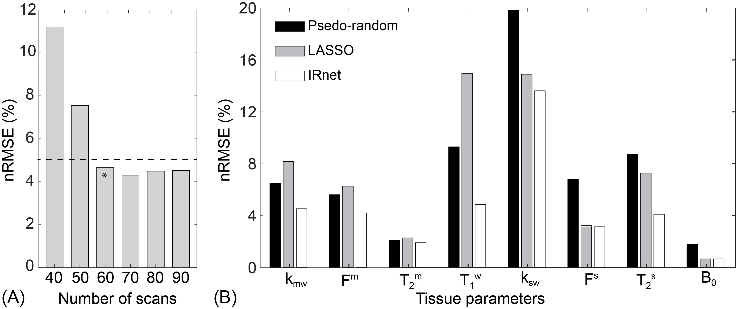
Fig. 5. (A) Average nRMSE values of in vivo tissue parameters obtained from IRnet. A very large improvement is observed when the number of dynamic scans increases from #50 to #60. However, no substantial improvement was found beyond #60 scans. (B) Comparison of the tissue quantification accuracy from different schedules with #60 dynamic scans. The nRMSE values were calculated with respect to the reference schedule.