1094
Frequency-selective inversion nulling ultra-short echo time (FINUTE) MRI for direct detection of lipids of the myelin bilayer1Department of Bioengineering, University of Pennsylvania, Philadelphia, PA, United States, 2Department of Radiology, University of Pennsylvania, Philadelphia, PA, United States
Synopsis
Keywords: CEST / APT / NOE, New Signal Preparation Schemes, Short T2, Lipids, Myelin
Motivation: Recent studies have shown the effects of lipid dyshomeostasis and demyelination in the pathology of neurodegenerative diseases, especially early-onset Alzheimer’s disease. Consequently, imaging methods to monitor these changes are necessary.
Goal(s): This study uses a novel sequence, termed FINUTE, to image short T2 lipids primarily associated with the myelin bilayer.
Approach: Simulations and experiments on ex-vivo spinal cord specimens are performed for validation of methodology and applied in-vivo to assess changes in myelination in a mouse model of AD.
Results: Results demonstrate FINUTE’s sensitivity to myelin lipids, with statistically significant white matter(corpus callosum) and visually-apparent gray matter(hippocampus) changes present in AD animals.
Impact: FINUTE presents a non-invasive MR-imaging technique that is sensitive to lipids primarily in the myelin bilayer as well as in gray matter, thus providing a method for assessing myelination and lipid dyshomeostasis in neurodegenerative diseases such as AD.
Introduction
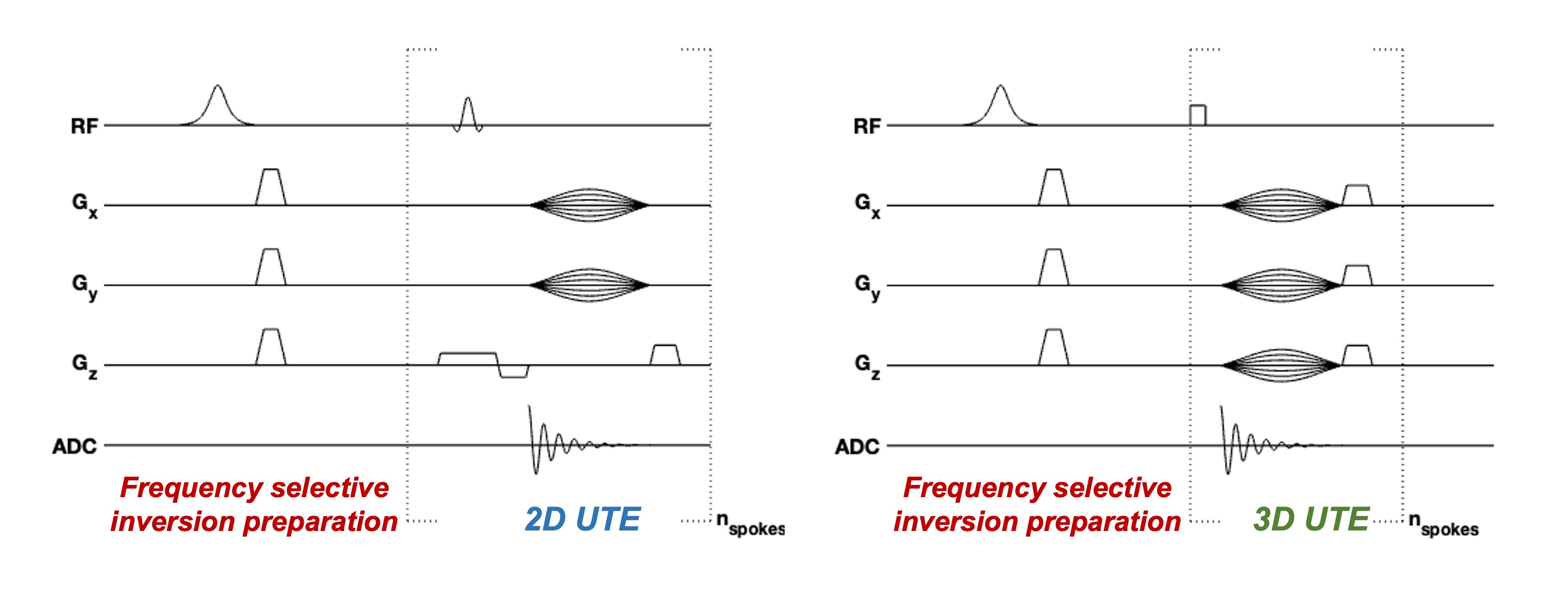
Myelin is a neuroprotective sheath comprised of a high-lipid low-protein bilayer (~70:30 lipid:protein) extending from the membrane of glial cells. It plays a critical role in enhancing neuronal signal conduction and providing axonal structural integrity1. Many neurodegenerative disease (e.g., Alzheimer’s disease (AD)), have impaired myelin sheath integrity/demyelination, and so it remains crucial to monitor changes in myelination for improved diagnosis/treatment. Existing MRI techniques sensitive to myelin rely on imaging the water trapped between the bilayers (i.e., myelin water) or myelin macromolecular content indirectly2,3. Recently, ultra-short echo time (UTE) MRI with adiabatic inversion preparation4 has been used to image myelin lipid protons directly but requires careful optimization of patient-specific parameters. This study uses a frequency selective inversion preparation, centered at the chemical shift of the methylene protons (~ -3.5ppm) of lipids, to null signal from lipid species with ultra-short transverse (T2) relaxation times5, followed by a UTE readout (Figure 1). Normalization with a reference image allows direct detection of these ultra-short T2 lipid species, which are primarily associated with the myelin bilayer. The technique is referred to as FINUTE.Methods
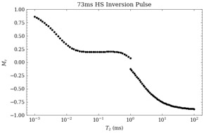
Simulation: A simulation was performed to determine the effect of an adiabatic hyperbolic secant (sech) pulse for T2 values ranging from 1µs to 1s (Figure 2).Ex-vivo phantoms: Fresh ovine spinal column (two pieces) was fixed in 4% paraformaldehyde solution and stored at -4°C prior to imaging. One of the fixed pieces underwent D2O-exchange (99.9% D2O for 24 hours prior to imaging to remove residual water.
In-vivo: Wild-type C57BL/6J(n = 6) and APPNL-F(AD mouse model, n = 6) mice were used for imaging experiments. The images were acquired with a 20mm 1H transceiver volume coil (m2m Imaging) on a 9.4T magnet interfaced with a Bruker Avance III console.
Imaging experiments: The FINUTE sequence involves a magnetization preparation using a 73ms sech pulse followed by a 50µs delay time. For 2D sampling, the preparation was performed prior to acquiring each half-spoke in k-space. For 3D sampling, five half-spokes were acquired after each preparation. The contrast-weighted image was calculated as follows: FINUTE(%)=(1–S-3.5ppm/S-100)x100%, where S-3.5 is an image with inversion preparation at -3.5ppm and S-100 is an image with preparation at -100ppm. The acquisition parameters were as follows: 2D – TR:5ms, TE:462µs, spokes:403, matrix size:128x128, slice thickness:1mm, in-plane resolution:0.156x0.156µm2, NA:4, 315 µs shaped-excitation-pulse; 3D – TR:5ms, TE:8µs, spokes:19149, under-sampling factor:1.5,matrix size:96x96x96, isotropic resolution:0.208µm3, NA:1, 2µs hard-pulse. 2D experiments were performed on ex-vivo and in-vivo specimens, while the 3D experiment was performed on one WT mouse.
Results
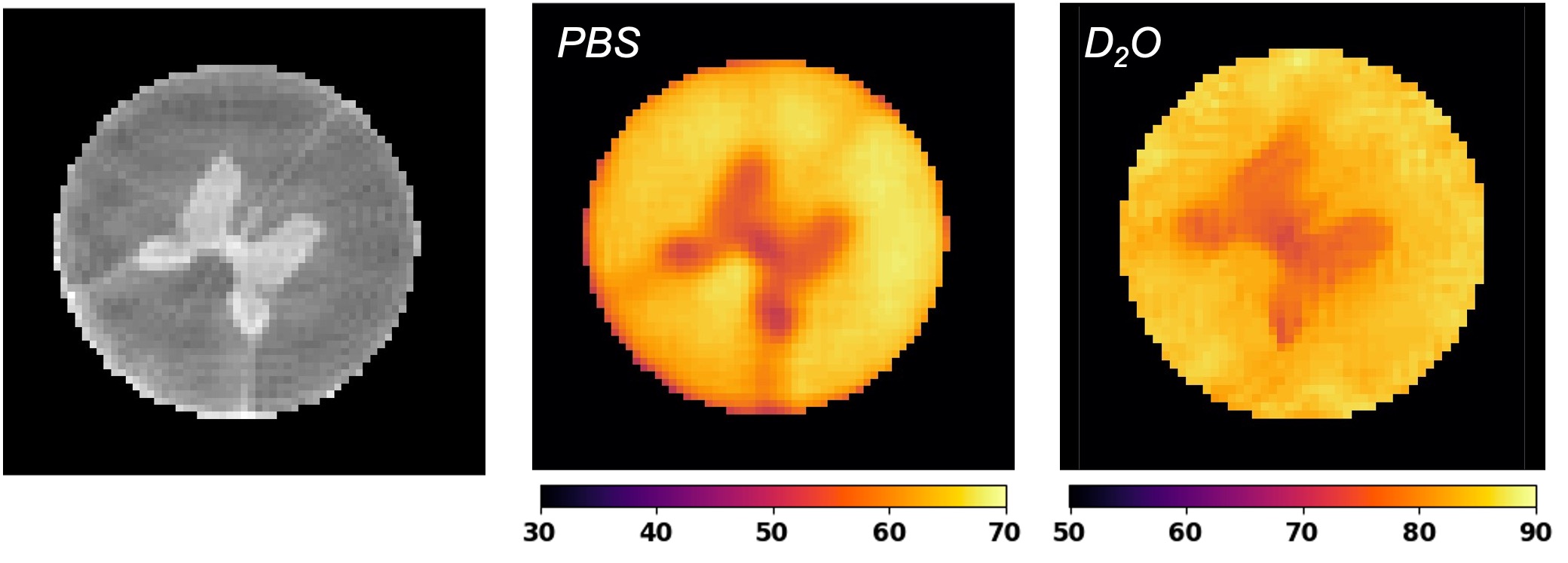
Simulation: Simulation results show that a 73ms adiabatic inversion pulse effectively nulls magnetization in the range of 10-2-101 ms.Ex-vivo: Spinal cords imaged in PBS and D2O (Figure 3) show similar contrast with clear differences between white matter (WM) and gray matter (GM) regions. For PBS, the mean WM contrast is 63±4% with a lower GM contrast of 56±2%. For D2O, the mean WM contrast is 82±5% with a lower GM contrast of 78±2%.
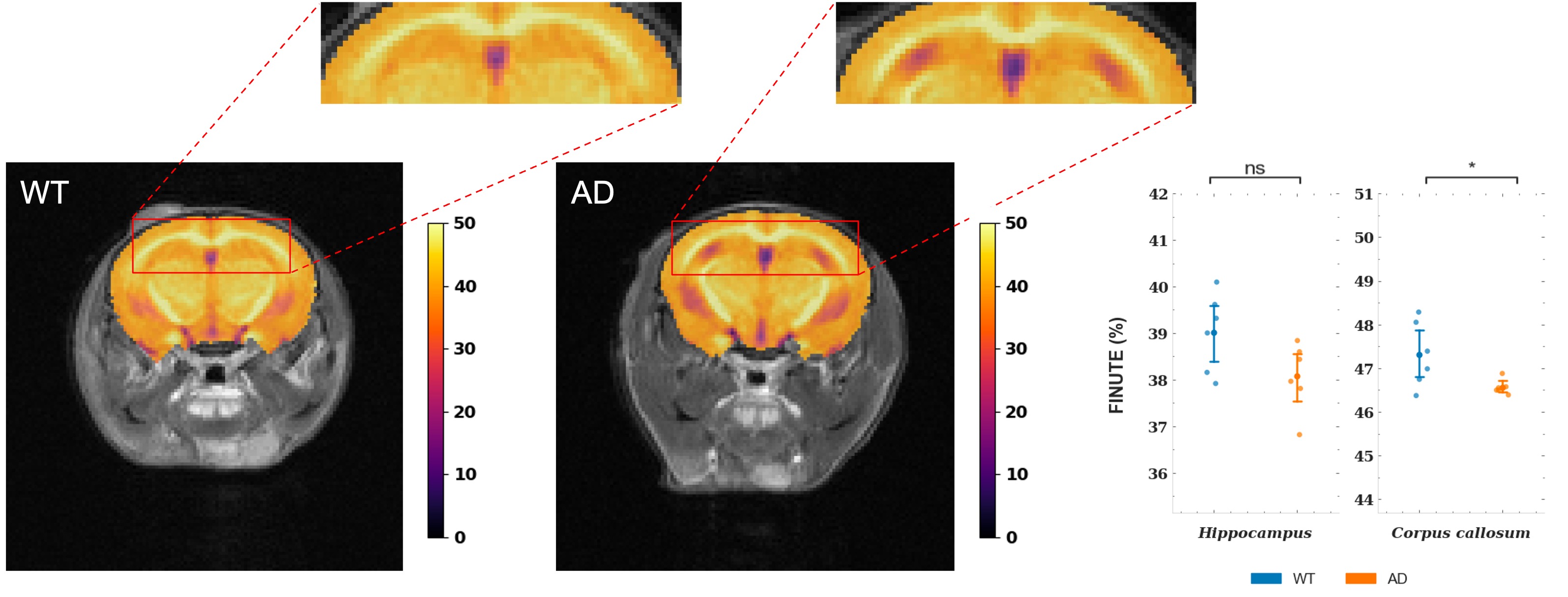
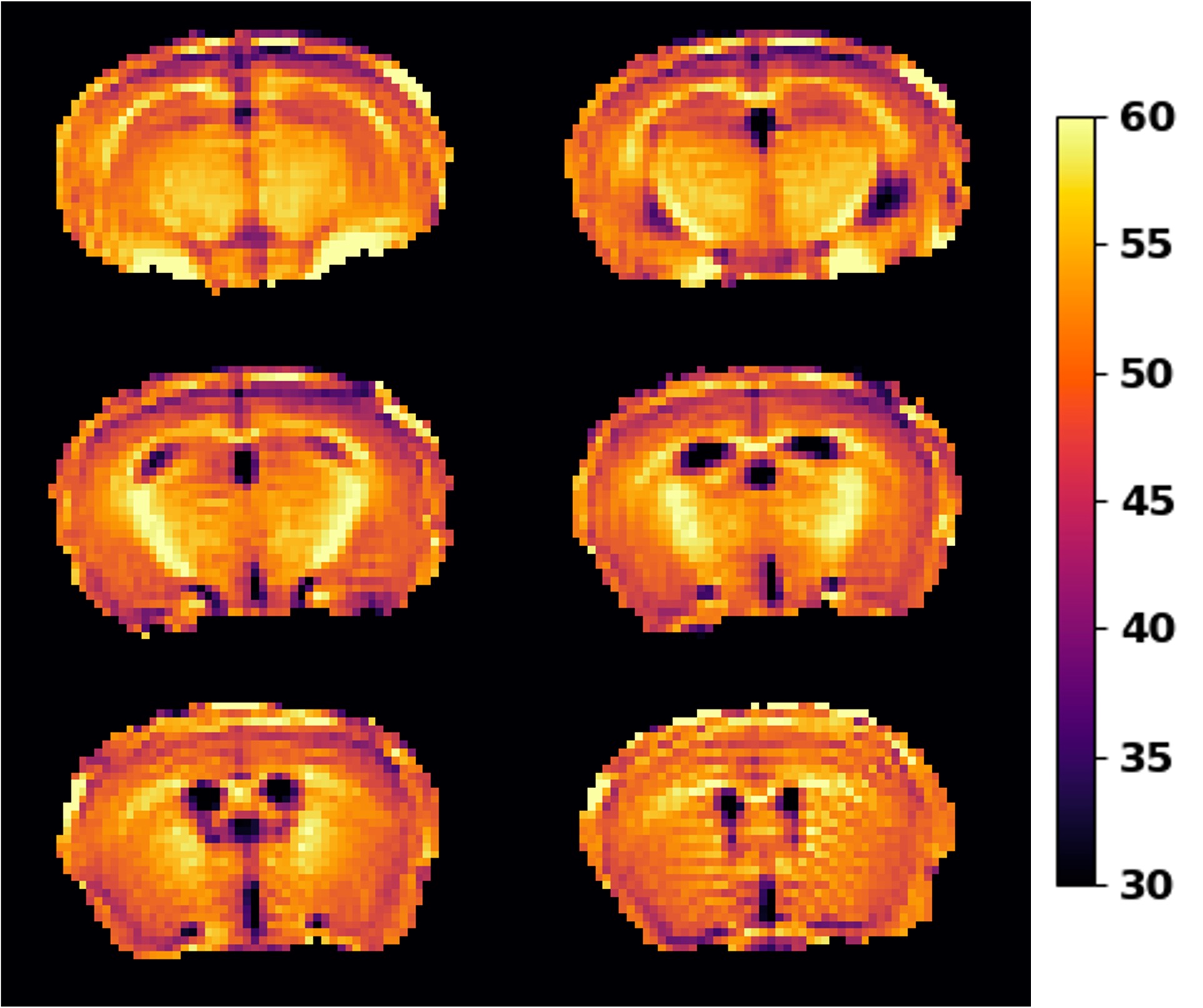
In-vivo: Compared to WT mice, AD mice show lower contrast in the corpus callosum (CC) (WT:47.3±0.7% v. AD:46.6±0.1%, p<0.05) and hippocampus (WT:39.0±0.8% v. AD:38±0.7%, p = 0.06), with the CC presenting a statistically significant change (Figure 4). Visually, the maps show a distinct decrease in the hippocampus, but a less discernable decrease in the CC. FINUTE with 3D sampling (Figure 5) shows good contrast between WM and GM regions with increased contrast (WM: 59±6%, GM: 50±5%) due to a shorter TE and thus increased sensitivity to nulled magnetization of extremely short T2 species (T2<10µs).
Discussion
FINUTE shows strong sensitivity to lipids with minimal contributions from myelin water and intra/extracellular water as observed from the D2O-exchanged spinal cord. The difference in contrast between the spinal cord in PBS v. D2O may stem from partially inverted water, which would increase signal intensity (due to magnitude reconstruction) in the -3.5ppm image. A frequency-dependent inversion spectrum may provide better insight into potential signal contributions. Comparing WT to AD mice, the significant decrease in in the CC reflects changes in a WM region known to be affected in early-stage AD6. Furthermore, the decrease in the hippocampus reflects a hallmark region affected in AD7. 3D FINUTE shows increased contrast as the extremely short TE provides increased sensitivity to T2 species on the order of tens of microseconds. However, simulation results show that an inversion pulse has minimal effect on extremely short T2 and prior literature places the majority of lipid T2 values in the 100-300µs range. Consequently, the TE used for 2D-FINUTE may be sufficient for detectable lipid species.Acknowledgements
Research reported in this publication was supported by the National Institute of Biomedical Imaging and Bioengineering of the National Institutes of Health under award Number P41EB029460, the National Institute of Aging of the National Institutes of Health under Award Number R01AG063869, and the national Institute of Diabetes and Digestive and Kidney Diseases of the National Institutes of Health under Award Number R01DK098656.References
1. Kister, A., & Kister, I. (2022). Overview of myelin, major myelin lipids, and myelin-associated proteins. Frontiers in Chemistry, 10, 1623. https://doi.org/10.3389/FCHEM.2022.1041961/BIBTEX
2. MacKay, A. L., & Laule, C. (2016). Magnetic Resonance of Myelin Water: An in vivo Marker for Myelin. Brain Plasticity, 2(1), 71. https://doi.org/10.3233/BPL-160033
3. Sled, J. G. (2018). Modelling and interpretation of magnetization transfer imaging in the brain. NeuroImage, 182, 128–135. https://doi.org/10.1016/J.NEUROIMAGE.2017.11.065
4. Ma, Y. J., Jang, H., Chang, E. Y., Hiniker, A., Head, B. P., Lee, R. R., Corey-Bloom, J., Bydder, G. M., & Du, J. (2020). Ultrashort echo time (UTE) magnetic resonance imaging of myelin: technical developments and challenges. Quantitative Imaging in Medicine and Surgery, 10(6), 1186. https://doi.org/10.21037/QIMS-20-541
5. Wilhelm, M. J., Ong, H. H., Wehrli, S. L., Li, C., Tsai, P.-H., Hackney, D. B., & Wehrli, F. W. (2012). Direct magnetic resonance detection of myelin and prospects for quantitative imaging of myelin density. Proceedings of the National Academy of Sciences, 109(24), 9605-9610. https://doi.org/doi:10.1073/pnas.1115107109
6. Papuc, E., & Rejdak, K. (2020). The role of myelin damage in Alzheimer’s disease pathology. Archives of Medical Science : AMS, 16(2), 345. https://doi.org/10.5114/AOMS.2018.76863
7. Rao, Y. L., Ganaraja, B., Murlimanju, B. V., Joy, T., Krishnamurthy, A., & Agrawal, A. (2022). Hippocampus and its involvement in Alzheimer's disease: a review. 3 Biotech, 12(2), 55. https://doi.org/10.1007/s13205-022-03123-4
Figures