1092
Highly Accelerated CEST Imaging with Stack-of-stars Acquisition using Unsupervised Implicit Neural Representation Networks1Shanghai Jiao Tong University, Shanghai, China
Synopsis
Keywords: CEST / APT / NOE, CEST & MT
Motivation: Reduction of scan time in CEST imaging is clinically meaningful.
Goal(s): Our goal is to develop an undersampled reconstruction algorithm to help vastly reduce the acquisition time.
Approach: A novel unsupervised deep-learning based algorithm is proposed to accelerate steady-state pulsed CEST imaging with golden-angle stack-of-stars trajectory using mixed-feature hash encoding implicit neural representation. Additionally, Imaging quality is further improved using the explicit prior knowledge of weighted joint sparsity in subtle structural features of CEST image domain. The low rankness and sparsity in the Z‐spectra domain are used to reduce acquisition time.
Results: It is possible to achieve a 30-fold acceleration for CEST imaging.
Impact: An unsupervised deep-learning algorithm is proposed to accelerate steady-state pulsed CEST imaging with golden-angle stack-of-stars trajectory using mixed-feature hash encoding implicit neural representation and weighted joint sparsity. It can vastly reduce the acquisition time and has potential for clinical applications.
Introduction
Chemical exchange saturation transfer (CEST) imaging[1] is a novel contrast mechanism in MRI with promising clinical applications. However, the acquisition is time-consuming due to the long saturation module and various frequency offsets of the saturation pulses. The combination of compressed sensing (CS) with parallel imaging (PI) allows us to achieve higher acceleration factors by exploiting data redundancy in the sparse transform domain[2-4]. The use of unsupervised learning helps to achieve highly accelerated imaging without using large numbers of training datasets. In this study, we propose an unsupervised deep-learning based algorithm for highly accelerated non-Cartesian CEST imaging using mixed-feature hash encoding implicit neural representation and weighted joint sparsity in CEST images.Theory
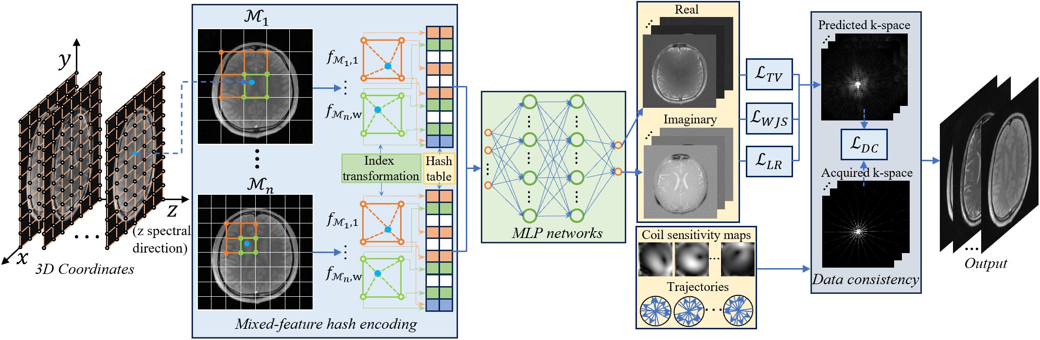
Implicit neural representations (INR) based algorithm[5] is applied to map the spatial and z-spectral 3D coordinates of CEST images to the corresponding intensity values using multilayer perceptron (MLP) due to the strong learning capabilities. Furthermore, the mixed-feature hash encoding is combined to improve imaging performance by exploiting the redundancy of different feature grids in multiresolution hash tables. The partial feature grids are adaptively mixed into a small number of hash tables using specified index transformation methods, allowing for higher quality reconstruction with fewer encoding parameters. In addition, the explicit prior knowledge of weighted joint sparsity[6] in subtle structural features of the CEST image domain is used to improved imaging quality. The low rankness and sparsity in the Z‐spectra domain are used to reduce the acquisition time. The proposed algorithm can be expressed as:$$\min _{ \mathcal{X}_{\theta}} \frac{1}{2}\|\boldsymbol{\Phi}(\mathcal{X}_{\theta})-\mathcal{Y}\|_{2}^{2}+\lambda_{1}\sum_{n}\left\|\mathcal{W}_{JS,n} \circ D_{n}\mathcal{X}_{\theta}\right\|_{2,1}+\lambda_{2}\left\|D_{n}\mathcal{X}_{\theta}\right\|_{1}+\lambda_{3}\left\|\mathcal{X}_{\theta}\right\|_{\star}$$ where $$$\mathcal{Y}$$$ represents k-space data, $$$\mathcal{X}_{\theta}$$$ is the corresponding CEST image series, $$$\theta $$$ represents the weights in MLP and hash tables to be optimized. $$$\Phi $$$ is the NUFFT operator. $$$ \mathcal{W}_{JS,n} (n=1,2)$$$and $$$ D_{3}$$$ are first-order difference matrices along different dimensions, and $$$ D_{n} (n=1,2)$$$ are weighted tensors of the weighted joint sparsity term[6]. $$$\lambda_{1}$$$, $$$\lambda_{2}$$$ and $$$\lambda_{3}$$$ are regularization parameters. The flowchart of our algorithm is illustrated in Figure.1. The spatial and z-spectral 3D coordinates are fed into the mixed-feature hash encoding block and MLPs to output the real and imaginary parts of CEST images. During the updating process, the coefficients in the MLPs and mixed-feature hash tables are simultaneously optimized by minimizing the data consistency loss $$$\mathcal{L}_{DC}$$$, the low-rank loss $$$\mathcal{L}_{LR}$$$, the weighted joint sparse loss $$$\mathcal{L}_{WJS}$$$, and the total variation loss $$$\mathcal{L}_{TV}$$$ to output CEST images.Methods
The human brain datasets were acquired from 10 healthy subjects (9 males, age 25.6±2.6; 1 female, age 23.7) on a 3T MRI scanner (United Imaging Healthcare, Shanghai, China). The steady-state pulsed CEST imaging with golden-angle stack-of-stars sequence with 32-channel head coil was used for acquisition. For CEST labeling, each Gaussian saturation pulse was applied with a duration tsat=50ms and an effective B1=1.5μT. Each radial stack (12 partitions) was acquired with center-out ordering. Imaging parameters were: FOV=220×220×42mm3, spatial resolution=1.15×1.15×3.5mm3, TR/TE=4.71/2.34ms, and FA=5° for readouts. The 30 saturation frequency offsets were: ±6,±5.5,±5,±4.5,±4,±3.5,±3.5,±3.5,±3,±2.5,±2.0,±1.5,±1,±0.5, and 0ppm. One prolonged acquisition S0 was conducted at the beginning (-300ppm) as reference. Three hundred radial stacks were acquired in each saturation frequency to obtain fully sampled data with a total scan time of approximately 16 minutes. Several acceleration rates of R=10/20/30 were evaluated. The performance of the proposed algorithm was compared to the state-of-the-art CS algorithms, including PBCS, kt-SLR[7], LRTES[8], and unsupervised deep learning algorithms including NeRP[9], and TDDIP[10].Results
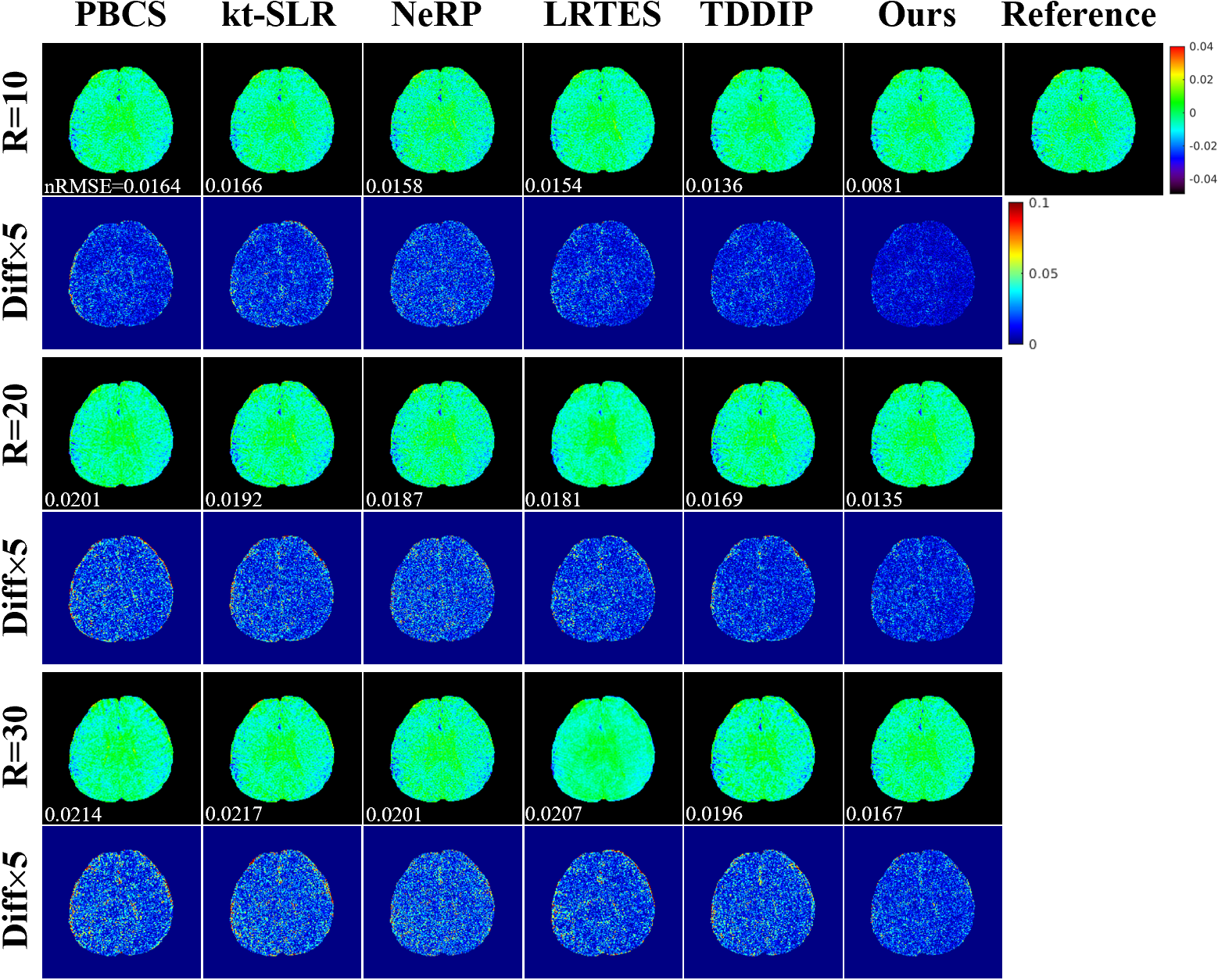
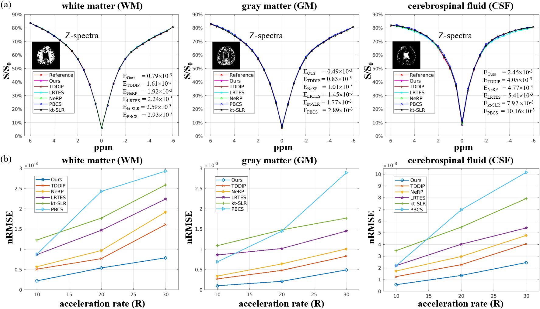
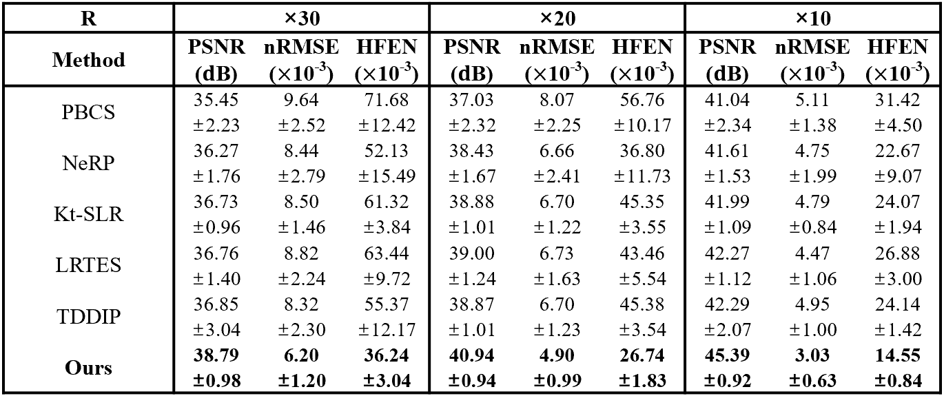
Figure.2 presents a comparison of the amide proton transfer weighted (APTw) images from a typical subject reconstructed with different algorithms at three accelerated rates R=10/20/30. The comparison of the Z-spectra from white matter (WM), gray matter (GM), and cerebrospinal fluid (CSF) with different algorithms at R=30 is shown in Figure.3(a). The plots of the nRMSE of the reconstructed Z‐spectra in different algorithms at three accelerated rates R=10/20/30 are shown in Figure.3(b). The means and standard deviations of the reconstruction metrics (PSNR/nRMSE/HFEN) in the human brain datasets are summarized in Table.1. Our algorithm generates a higher image quality and reduced error compared to other algorithms in both APTw images and Z-spectra.Discussion and Conclusion
An unsupervised deep-learning algorithm was presented to accelerate CEST imaging using implicit neural representation with mixed-feature hash encoding. Explicit prior knowledge of weighted joint sparsity in subtle structural features of the image domain was used to further improved image quality. The low rankness and sparsity in the Z‐spectra domain are also used to reduce the acquisition time. The proposed algorithm can achieve superior image quality compared to several state-of-the-art algorithms at R=30. It is possible to obtain a whole brain CEST imaging in 2 minutes using the proposed technique.Acknowledgements
This work was supported in part by the National Natural Science Foundation of China under Grant 81627901, in part by the Ministry of Science and Technology under Grant 2022YFB4702702, and in part by the Shanghai Science and Technology Commission Explorer Program under Grant 22TS1400300.References
[1] K. M. Ward et al., “A new class of contrast agents for MRI based on proton chemical exchange dependent saturation transfer (CEST),” J Magn Reson, vol. 143, no. 1, pp. 79-87, Mar, 2000.
[2] Y. Zhang et al., “Chemical exchange saturation transfer (CEST) imaging with fast variably-accelerated sensitivity encoding (vSENSE),” Magn Reson Med, vol. 77, no. 6, pp. 2225-2238, Jun, 2017.
[3] H. Y. Heo et al., “Accelerating chemical exchange saturation transfer (CEST) MRI by combining compressed sensing and sensitivity encoding techniques,” Magn Reson Med, vol. 77, no. 2, pp. 779-786, Feb, 2017.
[4] H. She et al., “Accelerating chemical exchange saturation transfer MRI with parallel blind compressed sensing,” Magn Reson Med, vol. 81, no. 1, pp. 504-513, Jan, 2019.
[5] B. Mildenhall et al., “Nerf: Representing scenes as neural radiance fields for view synthesis,” Communications of the ACM, vol. 65, no. 1, pp. 99-106, 2021.
[6] B. Liu et al. "Low-rank tensor subspace decomposition with weighted group sparsity for the acceleration of non-cartesian dynamic MRI." IEEE Transactions on Biomedical Engineering 70.2 (2022): 681-693.
[7] S. G. Lingala et al., “Accelerated dynamic MRI exploiting sparsity and low-rank structure: k-t SLR,” IEEE Trans Med Imaging, vol. 30, no. 5, pp. 1042-54, May, 2011.
[8] J. He et al., “Accelerated High-Dimensional MR Imaging With Sparse Sampling Using Low-Rank Tensors,” IEEE Trans Med Imaging, vol. 35, no. 9, pp. 2119-29, Sep, 2016.
[9] L. Shen et al., “NeRP: Implicit Neural Representation Learning With Prior Embedding for Sparsely Sampled Image Reconstruction,” IEEE Trans Neural Netw Learn Syst, vol. PP, Jun 3, 2022.
[10] J. Yoo et al., “Time-Dependent Deep Image Prior for Dynamic MRI,” IEEE Trans Med Imaging, vol. PP, May 27, 2021.
Figures