1084
Self-navigated Subspace Reconstruction for Real-time MRI Speech Tracking1Department of Diagnostic Radiology, The University of Hong Kong, Hong Kong, China, 2Department of Linguistics, The University of Hong Kong, Hong Kong, China
Synopsis
Keywords: Image Reconstruction, Motion Correction
Motivation: Real-time MRI offers a continuous and dynamic view of the object being imaged. Researchers have applied real-time MRI to speech tracking, which allows for the visualization of the vocal tract during speech production.
Goal(s): In this study, we propose applying self-navigated subspace reconstruction to real-time MRI for speech tracking.
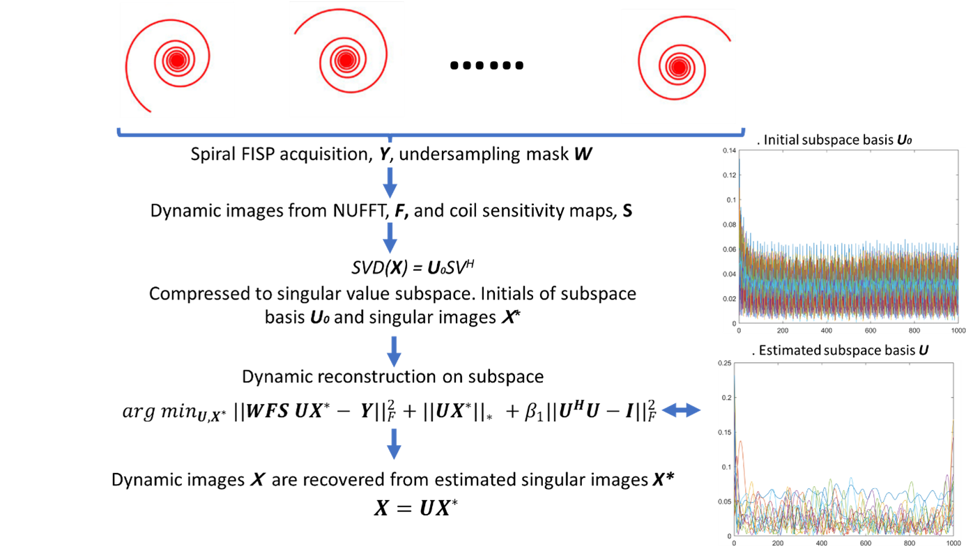
Approach: During reconstruction, 1000 frames were compressed to a few principal components, and iterative low-rank approximation was performed on compressed k-space, greatly reducing computation costs.
Results: The proposed method allows for the joint reconstruction of all time frames and provides the dynamic motion pattern of the vocal tract at a high frame rate.
Impact: Our study presented a subspace reconstruction technique that does not require a navigator echo, which can be used for real-time MRI, particularly in speech tracking applications.
INTRODUCTION
Real-time MRI captures a series of cross-sectional images of the subject's vocal tract, synchronized with audio recording [1]. Although real-time MRI was conventionally reconstructed using regularized iterative reconstruction [2, 3], advanced MRI reconstruction and acceleration methods could improve its speed further. Various algorithms are used for subspace reconstruction, such as spatial-temporal separable model, dictionary learning, and low-rank matrix completion [4]. Moreover, studies have shown the feasibility of using a multi-scale low rank approximation to reconstruct 100 gigabytes of dynamic volumetric image data [5]. Previously, subspace reconstruction with navigator echo was used for speech tracking [6, 7]. In addition, another previous study also showed the feasibility of updating the subspace basis in conjuncation with the iterative reconstruction [8]. Therefore, in this study, we propose applying self-navigated subspace reconstruction to real-time MRI for speech tracking. We performed experiments on a clinical 3T MRI using standard RF coils and rapid acquisition.METHODS
MRI acquisition, postprocessing, and subspace basis generation The in vivo experiments were approved by the local institutional research ethics committee. A healthy volunteer underwent a real-time MRI scan using the FIDALL sequence on a GE 3T system (General Electric Healthcare) with a clinical 21-channel head-neck coil for signal reception. Real-time MRI scan has parameters including: flip angle = 15⁰, FISP acquisition, constant echo time (TE)/ repetition time (TR) = 1.77/7 ms, slice thickness = 5 mm, matrix size = 128 × 128, field of view = 300 × 300 mm2, number of TRs = 1000, number of total spiral interleaves = 32, nominal temporal resolution = 7 ms/frame, golden angle rotation of spiral (56.25⁰), and scan time = 7 s/slice. The k-space data was compressed into singular value components. The error associated with the choice of 5% corresponds to around 10-20 singular values. Gradient descent for subspace image reconstruction. Noted that the images are compressed to singular images. All the computations in this study were performed in MATLAB (MathWorks, Natick) on a laptop computer.RESULTS
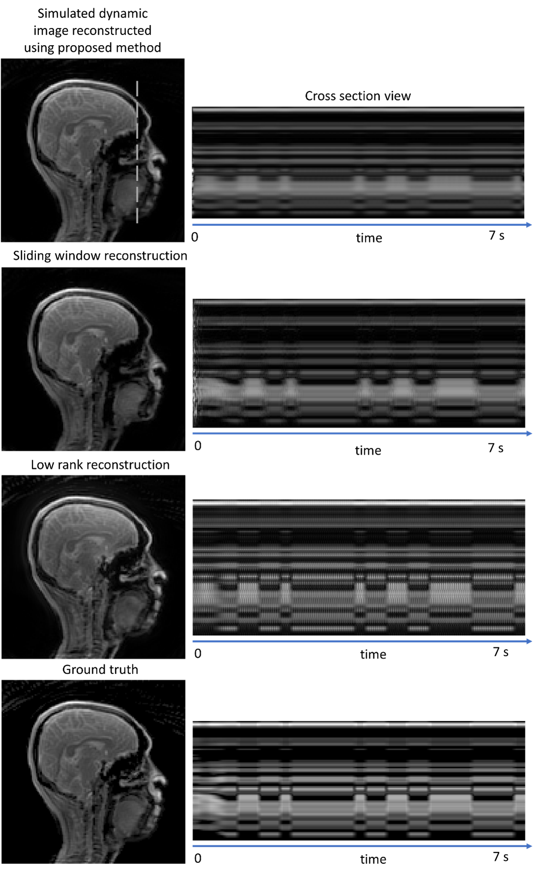
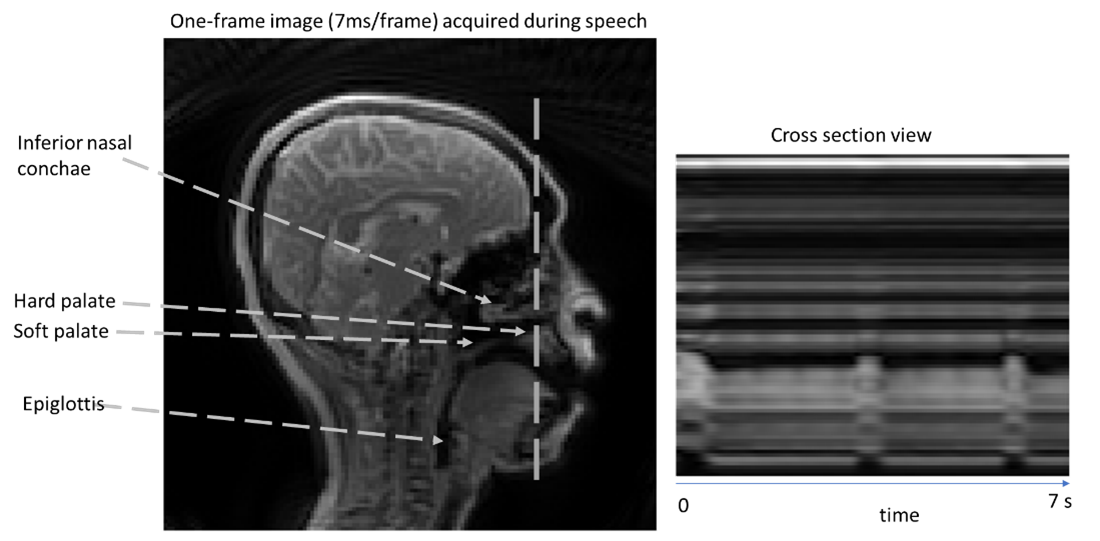
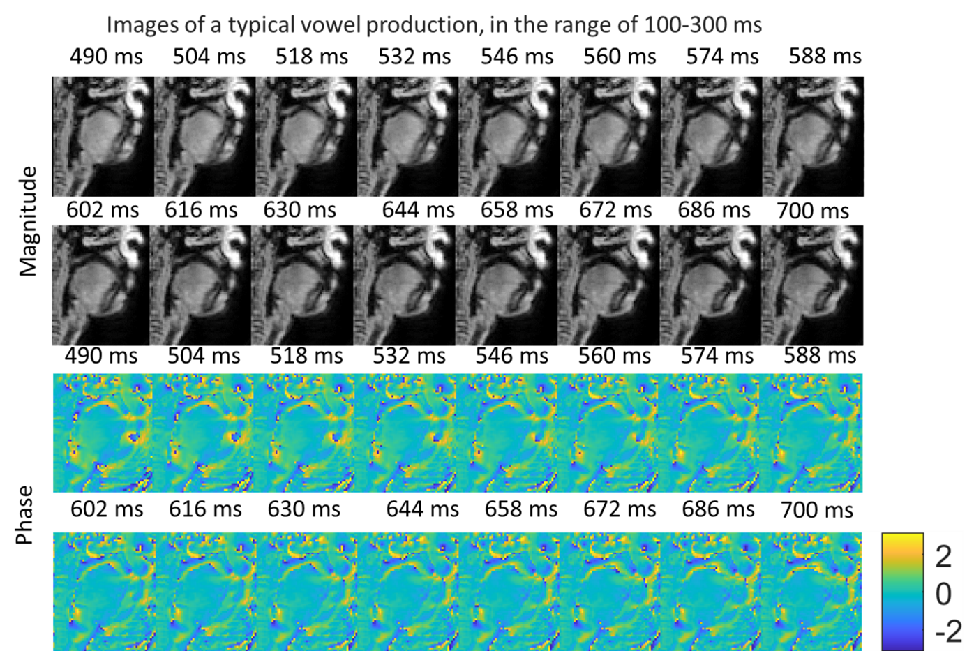
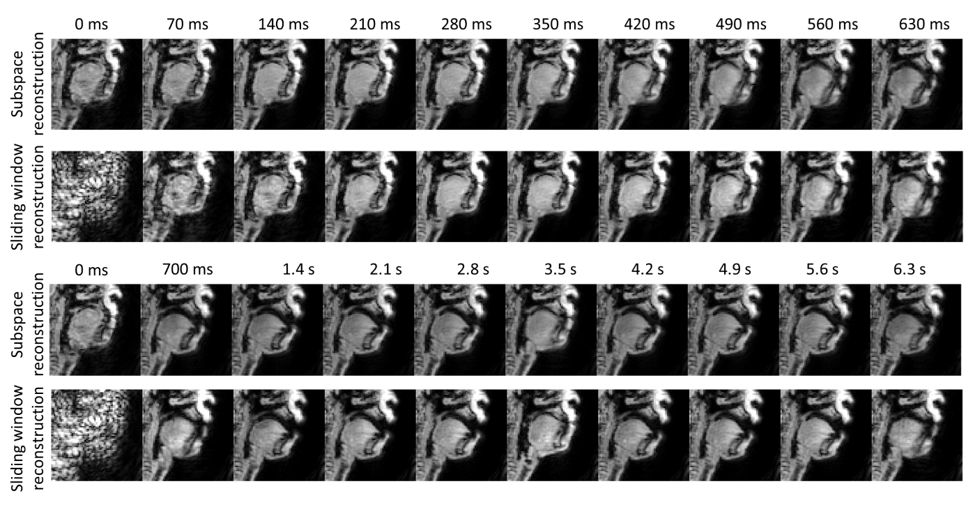
The results of a study that involved a 32-time acceleration simulation showed that the proposed method produced a reasonably small root mean square error (RMSE) of 0.154, compared to 0.278 for sliding window reconstruction, and 0.294 for low rank reconstruction. The study also presented in vivo images of a typical sagittal image with a temporal resolution of 7 ms/frame. The cross-sectional view from 1000 frames acquired over 7 seconds revealed the dynamics of the vocal tract, and several vowels were generated during this scan. In addition, the study displayed the magnitude and phase images of a typical vowel production in the range of 200 ms. The high temporal resolution of images from the proposed method enabled clear visualization of the soft palate. The phase maps showed a modest variation of the static magnetic field due to the airflow, which could cause phase cancellation if one summed these dynamics in a conventional sliding window approach. Furthermore, Figure 5 compared the proposed method and sliding window reconstruction. The image reconstructed from the proposed method had a high temporal resolution in resolving the motion of the soft palate in almost every frame.DISCUSSION
In this study, a subspace reconstruction method for real-time MRI is presented. The proposed method allows for the joint reconstruction of all time frames and provides the dynamic motion pattern of the vocal tract at a high frame rate. The proposed method delivers a nominal resolution of 7 ms/frame and enables the linguistic study of the position of the soft palate and the opening or closing of the passage between the nasopharynx and oropharynx.CONCLUSION
Our study presented a subspace reconstruction technique that does not require a navigator echo, which can be used for real-time MRI, particularly in speech tracking applications.Acknowledgements
No acknowledgement found.References
1. Ramanarayanan, V., et al., Analysis of speech production real-time MRI. Computer Speech and Language, 2018. 52: p. 1-22.
2. Lingala, S.G., et al., A fast and flexible MRI system for the study of dynamic vocal tract shaping. Magn Reson Med, 2017. 77(1): p. 112-125.
3. Lingala, S.G., et al., State-of-the-art MRI Protocol for Comprehensive Assessment of Vocal Tract Structure and Function. 17th Annual Conference of the International Speech Communication Association (Interspeech 2016), Vols 1-5, 2016: p. 475-479.
4. Shafieizargar, B., et al., Systematic review of reconstruction techniques for accelerated quantitative MRI. Magn Reson Med, 2023. 90(3): p. 1172-1208.
5. Ong, F., et al., Extreme MRI: Large-scale volumetric dynamic imaging from continuous non-gated acquisitions. Magn Reson Med, 2020. 84(4): p. 1763-1780.
6. Fu, M., et al., High-frame-rate full-vocal-tract 3D dynamic speech imaging. Magn Reson Med, 2017. 77(4): p. 1619-1629.
7. Fu, M.J., et al., High-Resolution Dynamic Speech Imaging with Joint Low-Rank and Sparsity Constraints. Magnetic Resonance in Medicine, 2015. 73(5): p. 1820-1832.
8. Bhave, S., et al., Accelerated whole-brain multi-parameter mapping using blind compressed sensing. Magn Reson Med, 2016. 75(3): p. 1175-86.
9. Cao, P., et al., Motion-resolved and free-breathing liver MRF. Magnetic Resonance Imaging, 2022. 91: p. 69-80.
Figures