1082
A (k,t)-RAKI Method for Interpolating Sparse Data in Accelerated MRSI Acquisitions1Department of Automation, Tsinghua University, Beijing, China, 2Department of Electrical and Computer Engineering, University of Illinois Urbana-Champaign, Urbana, IL, United States, 3Beckman Institute for Advanced Science and Technology, University of Illinois Urbana-Champaign, Urbana, IL, United States, 4Department of Bioengineering, University of Illinois Urbana-Champaign, Urbana, IL, United States
Synopsis
Keywords: Image Reconstruction, Spectroscopy
Motivation: To further accelerate high-resolution MRSI acquisitions leveraging parallel imaging.
Goal(s): While standard parallel imaging techniques such as (k,t)-GRAPPA can interpolate the sparsely sampled (k,t)-space in MRSI, learning-based nonlinear interpolation has demonstrated better performance in parallel MRI. But these methods have not effectively utilized the time/free induction decay (FID) dimension, which should be leveraged to improve interpolation accuracy.
Approach: We adapted and extended the RAKI method by incorporating the FID dimension, via a 3D, complex-valued convolutional network, for MRSI reconstruction.
Results: Our method effectively reconstructed data for different undersampling designs in in vivo MRSI, leading to improved subsequent spatiospectral processing results.
Impact: We presented a self-supervised learning-based (k,t)-space interpolation method, (k,t)-RAKI, that is useful for further accelerating MRSI acquisition, in combination with subspace methods.
Introduction
Parallel imaging has been used to accelerate the slow MRSI acquisition [1]. The commonly used linear methods, such as SENSE [2] and GRAPPA [3] based methods, still produce residual aliasing of water/lipid signals which are significant enough to disrupt spatiospectral processing for metabolite reconstruction. Nonlinear k-space interpolation methods have demonstrated success in parallel MRI [4]. RAKI (Robust Artificial neural networks for k‐space Interpolation) [5][6] has been designed to calibrate CNN from autocalibration signal (ACS) data which can later be employed on the whole undersampled k-space. However, RAKI has thus far been applied exclusively to MRI data. Formerly, MRSI reconstruction has also seen the integration of neural networks [7]. Nevertheless, these approaches focused on nonlinear relationships within the k-space domain, overlooking valuable information in the temporal dimension.In this work, we adapted and extended the self-supervised learning-based RAKI method by incorporating the FID dimension into a 3D, complex-valued convolutional network, for MRSI reconstruction. We improved the design by training a single network to handle multi-coil data simultaneously instead of the coil-by-coil interpolation in the original RAKI method. We demonstrate reduced aliasing by the proposed method and consequently improved spatiospectral processing results, using in vivo 1H-MRSI data.
Methods and Experiments
(k,t)-RAKI network design for MRSI:We considered 5D (ky, kx, kz, time, coil) input data. Specifically, we extract sampled z slices to process individually, reducing the input to 4-D (ky, kx, time, coil). To leverage the inherent full combination of channels in the CNN, we designate the coil dimension as the channel, and the (ky, kx, time)-shaped ACS data serves as the 3D input for the network. Simultaneously, we employ 3D convolutional kernels to maximize the utilization of information across all three dimensions. Our network comprises three convolutional layers, with the first two layers incorporating CReLU as the activation function to introduce nonlinearity. The 3D-CNN enables interpolation across all three dimensions (kx, ky, time) while maintaining continuous coil combination, thus maximizing the utilization of information inherent in parallel imaging. This approach leads to an enhancement in the overall quality of k-t-space reconstruction. We also improved the original RAKI design by training a single network to handle multi-coil data simultaneously instead of the coil-by-coil interpolation in the original RAKI method.
Furthermore, we customized the convolutional kernel in the first layer to function as a mask for the ACS data. Take (ky,t) CAIPIRINHA undersampling [8] as an example, we enforced all six points of the convolutional kernel in the first layer to be zero, except for the three points along the diagonal (see Figure 1). This design choice ensures that the convolutional kernel aligns exclusively with the acquired data under the undersampling pattern.
Evaluations using different (k,t)-space undersampling designs:
We evaluated (k,t)-RAKI on two different (k,t)-space undersampling designs: (a) three-fold ky undersampling, with uniform undersampling in the phase encoding (ky) dimension while kx, kz, and time dimensions fully sampled. (b) (ky, t) CAIPIRINHA undersampling, with specific diagonal points in both the ky and t directions undersampled. We used a high-resolution (ky=74, kx=74, kz=20, t=150, coil=16) MRSI dataset and limited ACS region. In both undersampling patterns, (k,t)-RAKI exhibited superior reconstruction quality in comparison to (k,t)-GRAPPA.
Source code available at https://github.com/Kyrrego/k-t-RAKI.git .
Results
Figure 2 shows that with 3-fold ky undersampling, our proposed (k,t)-RAKI successfully controls aliasing and reduces artifacts in reconstructed images while preserving spectral accuracy. Compared with (k,t)-GRAPPA, (k,t)-RAKI reduces the error of reconstructed image (compared to fully sampled image) for 36.67%. Figure 3 shows that with 3x3 (ky,t) CAIPIRINHA undersampling, (k,t)-RAKI reduces the error of reconstructed image (compared to fully sampled image) for 45.83%. Figure 4 shows in vivo 1H-MRSI results from a healthy volunteer, where (k,t)-RAKI achieves high resemblance with fully sampled data in both metabolite maps and spectra results. Figure 5 shows the robustness of (k,t)-RAKI network against noise in the input (k,t)-space.Discussion and Conclusion
We improve MRSI reconstruction significantly using customized 3D complex-valued CNN adapted from RAKI. For this, we capitalized on the fact that k-space interpolation has potential nonlinear relationships along all 5 dimensions (ky,kx,kz,time,coil), and can be well represented by a convolutional layer.In conclusion, our proposed self-supervised learning-based (k,t)-RAKI is useful for further accelerating MRSI acquisition and holds the promise of facilitating the integration of scan-specific deep learning into clinical scanners.
Acknowledgements
No acknowledgement found.References
[1] Banerjee, Suchandrima, Esin Ozturk-Isik, Sarah J. Nelson, and Sharmila Majumdar. ‘Fast Magnetic Resonance Spectroscopic Imaging at 3 Tesla Using Autocalibrating Parallel Technique’. Conference Proceedings: ... Annual International Conference of the IEEE Engineering in Medicine and Biology Society. IEEE Engineering in Medicine and Biology Society. Annual Conference 2006 (2006): 1866–69. https://doi.org/10.1109/IEMBS.2006.260659.
[2]Griswold, Mark A., Peter M. Jakob, Robin M. Heidemann, Mathias Nittka, Vladimir Jellus, Jianmin Wang, Berthold Kiefer, and Axel Haase. ‘Generalized Autocalibrating Partially Parallel Acquisitions (GRAPPA)’. Magnetic Resonance in Medicine 47, no. 6 (June 2002): 1202–10. https://doi.org/10.1002/mrm.10171.
[3] Pruessmann, K. P., M. Weiger, M. B. Scheidegger, and P. Boesiger. ‘SENSE: Sensitivity Encoding for Fast MRI’. Magnetic Resonance in Medicine 42, no. 5 (November 1999): 952–62.
[4] Chang, Yuchou, Dong Liang, and Leslie Ying. ‘Nonlinear GRAPPA: A Kernel Approach to Parallel MRI Reconstruction’. Magnetic Resonance in Medicine 68, no. 3 (2012): 730–40. https://doi.org/10.1002/mrm.23279.
[5] Akçakaya, Mehmet, Steen Moeller, Sebastian Weingärtner, and Kâmil Uğurbil. ‘Scan-Specific Robust Artificial-Neural-Networks for k-Space Interpolation (RAKI) Reconstruction: Database-Free Deep Learning for Fast Imaging’. Magnetic Resonance in Medicine 81, no. 1 (January 2019): 439–53. https://doi.org/10.1002/mrm.27420.
[6] Dawood, Peter, Felix Breuer, Jannik Stebani, Paul Burd, István Homolya, Johannes Oberberger, Peter M. Jakob, and Martin Blaimer. ‘Iterative Training of Robust K-Space Interpolation Networks for Improved Image Reconstruction with Limited Scan Specific Training Samples’. Magnetic Resonance in Medicine 89, no. 2 (2023): 812–27. https://doi.org/10.1002/mrm.29482.
[7] Nassirpour, Sahar, Paul Chang, and Anke Henning. ‘MultiNet PyGRAPPA: Multiple Neural Networks for Reconstructing Variable Density GRAPPA (a 1H FID MRSI Study)’. NeuroImage 183 (1 December 2018): 336–45. https://doi.org/10.1016/j.neuroimage.2018.08.032.
[8] Breuer, Felix A., Martin Blaimer, Matthias F. Mueller, Nicole Seiberlich, Robin M. Heidemann, Mark A. Griswold, and Peter M. Jakob. ‘Controlled Aliasing in Volumetric Parallel Imaging (2D CAIPIRINHA)’. Magnetic Resonance in Medicine 55, no. 3 (2006): 549–56. https://doi.org/10.1002/mrm.20787.
Figures
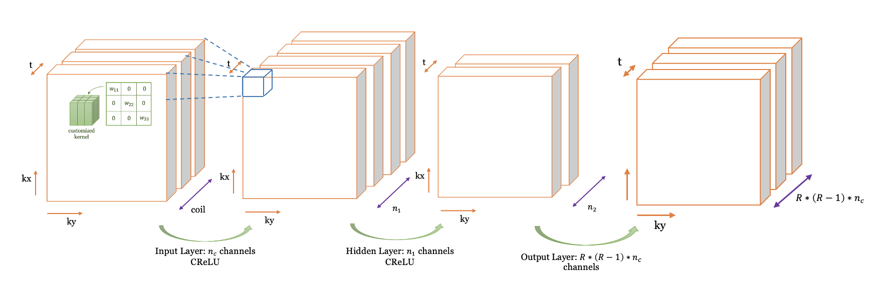
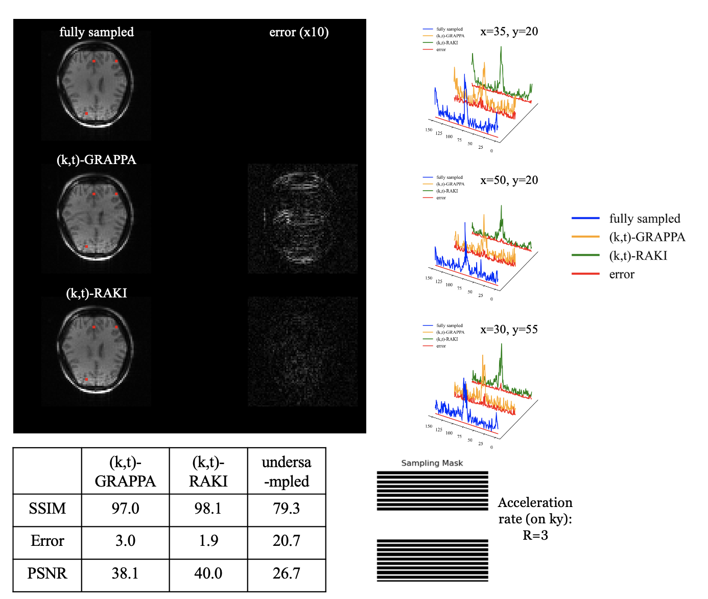
In this figure, only ky dimension is undersampled by R=3, the other dimensions (kx, kz, time) are fully sampled. Using dilated (by R) convolutional kernels in the first layer, RAKI successfully controls aliasing and outperforms GRAPPA in all time points. This figure gives an example of the reconstruction result at the 3rd time slice. The kernel sizes of the 3D-CNN are 2x5x2, 1x1x1, 1x5x1, respectively. The input data size is (ky=74, kx=74, kz=20, time=150, coil=16).
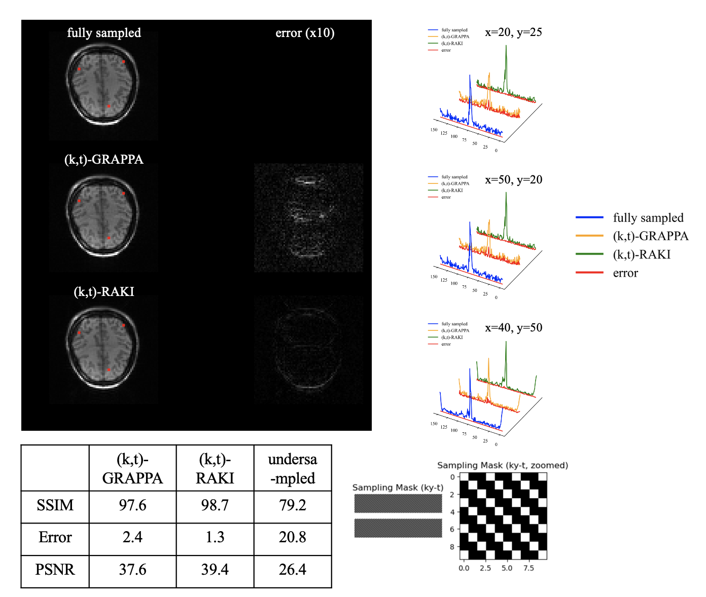
In this figure, ky and time dimension are undersampled by 3x3 CAIPIRINHA, the other dimensions (kx, kz) are fully sampled. Using customized convolutional kernels in the first layer, RAKI successfully controls aliasing and outperforms GRAPPA in all time points. This figure gives an example of the reconstruction result at the 3rd time slice. The kernel sizes of the 3D-CNN are 3x5x3, 1x1x1, 1x3x1, respectively. The input data size is (ky=74, kx=74, kz=20, time=150, coil=16).
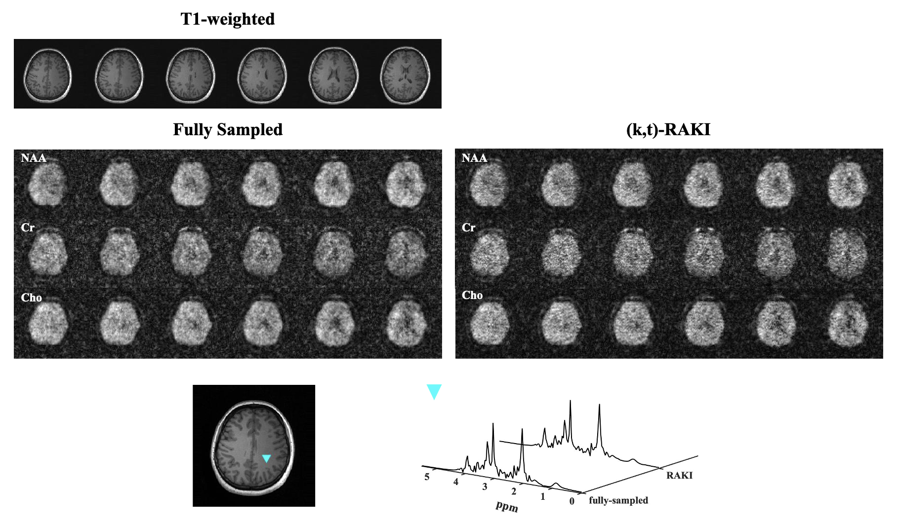
In vivo 1H-MRSI results from a healthy volunteer: The top panel compares metabolite maps of NAA, Cr and Cho from fully sampled data (left section) and (k,t)-RAKI reconstructed data (right section). The bottom panel compares reconstructed spectra, with voxel locations marked by the blue triangle shown in the T1 -weighted anatomical image. High resemblance between (k,t)-RAKI results and fully sampled (k,t)-space can be observed in our reconstruction.
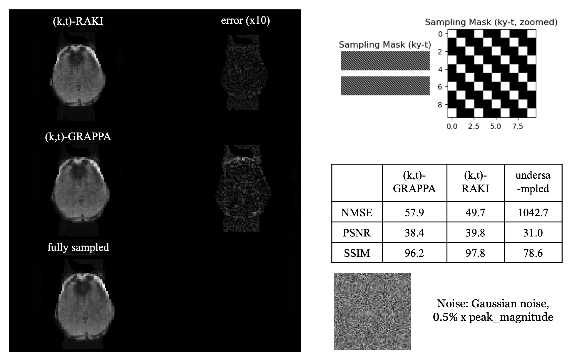
Gaussian noise equivalent to 0.5% of the peak magnitude of the (k,t)-space signal was intentionally introduced to the entire (k,t)-space for assessing network robustness. The results demonstrate that (k,t)-RAKI maintains excellent performance even in the presence of added noise, with minimal impact on reconstruction quality, surpassing (k,t)-GRAPPA. Furthermore, a comparison with t by t RAKI reveals that temporal information aids the network in mitigating noise-related effects.