1081
Navigator-free multi-shot EPI with shift-invariant kernel extraction in subspace1High-Field MR center, Max Planck Institute for Biological Cybernetics, Tuebingen, Germany, 2Institute of Biomedical Imaging, Graz University of Technology, Graz, Austria, 3Department for Biomedical Magnetic Resonance, University of Tuebingen, Tuebingen, Germany
Synopsis
Keywords: Image Reconstruction, Image Reconstruction
Motivation: In functional and diffusion MRI, multi-shot EPI enhances spatial resolution and minimizes distortion compared to single-shot scans. However, its vulnerability to shot-to-shot phase variations presents a significant challenge, with various proposed methods having drawbacks.
Goal(s): We propose a robust, navigator-free, computational efficient multi-shot method without SNR penalty.
Approach: In readout-segmented multi-shot EPI, we exploit the k-space overlapped regions between adjacent segments to extract relative phase fluctuations. This method, inspired by ESPIRiT and nonlinear gradient calibration, efficiently extracts shot-dependent phase variations in subspace.
Results: Our ex-vivo and in-vivo scans, including diffusion-weighted imaging, successfully achieves a high in-plane resolution of about 0.6mm without ghost artifacts.
Impact: Our proposed multi-shot technique eliminates the needs for time-consuming navigators, provides robust high-resolution diffusion and potentially functional imaging, and could be easily adapted for interleaved Cartesian and spiral EPI allowing robust phase error estimation from merely small k-space regions.
Introduction
For functional1–3 and diffusion4 MRI, multi-shot EPI5 offers enhanced spatial resolution and reduced geometric distortion compared to single-shot acquisitions; however, it introduces susceptibility to uncontrollable phase fluctuations between shots6,7, which could result from various factors (e.g., motions, respiration, heartbeat, scanner instability, diffusion gradients) and has attracted continuous research interests towards more sophisticated techniques.One established approach uses a 2D navigator8–11 to explicitly estimate the phase inconsistency, which inevitably prolongs the scan time and may fail in some cases. The phase fluctuations can also be estimated through pure post-processing, however, only with fully sampled k-space center12 or when the EPI interleave number is below the maximum parallel imaging acceleration factor13.
Alternatively, iteratively reconstruction with a low-rank penalty term, formulated in either k-space14 or local image-space15, can be applied without explicit phase estimation. Unfortunately, this could lead to substantially increased computational complexity, or image artifacts given highly undersampled data. Lastly, the inter-shot phase variations can be eliminated in a magnitude-based super-resolution scan with subpixel modulation16–19, but at the cost of inevitable SNR loss.
To address these issues, we propose a navigator-free multi-shot technique without significant constraints. This approach accurately and efficiently extracts inter-shot phase fluctuations in subspace, as inspired by studies of ESPIRiT20 and nonlinear gradient calibration21.
Methods
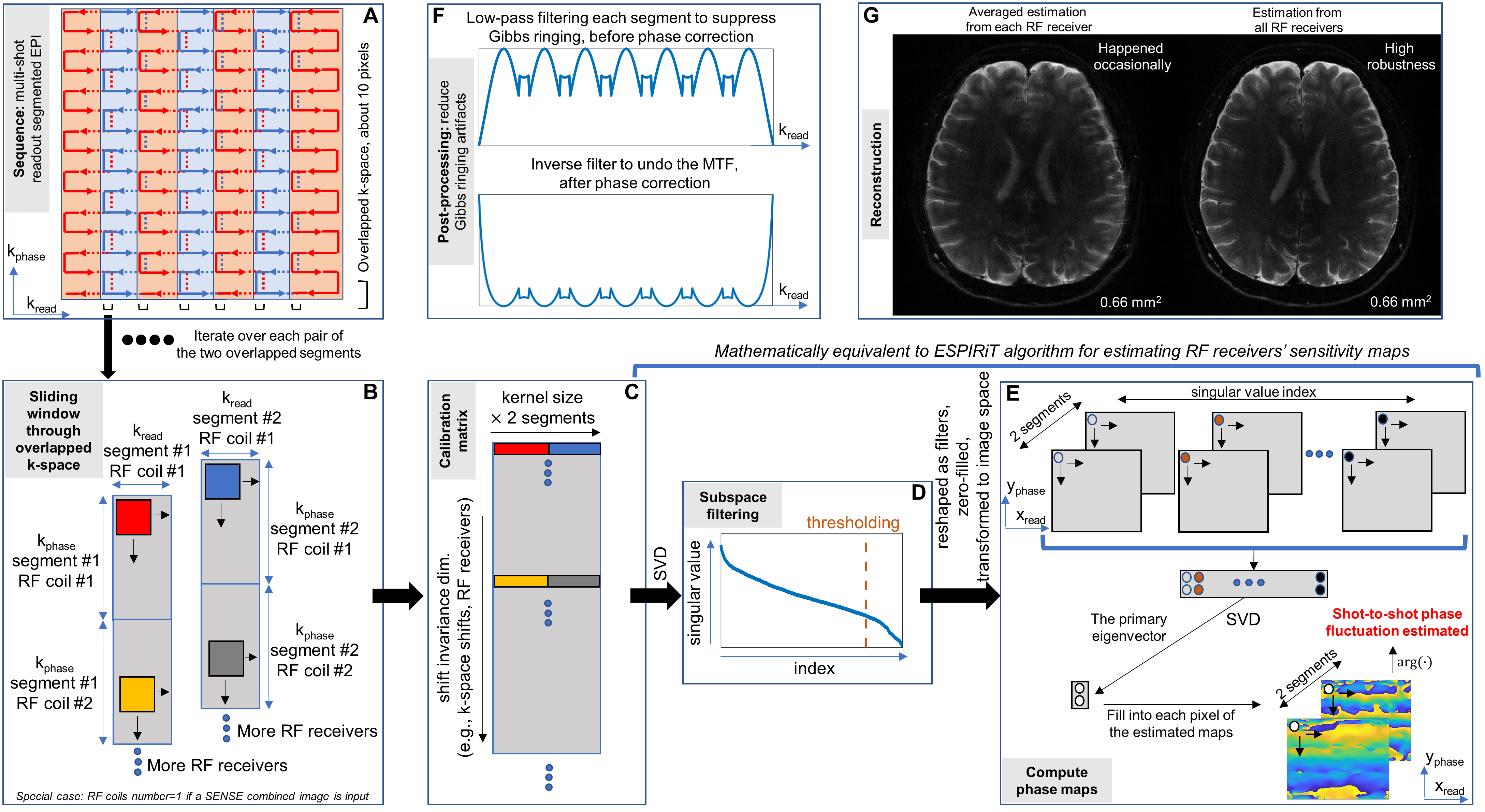
Mathematically, shot-dependent phase fluctuations in image space can be represented as shift-invariant kernels convolved with k-space signals, similar to the GRAPPA kernel22 in parallel imaging22–24. In parallel imaging, the RF receivers’ sensitivity maps can be directly estimated in subspace with the ESPIRiT algorithm based on central k-space calibration scans20, which allows efficient and robust reconstruction.A key insight for our technique is that, the calibration scans with commonly sampled (e.g., by distinct RF receivers, or different shots) k-space region need not be confined to the k-space center, thanks to the shift-invariant nature of the kernel across the entire k-space. For instance, in readout segmented EPI11 with a typical band overlapping ratio (e.g., 10%-20%) to avoid k-space holes, the kernel can be directly estimated using the subspace algorithm20 by treating the overlapped k-space pixels from different shots as from distinct RF receivers.
As illustrated in Figure 1, a multi-shot readout segmented EPI (A) is programmed in Pulseq25 and tested on a 3T Siemens Prisma scanner with a 64-element RF array. The opposite readout gradient polarity between neighboring segments ensures consistent T2 decay between overlapped k-space pixels. After pre-processing the data from the scanner (e.g., re-gridding, gradient calibration, or optional, parallel imaging), a calibration matrix (C) is constructed by selecting data in the overlapped regions between two adjacent k-space segments, using a sliding window with a specified kernel size (B). Notably, the data from different RF coils are treated analogously to the kernel data sliding across different k-space locations (C), as different than ESPIRiT for estimating RF sensitivity maps. Based on this calibration matrix, the phase maps between every two adjacent EPI segments can be estimated using a similar mathematical principle as ESPIRiT20, including subspace thresholding (D) and pixel-wise eigenvector extraction in image space along the shot dimension (E).
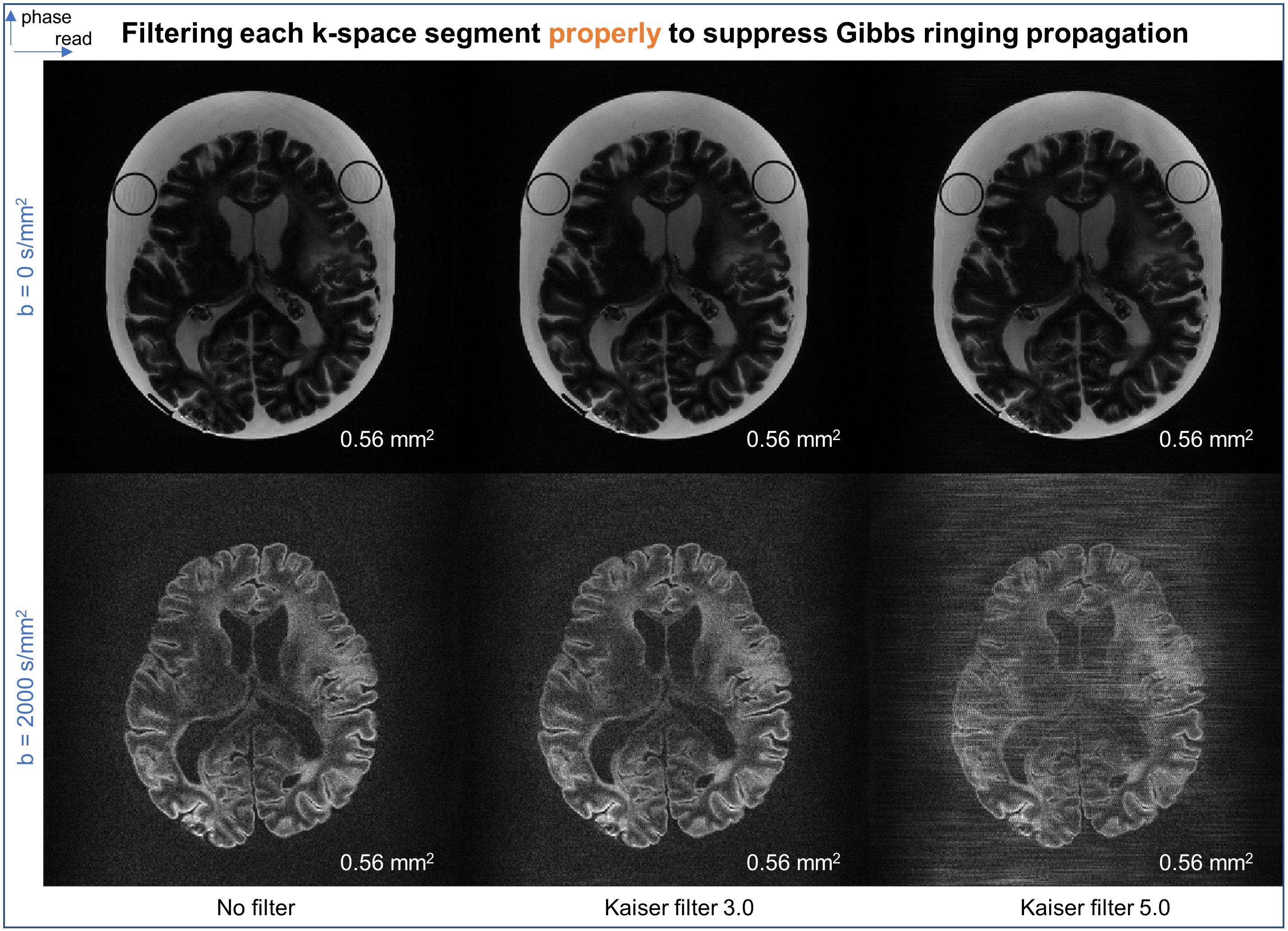
Additionally, when applying the estimated phase maps for correction, the Gibbs ringing in each low-resolution segment image can propagate to the finally reconstructed image. Hence, a Kaiser window is applied to filter each segment in k-space suppressing the ringing, and an inverse filter is utilized to undo the modulation transfer function (F), as a reconstruction treatment used in phaseless encoding MRI18,19.
Results
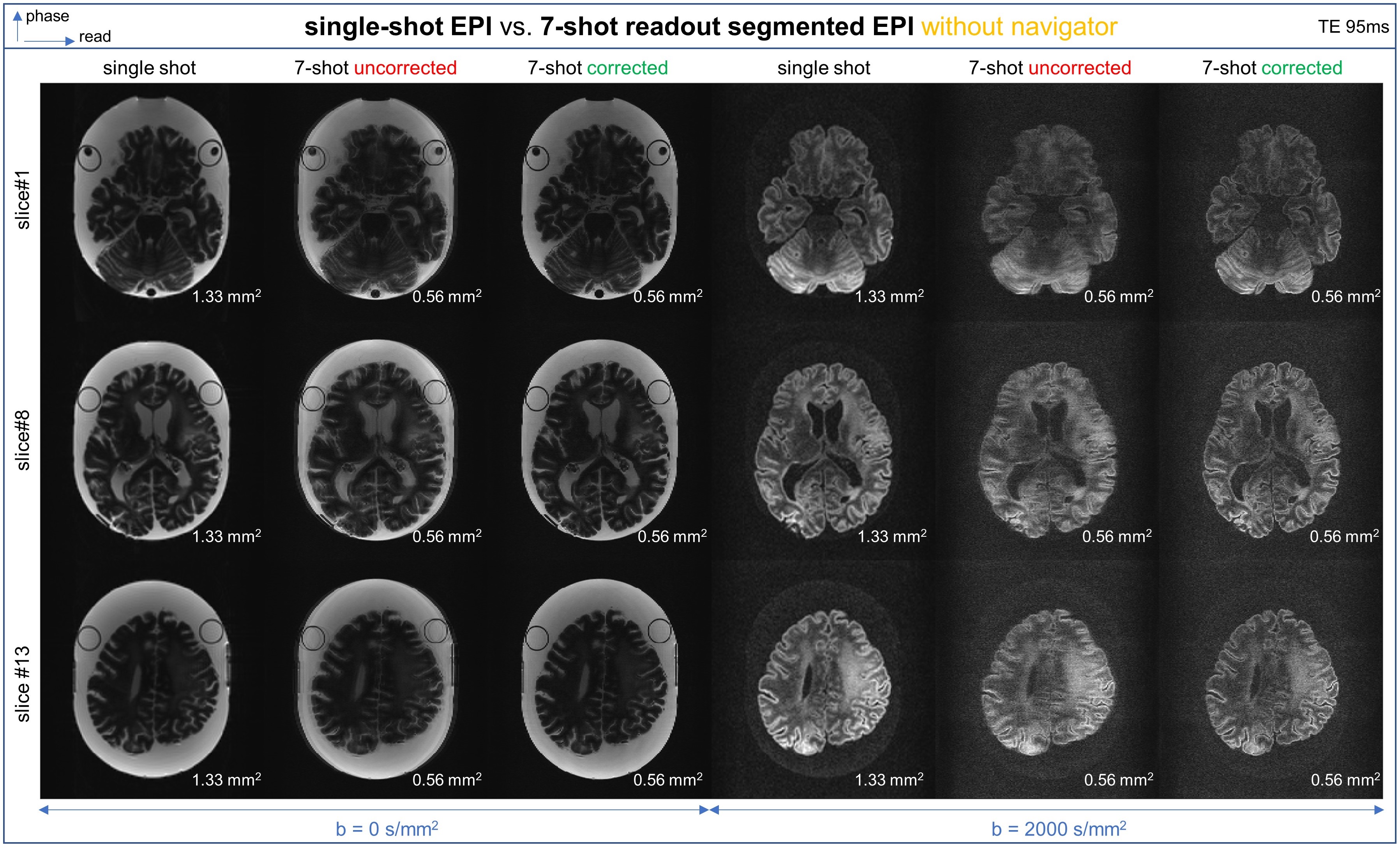
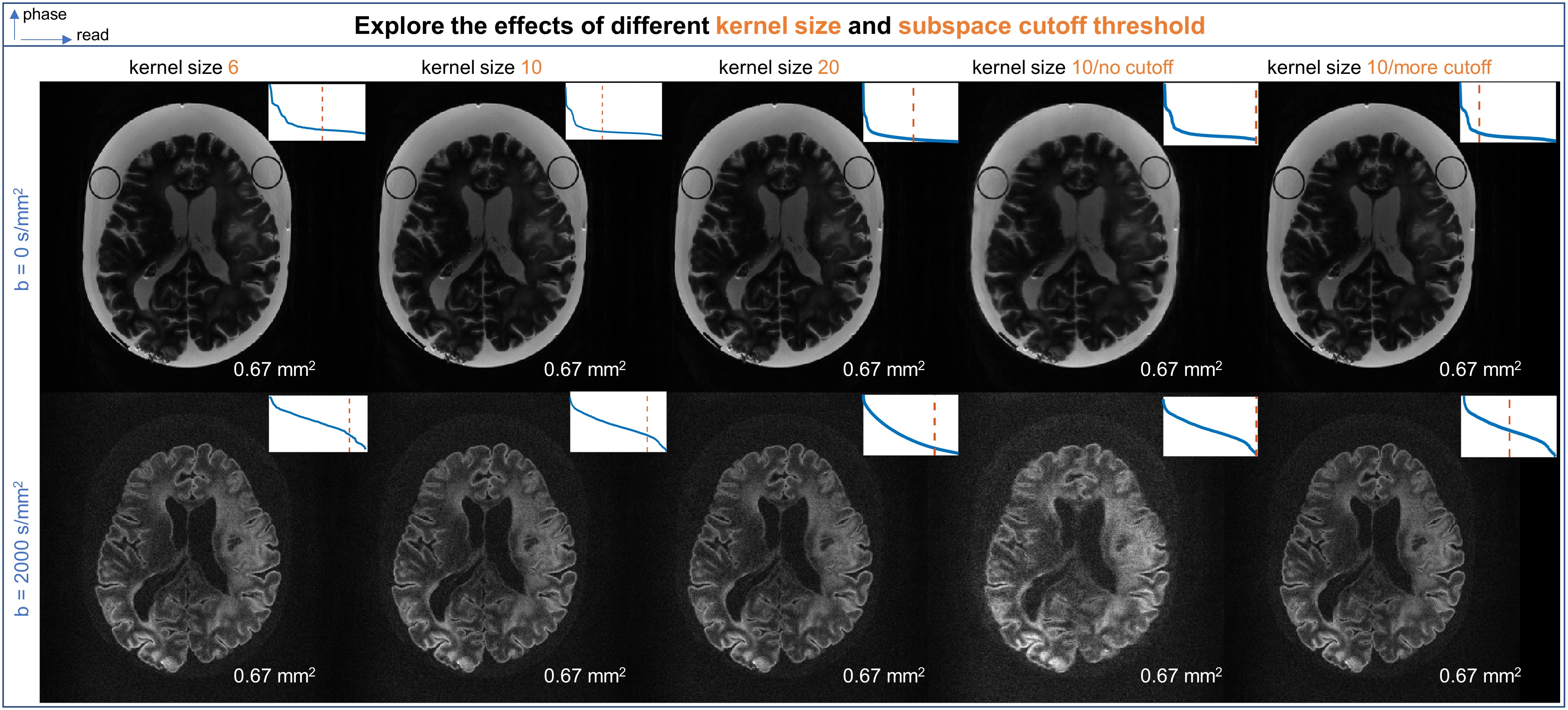
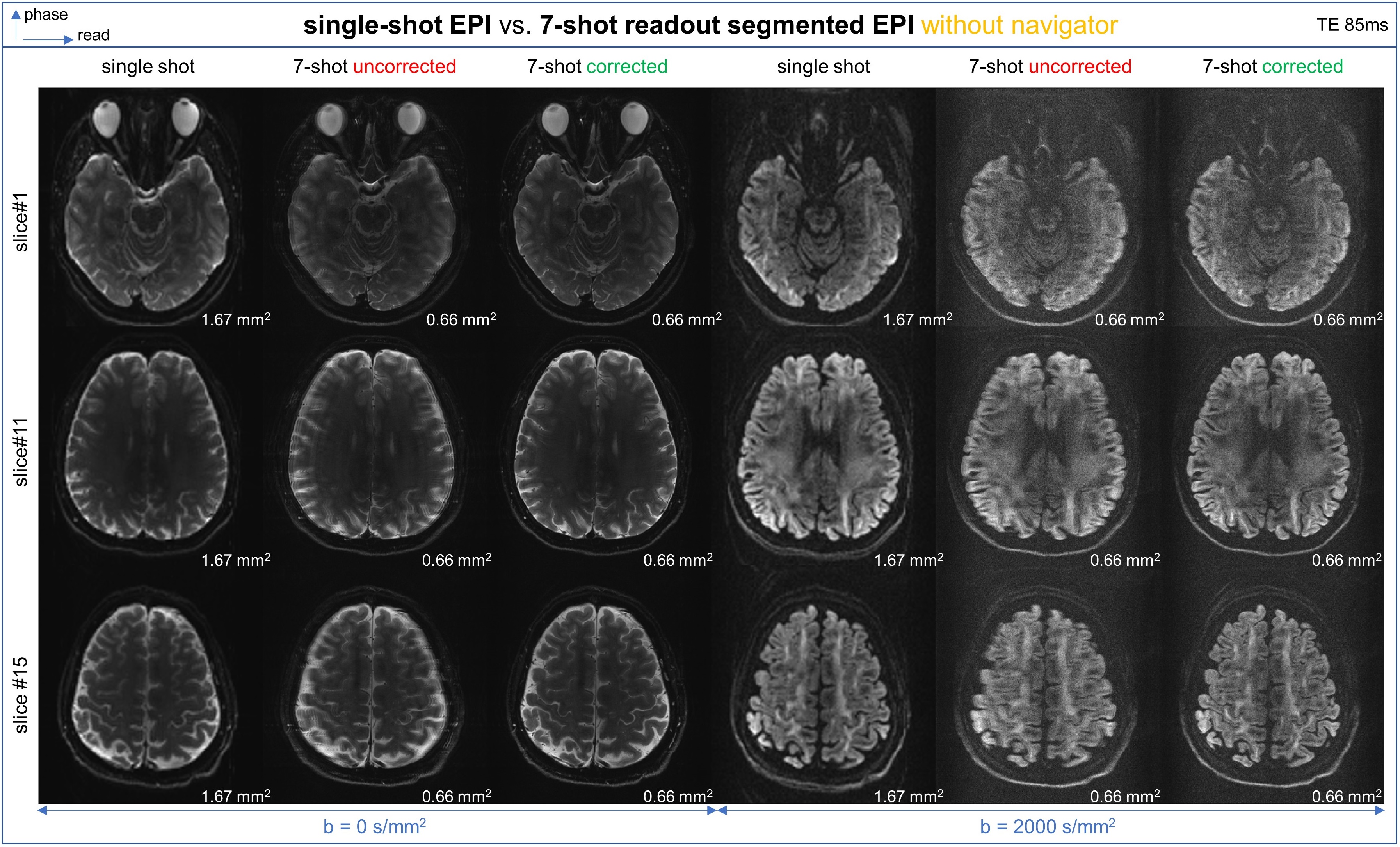
In Figures 2 and 5, our technique is experimentally validated with multi-slice ex-vivo phantom26,27 and in-vivo brain scans. Compared to uncorrected images, the corrected multi-shot (here, 7-shot) reconstruction achieves significant resolution enhancement (0.56 mm2/0.66mm2) than the single-shot acquisitions (1.33 mm2/1.67mm2) without ghost artifacts.Additionally, in Figure 3, a moderate apodization for each segment proves useful for images with sharp edges, but appeared unnecessary for high b-value scans. Furthermore, in Figure 4, different kernel sizes and subspace cutoff thresholds are investigated, and empirically, a kernel size of 6x6 to 10x10 pixels with moderate thresholding will be sufficient to correct the inter-shot phase fluctuations.
Discussions/Conclusion
Our subspace estimation technique successfully achieves high-resolution non-diffusion and diffusion-weighted imaging with multi-shot readout-segmented EPI without navigators, by extracting the phase fluctuations as shift-invariant kernel convolved in k-space, from only about 90 (phase) x10 (read) overlapped k-space pixels between adjacent segments. This technique can also be adapted for interleaved Cartesian or spiral EPI to achieve robust phase error estimation from merely a small k-space region commonly sampled during different shots.Acknowledgements
This study is supported by ERC Advanced Grant No 834940.
The ex-vivo brain phantom was with courtesy of the Institute of Clinical Anatomy and Cell Analysis, Department of Anatomy, Eberhard Karls University of Tübingen. The first author thanks Dr. Thomas Shiozawa (Institute of Clinical Anatomy and Cell Analysis) for assistance with sample preparation, and Dr. Gisela Hagberg for assistance in scanning this phantom.
The first author thanks Dr. Sebastian Mueller, Dr. Felix Glang, Praveen Iyyappan Valsala, and Dana Ramadan for experimental assistance.
References
1. Ogawa S, Tank DW, Menon R, et al. Intrinsic signal changes accompanying sensory stimulation: functional brain mapping with magnetic resonance imaging. Proc Natl Acad Sci. 1992;89(13):5951-5955. doi:10.1073/pnas.89.13.5951
2. Kwong KK, Belliveau JW, Chesler DA, et al. Dynamic magnetic resonance imaging of human brain activity during primary sensory stimulation. Proc Natl Acad Sci. 1992;89(12):5675-5679. doi:10.1073/pnas.89.12.5675
3. Bandettini PA, Wong EC, Hinks RS, Tikofsky RS, Hyde JS. Time course EPI of human brain function during task activation. Magn Reson Med. 1992;25(2):390-397. doi:10.1002/mrm.1910250220
4. Basser PJ, Mattiello J, LeBihan D. MR diffusion tensor spectroscopy and imaging. Biophys J. 1994;66(1):259-267. doi:10.1016/S0006-3495(94)80775-1
5. Mansfield P. Multi-planar image formation using NMR spin echoes. J Phys C Solid State Phys. 1977;10(3):L55-L58. doi:10.1088/0022-3719/10/3/004
6. Van de Moortele PF, Pfeuffer J, Glover GH, Ugurbil K, Hu X. Respiration-induced B0 fluctuations and their spatial distribution in the human brain at 7 Tesla. Magn Reson Med. 2002;47(5):888-895. doi:10.1002/mrm.10145
7. Norris DG. Implications of bulk motion for diffusion-weighted imaging experiments: Effects, mechanisms, and solutions. J Magn Reson Imaging. 2001;13(4):486-495. doi:10.1002/jmri.1072
8. Atkinson D, Porter DA, Hill DLG, Calamante F, Connelly A. Sampling and reconstruction effects due to motion in diffusion-weighted interleaved echo planar imaging. Magn Reson Med. 2000;44(1):101-109. doi:10.1002/1522-2594(200007)44:1<101::AID-MRM15>3.0.CO;2-S
9. Atkinson D, Counsell S, Hajnal JV, Batchelor PG, Hill DLG, Larkman DJ. Nonlinear phase correction of navigated multi-coil diffusion images. Magn Reson Med. 2006;56(5):1135-1139. doi:10.1002/mrm.21046
10. Holdsworth SJ, Skare S, Newbould RD, Guzmann R, Blevins NH, Bammer R. Readout-Segmented EPI for Rapid High Resolution Diffusion Imaging at 3T. Eur J Radiol. 2008;65(1):36-46. doi:10.1016/j.ejrad.2007.09.016
11. Porter DA, Heidemann RM. High resolution diffusion-weighted imaging using readout-segmented echo-planar imaging, parallel imaging and a two-dimensional navigator-based reacquisition. Magn Reson Med. 2009;62(2):468-475. doi:10.1002/mrm.22024
12. Uecker M, Karaus A, Frahm J. Inverse reconstruction method for segmented multishot diffusion-weighted MRI with multiple coils. Magn Reson Med. 2009;62(5):1342-1348. doi:10.1002/mrm.22126
13. Chen N kuei, Guidon A, Chang HC, Song AW. A robust multi-shot scan strategy for high-resolution diffusion weighted MRI enabled by multiplexed sensitivity-encoding (MUSE). NeuroImage. 2013;72:41-47. doi:10.1016/j.neuroimage.2013.01.038
14. Mani M, Jacob M, Kelley D, Magnotta V. Multi-shot sensitivity-encoded diffusion data recovery using structured low-rank matrix completion (MUSSELS). Magn Reson Med. 2017;78(2):494-507. doi:10.1002/mrm.26382
15. Hu Y, Levine EG, Tian Q, et al. Motion-robust reconstruction of multishot diffusion-weighted images without phase estimation through locally low-rank regularization. Magn Reson Med. 2019;81(2):1181-1190. doi:10.1002/mrm.27488
16. Hennel F, Pruessmann KP. MRI with phaseless encoding: MRI with Phaseless Encoding. Magn Reson Med. 2017;78(3):1029-1037. doi:10.1002/mrm.26497
17. Hennel F, Tian R, Engel M, Pruessmann KP. In-plane “superresolution” MRI with phaseless sub-pixel encoding: Hennel et al. Magn Reson Med. 2018;80(6):2384-2392. doi:10.1002/mrm.27209
18. Tian R, Hennel F, Pruessmann KP. Low-distortion diffusion tensor MRI with improved phaseless encoding. J Magn Reson. 2019;309:106602. doi:https://doi.org/10.1016/j.jmr.2019.106602
19. Tian R, Hennel F, Bianchi S, Pruessmann KP. Superresolution MRI with a Structured-Illumination Approach. Phys Rev Appl. 2023;19(3):034074. doi:10.1103/PhysRevApplied.19.034074
20. Uecker M, Lai P, Murphy MJ, et al. ESPIRiT-an eigenvalue approach to autocalibrating parallel MRI: Where SENSE meets GRAPPA. Magn Reson Med. 2014;71(3):990-1001. doi:10.1002/mrm.24751
21. Tian R, Uecker M, Davids M, et al. Accelerated 2D Cartesian MRI with an 8-channel local B0 coil array combined with parallel imaging. Magn Reson Med. n/a(n/a). doi:10.1002/mrm.29799
22. Griswold MA, Jakob PM, Heidemann RM, Nittka M, Jellus V, Wang J, Kiefer B, Haase A. (2002) “Generalized autocalibrating partially parallel acquisitions (GRAPPA),” Magnetic Resonance in Medicine, 47(6), pp. 1202–1210. Available at: https://doi.org/10.1002/mrm.10171.
23. Sodickson DK, Manning WJ. Simultaneous acquisition of spatial harmonics (SMASH): Fast imaging with radiofrequency coil arrays. Magn Reson Med. 1997;38(4):591-603. doi:10.1002/mrm.1910380414
24. Pruessmann KP, Weiger M, Scheidegger MB, Boesiger P. SENSE: Sensitivity encoding for fast MRI. Magn Reson Med. 1999;42(5):952-962. doi:10.1002/(SICI)1522-2594(199911)42:5<952::AID-MRM16>3.0.CO;2-S
25. Layton KJ, Kroboth S, Jia F, et al. Pulseq: A rapid and hardware-independent pulse sequence prototyping framework. Magn Reson Med. 2017;77(4):1544-1552. doi:10.1002/mrm.26235
26. Hagberg GE, Shiozawa-Bayer T, Mirkes C, et al. MRI of whole brain formalin-fixed samples at 9.4T: influence of the fixation agent and its dielectric properties on image quality. In Proceedings of the joint annual meeting of ISMRM-ESMRMB, Paris, France, June 2018. Abstract #3310.
27. Nazemorroaya A, Aghaeifar A, Shiozawa T, et al. Developing formalin-based fixative agents for post mortem brain MRI at 9.4 T. Magn Reson Med. 2022;87(5):2481-2494. doi:10.1002/mrm.29122
Figures