1077
High-Fidelity Intravoxel Incoherent Motion (HIFIVIM) Parameter Mapping Using Locally Low-Rank with Temporal Subspace Constraint1Department of Biomedical Engineering, University of Rochester, Rochester, NY, United States, 2Department of Radiology, Stanford University, Stanford, CA, United States, 3University of Iowa, Iowa City, IA, United States, 4Department of Neurology, University of Rochester, Rochester, NY, United States, 5Department of Imaging Sciences, University of Rochester, Rochester, NY, United States, 6Department of Electrical and Computer Engineering, University of Rochester, Rochester, NY, United States, 7Department of Physics and Astronomy, University of Rochester, Rochester, NY, United States
Synopsis
Keywords: Image Reconstruction, Diffusion/other diffusion imaging techniques, Intravoxel Incoherent Motion, Model-Based Reconstruction, Subspace Reconstruction
Motivation: Intravoxel incoherent motion (IVIM) is a measure in MRI to quantify tissue perfusion. However, clinical applications are limited by noisy parameter estimates for the perfusion fraction (f) and pseudodiffusion coefficient (D*).
Goal(s): We sought to improve IVIM parameter estimation using a model-based reconstruction
Approach: We combined locally low-rank (LLR) and temporal subspace constraints to reliably perform joint reconstruction of IVIM images before fitting while correcting shot-to-shot phase variations between each b-value.
Results: Our method resulted in smoother signal decay curves before fitting and improved the estimation of IVIM parameter maps with less noise and fewer outliers.
Impact: A model-based reconstruction with low rank and temporal constraints improved IVIM image reconstruction, reducing noise and outliers in parameter estimates. Spline interpolation further facilitated reliable estimation of IVIM maps from just 5 b-values, benefiting clinical situations like stroke.
Introduction
Intravoxel incoherent motion (IVIM) is a quantitative method that measures perfusion properties of tissue at low b-values1. Conventionally, parametric mapping is achieved with a separate magnitude-based reconstruction of each b-value, followed by parameter fitting. Downstream parameter maps are often noisy and unreliable due to shot-to-shot phase variations between each b-value, a Rician noise distribution, and low SNR. Additionally, more b-values are needed to reliably estimate the IVIM signal decay curve, resulting in longer acquisition times and limiting its clinical utility in acute settings. Conversely, model-based reconstructions have been used to reconstruct dynamic MRI data, such as inversion-recovery FLASH for T1 mapping2 and MR Fingerprinting (MRF)3. Model-based reconstructions are a flexible framework for non-linear mapping, ensuring data consistency, that can incorporate a signal model2 and regularization terms to constrain the model, reducing noise. This work utilized a locally low-rank (LLR) constraint and a concomitant temporal subspace constraint based on an IVIM signal model. Our approach showed improved SNR and a smoother IVIM signal decay curve before fitting. IVIM parameters were estimated using a Bayesian fitting approach4. Our proposed method reduced noise in tissue parameter maps, D, f, and D*. Further, we showed that our approach facilitated a reduction in the number of b-values from 15 to 5 using linear spline interpolation before subspace reconstruction, with substantial acquisition time reduction and no appreciable loss in fidelity in the estimated maps.Methods
Pulse sequence: Data were acquired using a 3T Siemens Prisma scanner (Erlangen, Germany) with a 64-channel head coil. IVIM-DWI was performed using 15 b-values (0,5,7,10,15,20,30,40,50,60,100,200,400,700 and 1000 s/mm2) with three orthogonal directions for each b-value (TR/TE = 4300/69 ms, 1.5 mm isotropic resolution).Reconstruction and fitting: A dictionary of 180,000 entries was simulated (for f=0-0.5, D*=0.001-0.1 s/mm2, and D=0.0005-0.0035 s/mm2) for the same 15 b-values used in vivo based on the following biexponential model:
S(b)/S(0) = fe-bD* + (1-f)e-bD
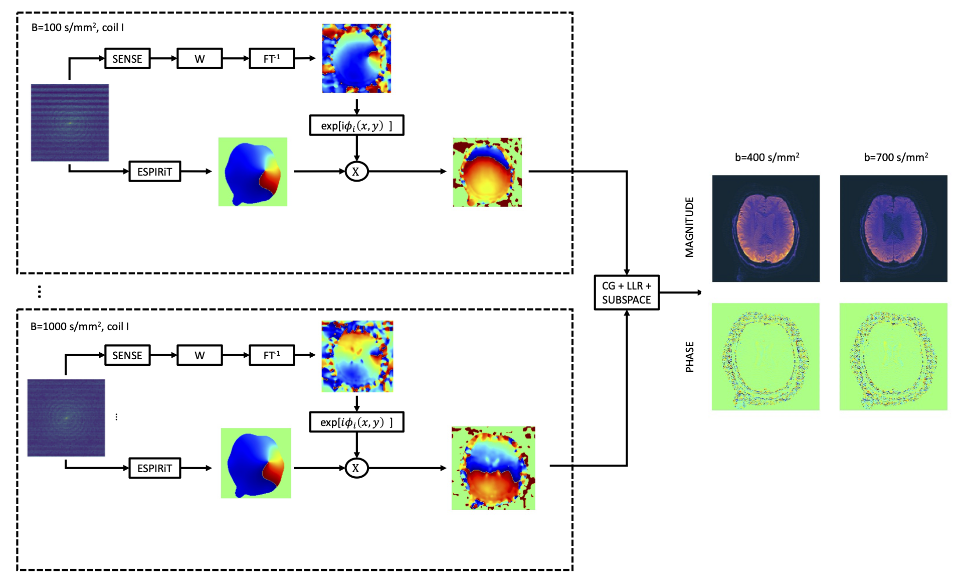
Singular value decomposition was performed, and the top 2 basis functions, explaining more than 95% of the variance, were used as the subspace. Coefficient maps were calculated using subspace reconstruction with LLR regularization using the BART toolbox5 and projected back to the temporal domain, yielding denoised IVIM signal decay curves. Sensitivity maps were estimated using ESPiRIT6 and corrected for low-resolution phase errors to account for phase variations before reconstruction (Figure 1). IVIM parameters (D, f, and D*) were estimated using data obtained with the proposed method and conventional magnitude images. For each data set, voxels were fit using Bayesian inference.
Interpolation: The 15 b-value data was downsampled to 5 (0, 15, 60, 200, and 1000 s/mm2) and 10 (0,7,15,30,50, 60, 100, 200, 400, 1000 s/mm2) b-values, which were then upsampled back to 15 b-values using linear spline interpolation in python, before subspace reconstruction.
Results
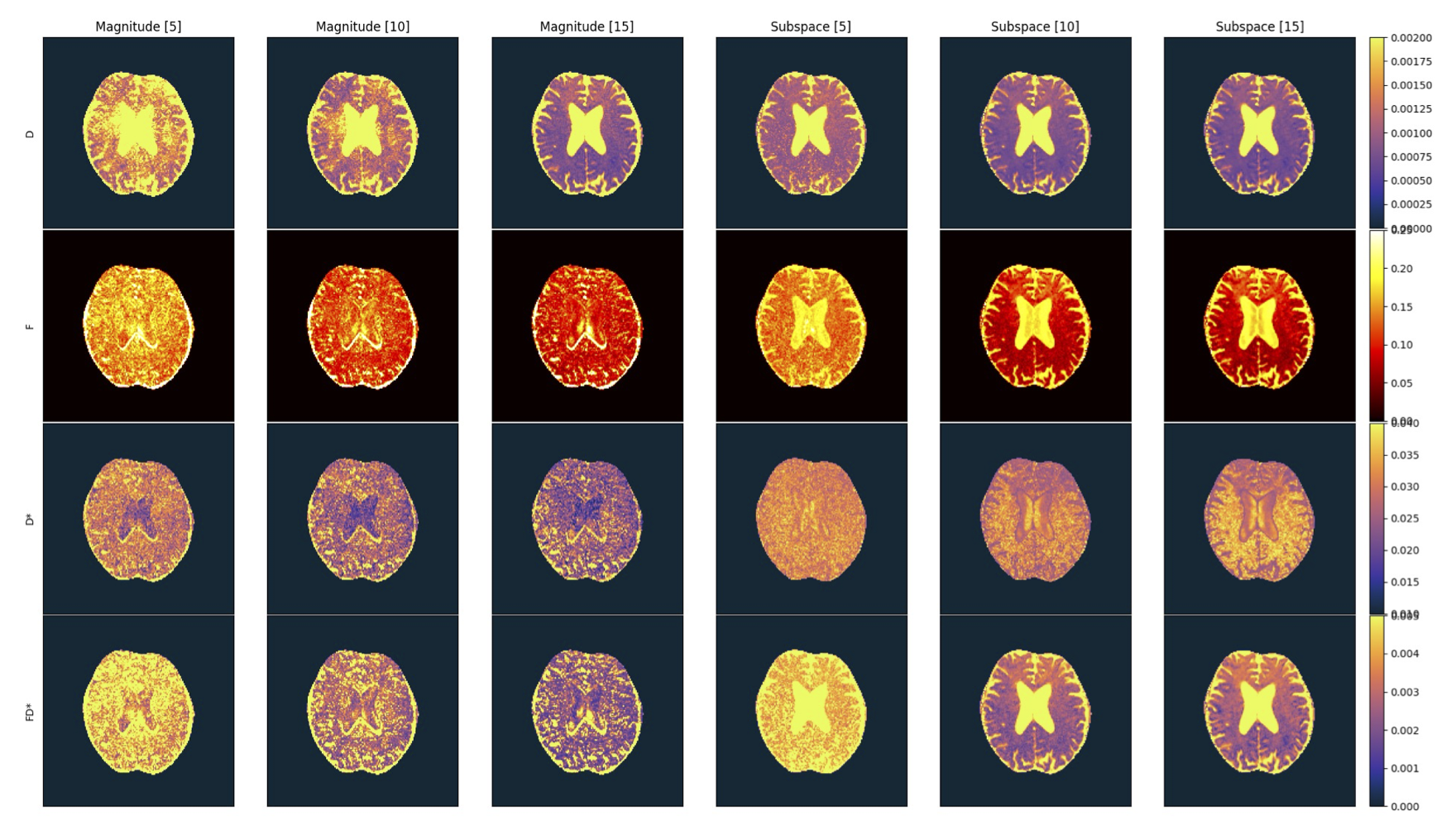
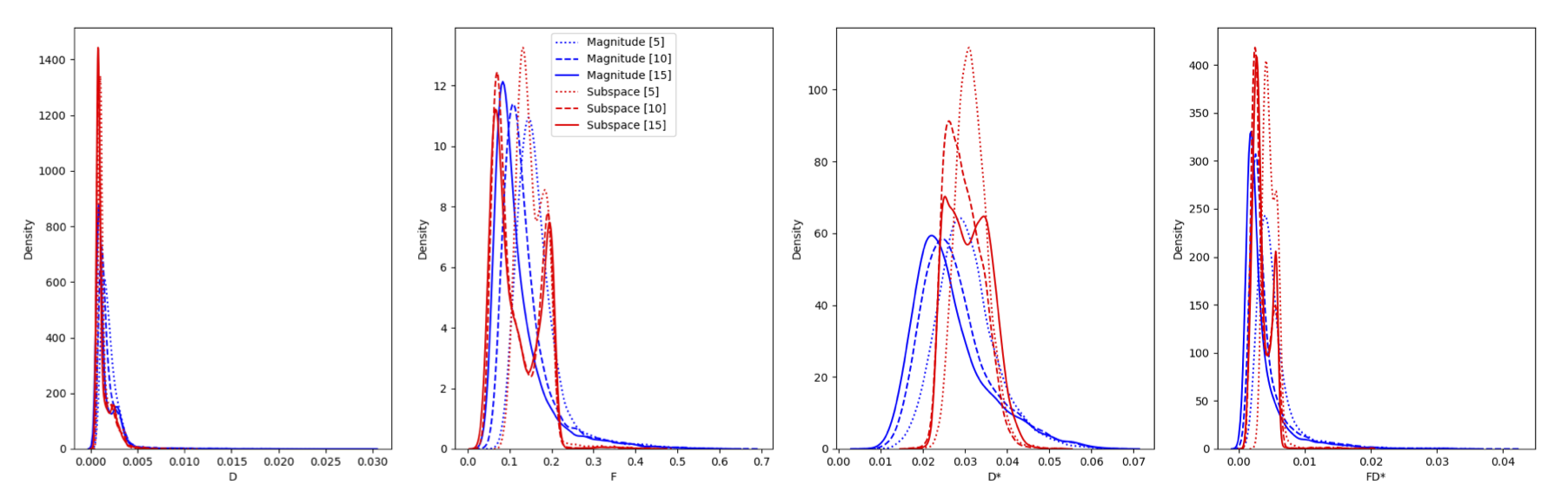
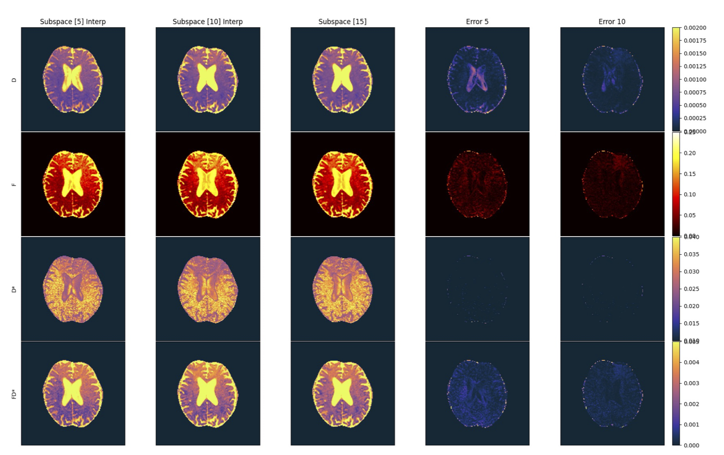
Figure 2 shows IVIM parameter estimates (D, f, D*, and fD*) using 5, 10, and 15 b-values for the conventional magnitude and proposed reconstructions without interpolation derived from in vivo data. Figure 3 shows a histogram comparison of the IVIM parameter estimates for the magnitude-based and proposed reconstruction methods for 5, 10, and 15 b-values, illustrating that our proposed method resulted in fewer outliers than the conventional magnitude reconstruction. Linear spline interpolation, in combination with the proposed reconstruction method, facilitated reliable estimation of IVIM parameters using only 5 and 10 b-values with minimal error (Figure 4).Discussion
This work leveraged a model-based reconstruction that used LLR and subspace constraints, simultaneously incorporating composite sensitivity maps correcting for phase variations. This led to higher SNR diffusion data and IVIM decay curves that are more reliably fit. As a result, the proposed subspace reconstruction method resulted in less noisy and more reliable parameter estimations for D, f, and D* compared to the conventional magnitude reconstruction. The magnitude reconstructions were noisier (Figure 2), resulting in more outliers than the proposed method (Figure 3). Further, this method allowed for better differentiations between gray matter and white matter, consistent with anatomy and estimates of the diffusion coefficient, which is more reliably estimated (Figure 2). Additionally, our method, in combination with linearly spline interpolation, enabled reliable reconstruction of parameter estimates using only 5 b-values (Figure 4). Overall, subspace reconstructions provided less noisy and more anatomically accurate f and D*estimates.Conclusion
We implemented a model-based reconstruction with temporal subspace and LLR constraints for improved IVIM reconstruction, reducing noise and improving parameter estimates. We showed that our method could be used with linear spline interpolation using only 5 b-values. Future work will investigate isotropic diffusion gradients7 and deep learners for accelerated high-fidelity parameter mapping.Acknowledgements
No acknowledgement found.References
1. Le Bihan D. What can we see with IVIM MRI? Neuroimage. Feb 15 2019;187:56-67. doi:10.1016/j.neuroimage.2017.12.062
2. Wang X, Tan Z, Scholand N, Roeloffs V, Uecker M. Physics-based reconstruction methods for magnetic resonance imaging. Philos Trans A Math Phys Eng Sci. Jun 28 2021;379(2200):20200196. doi:10.1098/rsta.2020.0196
3. Zhao B, Setsompop K, Adalsteinsson E, et al. Improved magnetic resonance fingerprinting reconstruction with low-rank and subspace modeling. Magn Reson Med. Feb 2018;79(2):933-942. doi:10.1002/mrm.26701
4. Gurney-Champion OJ, Klaassen R, Froeling M, et al. Comparison of six fit algorithms for the intra-voxel incoherent motion model of diffusion-weighted magnetic resonance imaging data of pancreatic cancer patients. PLoS One. 2018;13(4):e0194590. doi:10.1371/journal.pone.0194590
5. Tamir JI, Ong F, Cheng JY, Uecker M, Lustig M. Generalized magnetic resonance image reconstruction using the Berkeley advanced reconstruction toolbox. 2016:
6. Uecker M, Lai P, Murphy MJ, et al. ESPIRiT--an eigenvalue approach to autocalibrating parallel MRI: where SENSE meets GRAPPA. Magn Reson Med. Mar 2014;71(3):990-1001. doi:10.1002/mrm.24751
7. Yang G, McNab JA. Eddy current nulled constrained optimization of isotropic diffusion encoding gradient waveforms. Magn Reson Med. Mar 2019;81(3):1818-1832. doi:10.1002/mrm.27539
Figures