1073
High-Resolution Free-breathing Perfusion MRI with High Slice Coverage via SAM in Self-Supervised Learning with Self-Regularization1Chemical and Biomedical Engineering, University of Missouri Columbia, Columbia, MO, United States, 2Radiology, University of Missouri Columbia, Columbia, MO, United States, 3Medicine-Cardiology, University of Missouri Columbia, Columbia, MO, United States
Synopsis
Keywords: Image Reconstruction, Perfusion
Motivation: Enhancing myocardial perfusion MRI with self-supervised learning is key to achieving higher image quality and fidelity, especially in patients with varying image matrix sizes and asymmetric echo.
Goal(s): To enhance perfusion MRI by increasing resolution and slice coverage using self-supervised learning, self-regularization, and spatial attention, tailored for varied image sizes and asymmetric echo.
Approach: Implemented an accelerated perfusion MRI sequence with asymmetric echo; collected data from 20 patients; developed self-LR with SAM to enhance image quality.
Results: Self-LR with SAM yielded superior image quality and fewer artifacts in varied sizes and asymmetric echo, outperforming other methods, confirmed by expert evaluations.
Impact: The integration of Spatial Attention Module (SAM) with Self-Supervised Learning and Self-Regularization significantly enhances myocardial perfusion MRI, enriching spatial resolution and slice coverage. This development could potentially improve diagnostic accuracy, facilitating non-invasive whole-heart assessments with improved image quality.
Background
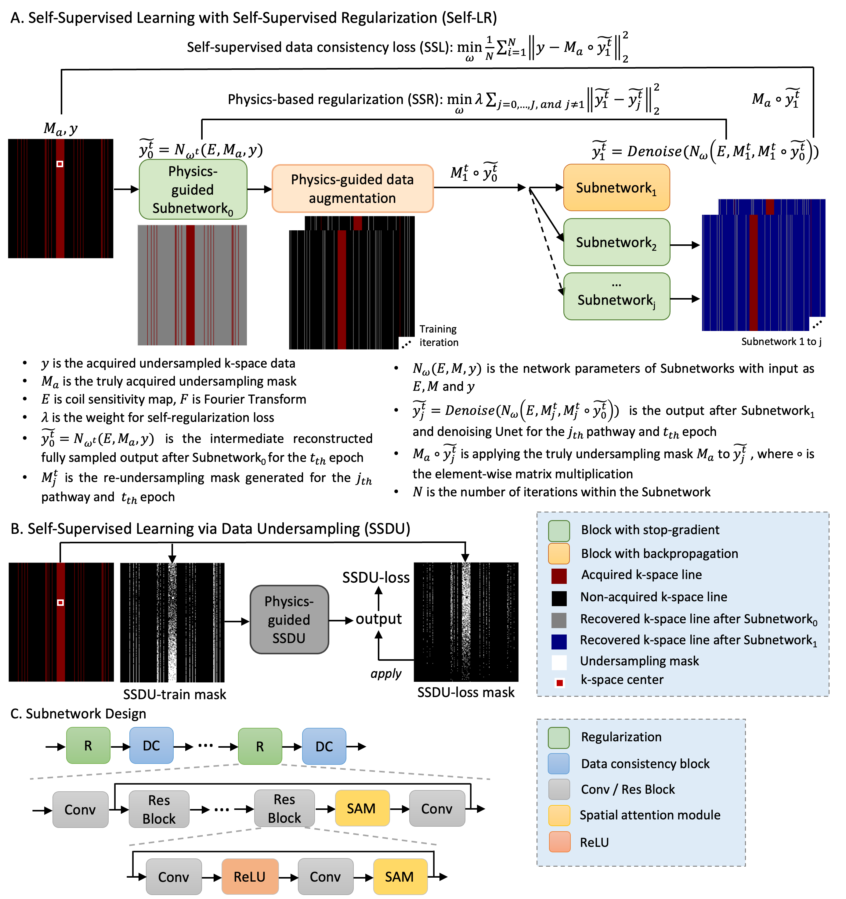
Myocardial first-pass perfusion MRI (1) is crucial for identifying coronary artery disease and coronary microvascular dysfunction (CMD). High resolution and slice coverage can improve accuracy in detecting vascular dysfunction. However, this requires high acceleration rates, risking spatiotemporal fidelity loss due to motion and preset temporal constraints. Asymmetric echo and varied image sizes are used clinically, which can challenge methods like self-supervised learning via data undersampling (SSDU) (2). We recently developed a self-supervised learning method, self-supervised Learning with self-supervised Regularization (self-LR) (3), which may improve image quality in high resolution perfusion MRI, especially for asymmetric echo data, as it does not virtually split data along the readout dimension. Here, we develop self-LR method with spatial attention modules (SAM) incorporating (4) in the convolutional residual blocks for further improving the image fidelity. We modified a highly accelerated first-pass perfusion MRI sequence with asymmetric echo and varied image sizes, acquired free-breathing rest perfusion data on 20 patients, and compared self-LR to other methods.Methods
We collected 20 datasets (9800 multicoil k-space) on a 1.5T scanner (Aera, Siemens) using a modified saturation-recovery gradient-echo sequence with asymmetric echo and Poisson disc undersampling. The undersampling resulted in about 18 centrally located fully sampled lines, with an effective undersampling rate of over 11-fold applied to regions outside the center (Figure 1). The image sizes of the 20 datasets range from 256-264 x 200-246. The 20 datasets were divided into two groups: 15 datasets were used for training the model, and the remaining 5 were used for model inference. Each dataset was acquired with 6-8 slices and 70 measurements. Prospective datasets were reconstructed using CG-SENSE, SSDU and self-LR. The image quality for deep learning methods was scored by a cardiologist and a radiologist based on SNR, sharpness, artifacts, and overall image quality. For each training epoch, one measurement is randomly selected from a set of five successive measurements in each dataset. We designed a residual block with SAM for improving the spatial fidelity of the images, which is compared to self-LR without SAM. The self-LR model was trained for 60 epochs with learning rate of 5e-5. Three subnets were used in self-LR training, but only the first subnet was used in inference, so that there is no significant additional model parameters compared to SSDU. Self-LR was implemented in PyTorch using stop-gradient (5) and a combination of normalized l1 and l2 loss for both self-supervised data loss and self-regularization loss. SSDU was trained for 100 epochs, and the learning rate of SSDU was experimented and compared between 5e-5 and 5e-4, and 5e-5 was selected due to fewer artifacts. The prospectively acquired data, obtained with clinically used asymmetric echo and varied data matrix size, may not be optimal for data splitting in SSDU. For comparing the performance of different reconstruction methods, late gadolinium enhancement (LGE) images from one patient with infarction were used as reference.Results
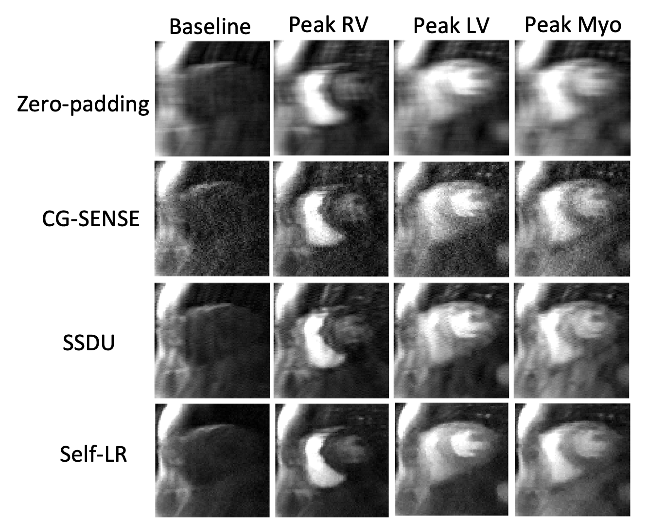
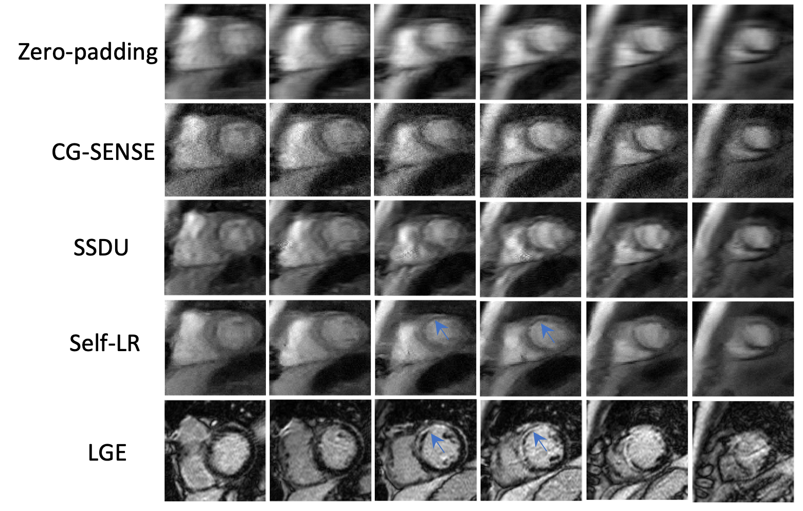
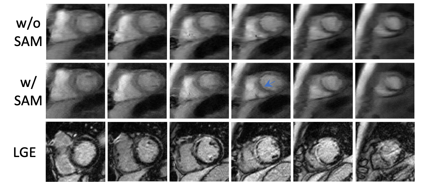
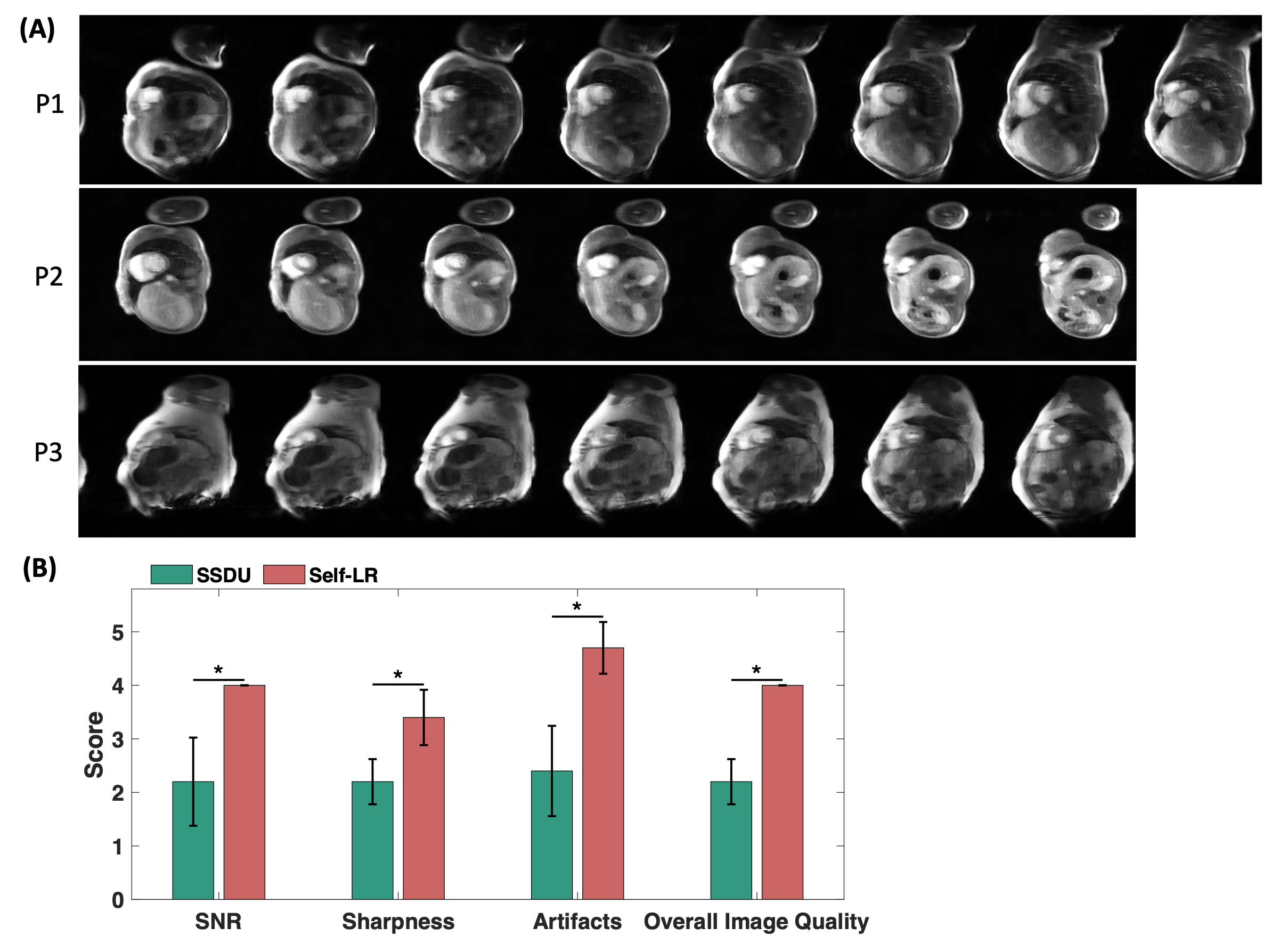
Figure 2 compares zero-padding, CG-SENSE, SSDU, and self-LR at various contrast phases from one patient. Self-LR consistently outperforms other methods, producing images with higher SNR, less artifacts, and better overall quality across all dynamic phases. Figure 3 compares images reconstructed using zero-padding, CG-SENSE, SSDU, and self-LR at various slice locations from one patient. Self-LR outperforms other methods, producing higher quality images with higher fidelity when LGE images are used as reference for perfusion defects. Figure 4 shows all slices from the one testing data, serving as an ablation study for comparing the performance of self-LR reconstruction with and without SAM. Self-LR with SAM improves the image fidelity compared to LGE images from the patient. Figure 5 shows three additional patient examples, illustrating the performance of self-LR reconstruction across the entire field-of-view for all slices. This superiority is further confirmed by reader evaluations, which indicates significantly better performance of self-LR over SSDU in terms of SNR, sharpness, artifacts, and overall image quality (Figure 3). A Wilcoxon signed rank test was employed for statistical analysis, with significant difference at p<.05 for all terms.Conclusions
Self-LR, with over 11-fold accelerated high-resolution perfusion MRI, provides a promising and efficient approach for acquiring and reconstructing high resolution and high slice coverage free-breathing first-pass perfusion MRI compared to CG-SENSE and SSDU. Self-LR with SAM is promising for improving image fidelity compared to self-LR without SAM using LGE as reference. The self-LR method potentially enables inline reconstruction of high resolution and high slice coverage myocardial perfusion MRI. Self-LR is a promising method for improving spatiotemporal resolution and slice coverage while maintaining spatiotemporal fidelity and high reconstruction speed.Acknowledgements
Thanks for the collaboration of the UVA team.References
[1] Sun C, Robinson A, Wang Y, Bilchick KC, Kramer CM, Weller D, Salerno M, Epstein FH. A Slice-Low-Rank Plus Sparse (slice-L+S) Reconstruction Method for k-t Undersampled Multiband First-Pass Myocardial Perfusion MRI. Magn Reson Med 2022;88(3):1140-1155.
[2] Yaman B, Hosseini SAH, Moeller S, Ellermann J, Ugurbil K, Akcakaya M. Self-supervised learning of physics-guided reconstruction neural networks without fully sampled reference data. Magn Reson Med 2020;84(6):3172-3191.
[3] Sun C, Bilchick K, Salerno M, Altes T. Self-supervised learning with self-supervised regularization reconstruction method for high spatiotemporal fidelity of accelerated myocardial perfusion MRI. 26th Society for Cardiovascular Magnetic Resonance (SCMR) Annual Scientific Sessions 2023:Abstract 1356037.
[4] Woo SH, Park J, Lee JY, Kweon IS. CBAM: Convolutional Block Attention Module. Computer Vision - Eccv 2018, Pt Vii 2018;11211:3-19.
[5] Chen XL, He KM. Exploring Simple Siamese Representation Learning. Proc CVPR IEEE 2021:15745-15753.
Figures