1069
Accelerating Longitudinal Dynamic MRI by Exploiting Multi-Session Temporal Correlations1Bernard and Irene Schwartz Center for Biomedical Imaging, Department of Radiology, New York University Grossman School of Medicine, New York, NY, United States, 2Center for Advanced Imaging Innovation and Research (CAI2R), Department of Radiology, New York University Grossman School of Medicine, New York, NY, United States
Synopsis
Keywords: Image Reconstruction, Image Reconstruction
Motivation: Longitudinal MRI scans performed on the same patient offer valuable temporal redundancy that can be exploited for image reconstruction. However, this wealth of information is usually ignored in current clinical practice, with data from different sessions typically reconstructed separately.
Goal(s): This study introduces a longitudinal dynamic MRI framework that leverages temporal correlations across multiple imaging sessions to improve image reconstruction.
Approach: Our reconstruction approach aims to reconstruct multi-session data jointly as a dynamic image series employing a combination of low-rank subspace and spatiotemporal constraints.
Results: The initial results demonstrate that joint longitudinal reconstruction outperforms standard separate reconstructions, which may allow for additional acceleration.
Impact: By exploiting image correlations across multiple sessions, our longitudinal dynamic MRI framework can improve image reconstruction and enable higher acceleration compared to standard separate reconstruction.
Introduction
In current clinical practice, many patients often undergo serial MRI exams (also known as longitudinal MRI exams) to assess disease risk, monitor disease progression, or evaluate treatment response. These longitudinal exams offer rich information over an extended period. However, current MRI reconstruction techniques rarely capitalize on this wealth of longitudinal information.Dynamic MRI reconstruction has long been a vibrant research area in our MRI community. In a typical dynamic MRI reconstruction task, image correlations over seconds or minutes are commonly exploited by enforcing a temporal constraint. Building on this concept, we hypothesized that longitudinal MRI reconstruction can also be performed in a similar manner, considering an extended temporal scale spanning days or months rather than seconds or minutes. This perspective enables a rethinking of longitudinal image reconstruction, leveraging decades of experience in standard rapid dynamic MRI.
To explore our hypothesis, we developed an acquisition and reconstruction framework tailored for accelerated longitudinal imaging. We then demonstrated the performance of this new framework for dynamic liver MRI in a longitudinal MRI setting.
Methods
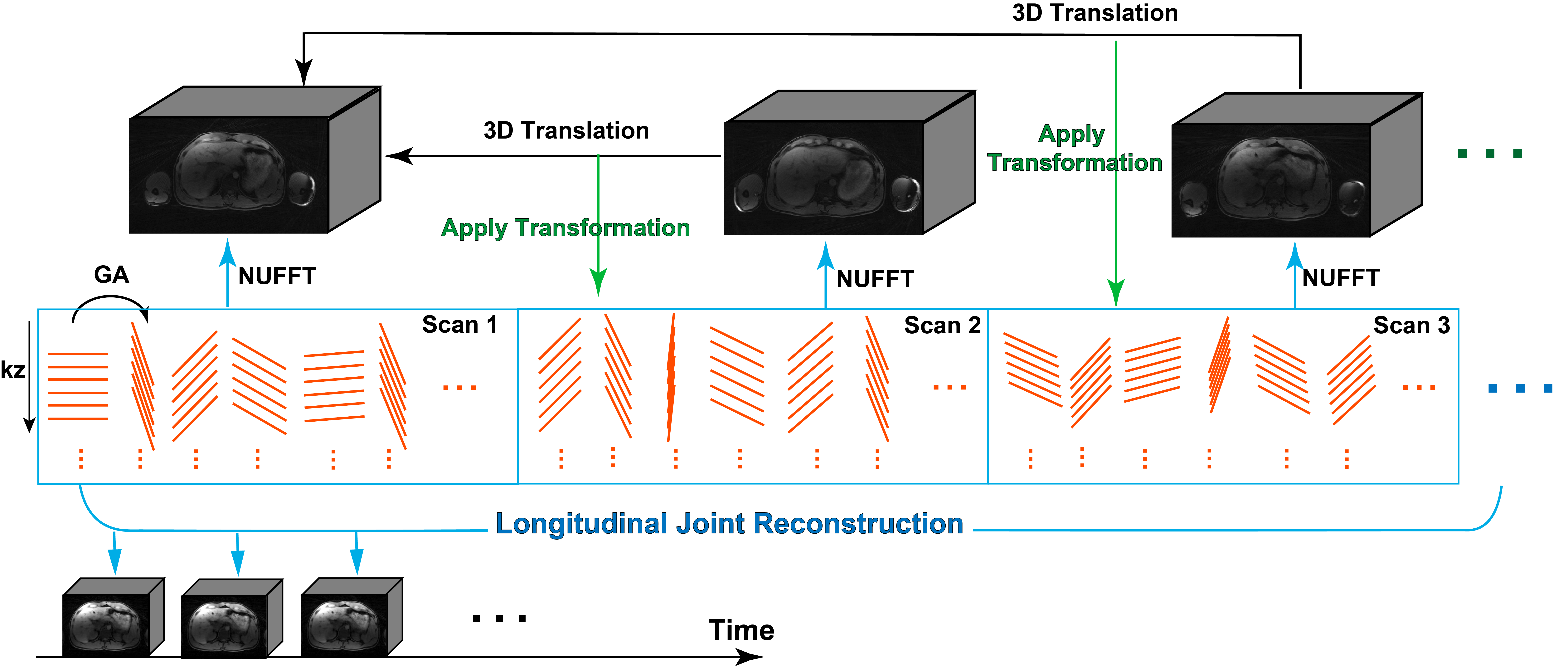
To test the proposed longitudinal reconstruction strategy, three successive free-breathing 3D liver examinations were performed on each of 8subjects using a GRASP sequence[1,2]. Individual sessions were separated by at least 7 days and at most 1 year. During acquisition, 2D navigators were acquired intermittently every two imaging spokes to track respiratory motion during the scan, and also to record the change in body position between scans at different dates. Imaging parameters for each session were as follows: B0=2.98T, spatial resolution=1.4x1.4x6mm3, TR/TE=2.51/1.16ms, flip angle=10o, partial Fourier=75%, number of spokes=1500 (including 500 2D navigators), and total acquisition time=2min21sec.Before longitudinal reconstruction, 3D images from each scan were reconstructed using NUFFT and follow-up scans were registered to the initial scan through a simple 3D translation (Figure 1) to correct misalignment of the imaging volume. The translation operation was then applied to the raw k-space data. A subspace-based low-rank reconstruction was used for the final reconstruction[2,3] with spatiotemporal total variation constraints. The 2D navigators helped to establish a reliable subspace basis containing not only respiratory motion but also bulk displacement across different exam dates.
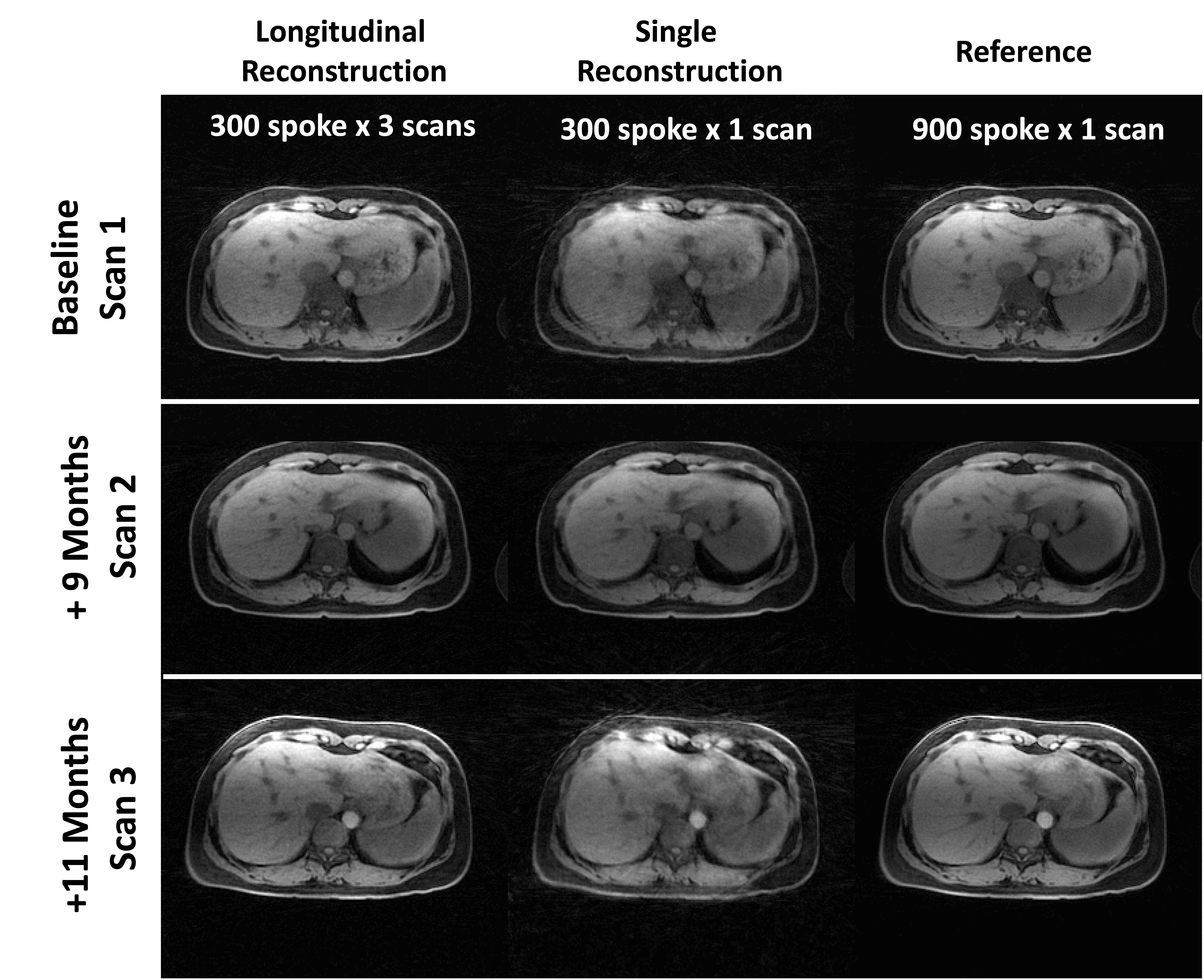
Nine hundred imaging spokes from each scan were used to reconstruct reference dynamic volumes with temporal resolution of 0.14s/frame. From each scan, 300 consecutive imaging spokes were then selected and concatenated for longitudinal reconstruction, to generate 450 frames in total. The same set of spokes for each scan was used for single-session reconstruction as a comparison.
Results
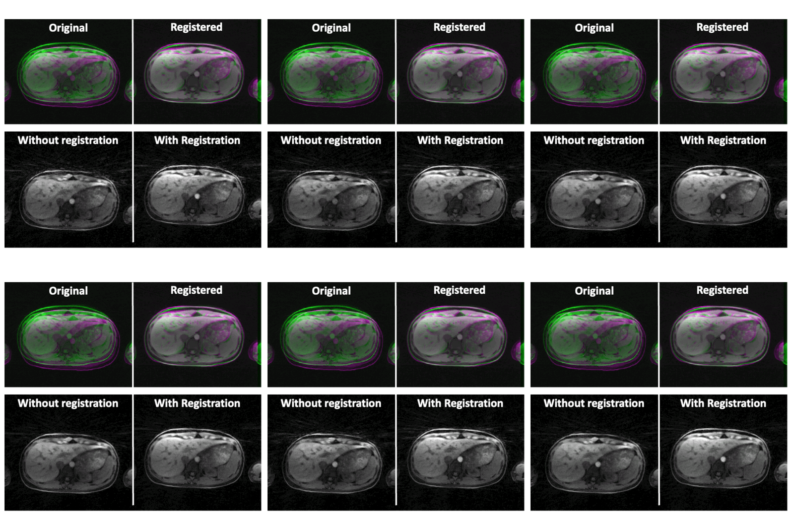
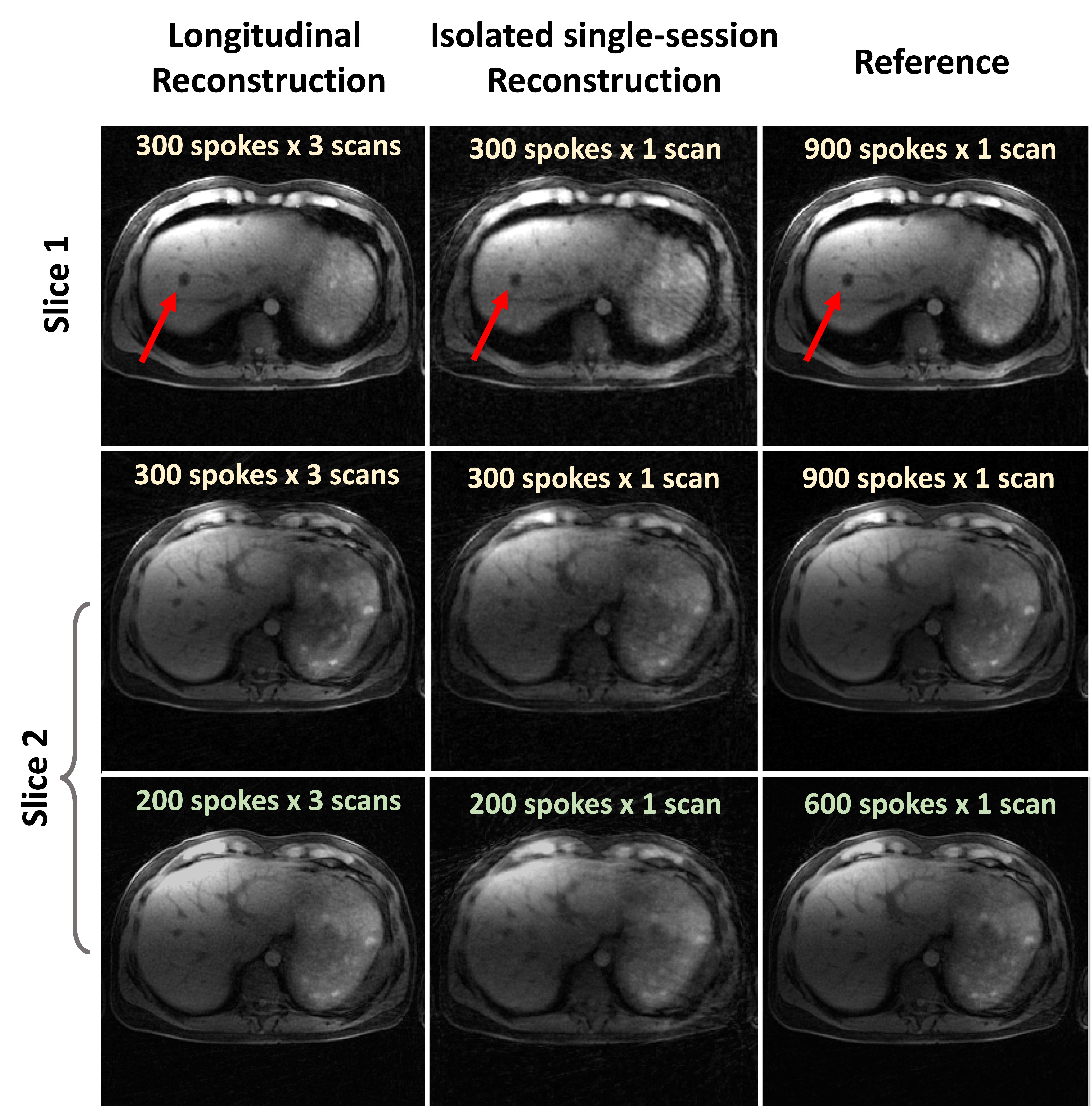
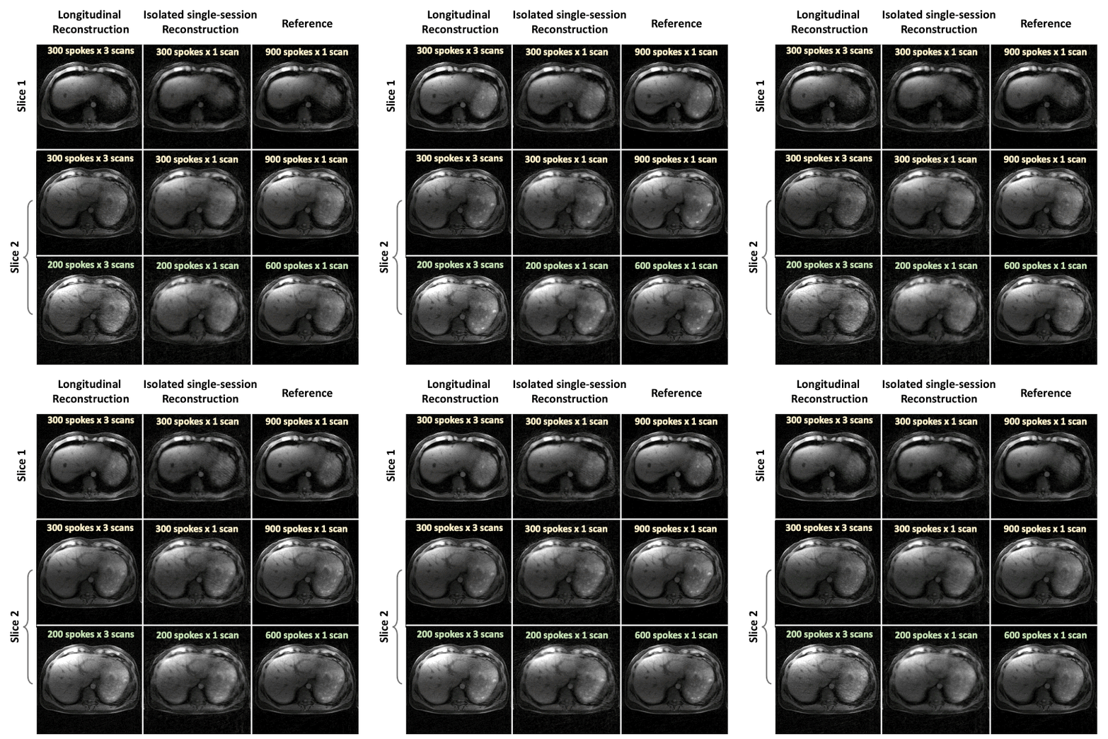
Figure 2 shows one frame of dynamic liver images reconstructed from one subject. The second and third scans were performed 9 and 11 months, respectively, after the first scan. Longitudinal image reconstruction using a limited number of spokes yielded image quality comparable to that of reference images. Meanwhile, using the same set of limited data for conventional isolated session-by-session reconstruction resulted in artifacts and blurring, especially in scans 1 and 3. Figure 3 shows that registration is necessary when the field-of-view is poorly aligned from session to session. Longitudinal reconstruction for a subject with a liver lesion presented in all three imaging sessions is shown in Figure 4 and the corresponding dynamical movie in Figure 5. Longitudinal reconstruction preserved high image quality with as few as 200 spokes, while separate reconstruction showed blurring and under-sampling artifacts. A liver lesion close to the diaphragm was delineated clearly even while moving substantially during breathing.Discussion
Temporal correlations are routinely used in dynamic image reconstruction, but longitudinal correlations over multiple imaging sessions from the same subject are not commonly used. We have shown that this rich longitudinal information can be leveraged to improve image reconstruction and to push for higher accelerations. We demonstrated the performance of longitudinal reconstruction in free-breathing dynamic liver MRI. Even with high acceleration and only 200 imaging spokes, the longitudinal reconstruction provides sufficient image quality for each scan while preserving the visual anatomical and pathological features from each imaging session. Further, a registration step using 3D translation can correct for the misalignment of imaging volumes acquired in different sessions, thereby improving reconstruction quality. Though this work used a fixed acceleration factor at all time points for purposes of demonstration, longitudinal MRI reconstruction could also allow for progressive acceleration of data acquisition as longitudinal information accumulates, ultimately extending acceleration limits beyond what can be achieved using only data from a single scan.Acknowledgements
This work was supported by the NIH (R01EB030549, R21EB032917, and P41EB017183) and was performed under the rubric of the Center for Advanced Imaging Innovation and Research (CAI2R), a National Center for Biomedical Imaging and Bioengineering supported by NIBIB.References
(1) Feng, L.; Grimm, R.; Block, K. T.; Chandarana, H.; Kim, S.; Xu, J.; Axel, L.; Sodickson, D. K.; Otazo, R. Golden-Angle Radial Sparse Parallel MRI: Combination of Compressed Sensing, Parallel Imaging, and Golden-Angle Radial Sampling for Fast and Flexible Dynamic Volumetric MRI. Magnetic Resonance in Medicine 2014, 72 (3), 707–717. https://doi.org/10.1002/mrm.24980.
(2) Feng, L.; Wen, Q.; Huang, C.; Tong, A.; Liu, F.; Chandarana, H. GRASP-Pro: imProving GRASP DCE‐MRI through Self-Calibrating Subspace-Modeling and Contrast Phase Automation. Magn Reson Med 2020, 83 (1), 94–108. https://doi.org/10.1002/mrm.27903.
(3) Benkert, T.; Tian, Y.; Huang, C.; DiBella, E. V. R.; Chandarana, H.; Feng, L. Optimization and Validation of Accelerated Golden-Angle Radial Sparse MRI Reconstruction with Self-Calibrating GRAPPA Operator Gridding. Magnetic Resonance in Medicine 2018, 80 (1), 286–293. https://doi.org/10.1002/mrm.27030.
Figures