1064
Impact of Spinal Cord Injury (SCI) level on Cortical Reorganization1Russell H. Morgan Department of Radiology and Radiological Science, Johns Hopkins University School of Medicine, Baltimore, MD, United States, 2F.M. Kirby Research Center for Functional Brain Imaging, Kennedy Krieger Institute, Baltimore, MD, United States, 3Department of Biostatistics, Johns Hopkins Bloomberg School of Public Health, Baltimore, MD, United States, 4International Center for Spinal Cord Injury, Kennedy Krieger Institute, Baltimore, MD, United States, 5Department of Physical Medicine and Rehabilitation, Johns Hopkins School of Medicine, Baltimore, MD, United States, 6The Malone Center for Engineering in Healthcare, Whiting School of Engineering, Johns Hopkins University, Baltimore, MD, United States
Synopsis
Keywords: Spinal Cord, Brain Connectivity, Spinal Cord, Graph Theory
Motivation: We aimed to fill the knowledge gap regarding the impact of spinal cord injury (SCI) level on cortical reorganization.
Goal(s): We sought to investigate cortical reorganization patterns in chronic SCI patients, specifically differentiating between cervical and thoracic injuries.
Approach: Employing graph theory analysis of functional connectivity, we analyzed data from 32 chronic SCI patients and 32 healthy controls.
Results: Significant alterations in somatomotor and visual networks in SCI cohort was observed. Notably, those with thoracic injuries exhibited more pronounced functional segregation within the somatomotor network, dividing it into dorsolateral and paramedian SMN regions and a ventrolateral SMN region.
Impact: Our findings on the influence of SCI level on brain reorganization may impact clinicians, researchers, and rehabilitation specialists, guiding tailored interventions and raising new questions about optimizing SCI recovery.
Introduction
Neurological recovery in spinal cord injury (SCI) patients is linked to cortical reorganization1. This process can be investigated using graph theory analysis of inter-regional functional connectivity (FC), which is derived from resting-state fMRI (rsfMRI) data2. In prior research, we noted changes in mesoscale graph measures across somatomotor, visual, and other functional networks in individuals with chronic SCI. These changes suggest compensatory adaptations due to the disrupted spinal cord-brain communication. Notably, within the somatomotor network, we detected a pattern of segregation that correlates with the anatomical divisions of upper and lower body representations, as well as orofacial regions.Purpose
The primary aim of this study was to explore if the segregation observed within the somatomotor network is influenced by the level of SCI (i.e., cervical vs. thoracic SCI).Methods
In our study, we enrolled 32 individuals with chronic SCI, including 16 with cervical injuries (SCIc) and 16 with thoracic injuries (SCIt). For comparison, 32 demographically matched healthy controls (HC)3 was considered. We used mesoscale graph measures, including recruitment and integration coefficients derived from parcel-level module allegiance matrices, to analyze and differentiate cortical network alterations among the HC, SCIc, and SCIt cohorts. All study participants were scanned on a 3T Philips Achieva scanner. The imaging protocol included a T1 weighted (T1w) structural run (acquisition time=6 min, TR/TE/TI=6.7/3.1/842 ms, resolution=1x1x1.2 mm3, SENSE factor=2, flip angle=8°) and two rsfMRI runs. The rsfMRI data was acquired using a multi-slice SENSE-EPI pulse sequence (TR/TE=2000/30 ms, SENSE factor=2, flip angle=75°, 37 axial slices, nominal resolution=3x3x3 mm3, 1 mm gap, 32 channel head coil, number of dynamics=200). Preprocessing was performed using the Analysis of Functional NeuroImages (AFNI) software4. We followed the preprocessing detailed by Jo et al5. The preprocessed fMRI data set was then parcellated into 200 regions of interest (ROIs) using Schaefer-Yeo atlas6, and corresponding time courses from each ROI were extracted to compute the weighted connectivity matrices. Each node was allocated to one of the following systems: visual (VN), sensorimotor (SMN), dorsal attention (DAN), salience/ventral attention (VAN), limbic (LN), frontoparietal (FPN), or default mode networks (DMN). We then used a multi-layer community detection approach7, where each layer represents an individual’s connectivity matrix, to compare the modular structure of the brain networks between the HC and SCI groups. Subsequently, a set of mesoscale graph measures, specifically the recruitment and integration coefficients, was calculated from the module allegiance matrix. The module allegiance matrix was determined by quantifying the probability that pairs of brain regions engage in the same functional network community across subjects. The module allegiance matrix was calculated for HC, SCI, SCIc (cervical SCI), and SCIt (thoracic SCI) cohorts. This analysis approach provided quantitative outcome measures of, across subjects, how stably the brain regions are recruited (recruitment) and how consistently the regions interact with other functional networks (integration).Results
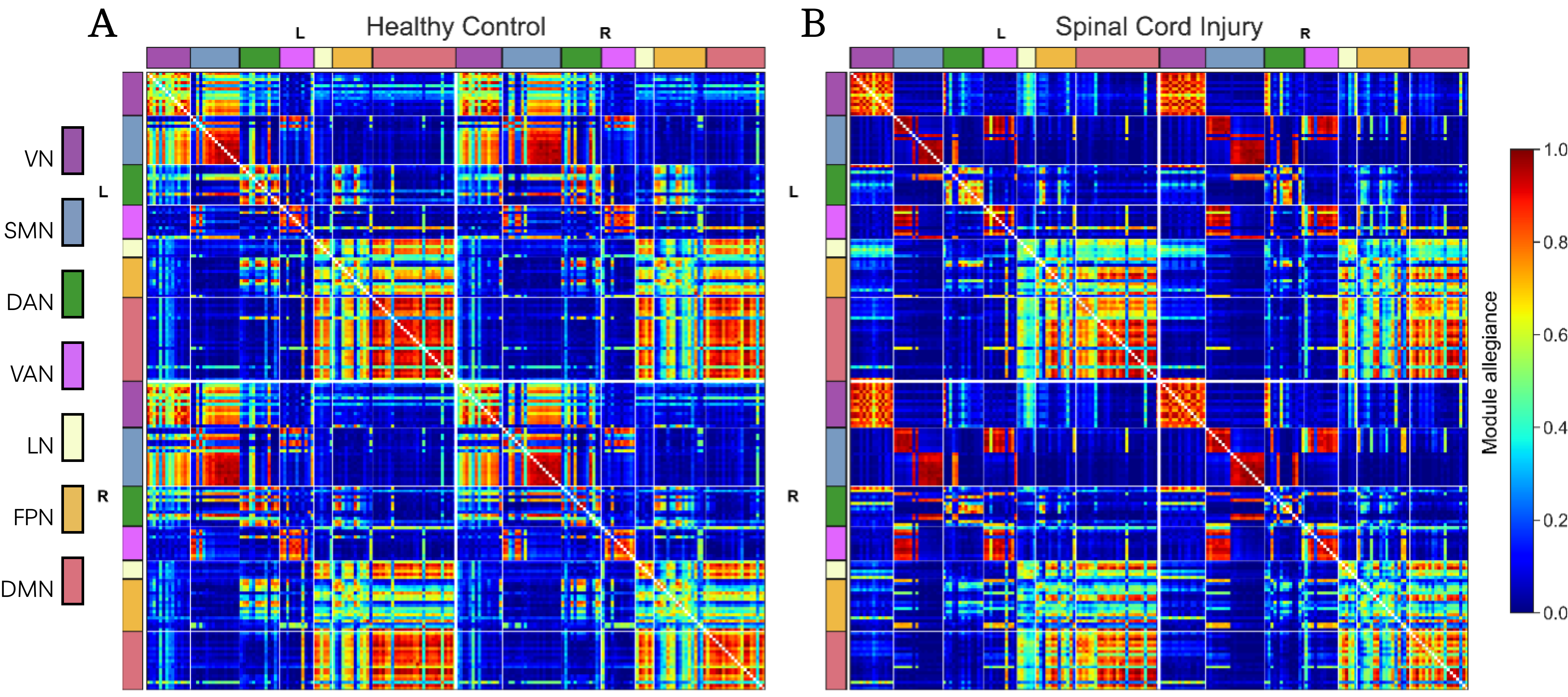
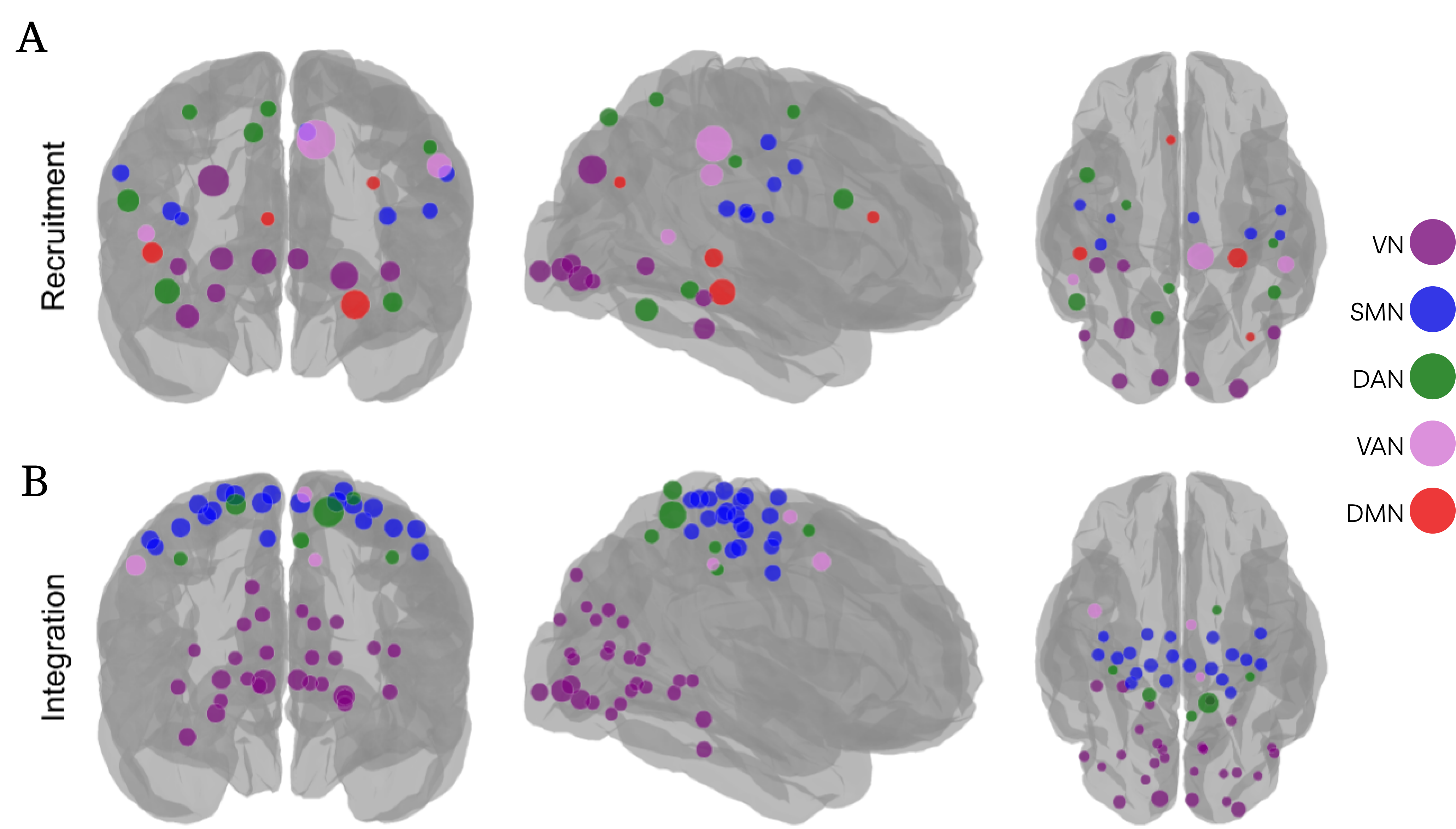
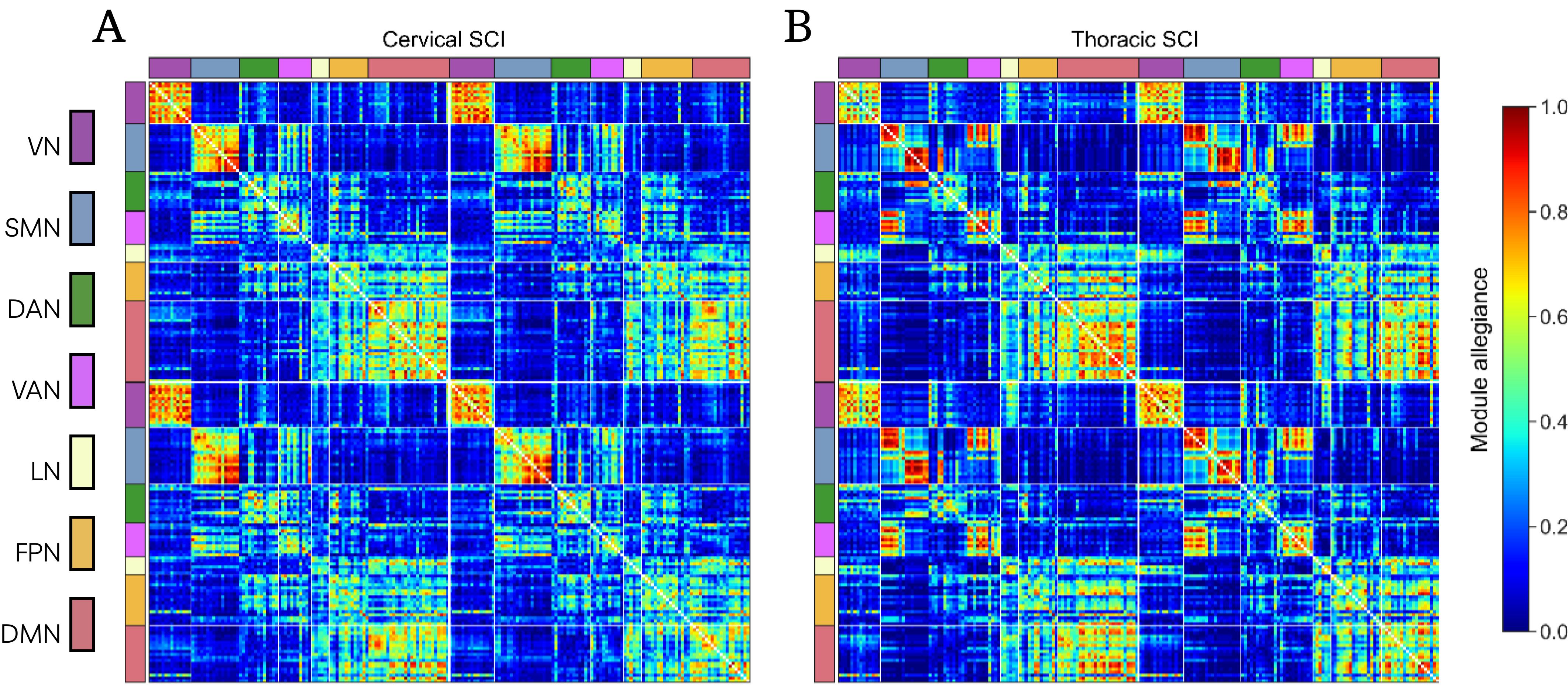
Figure 1 displays the parcel-level module allegiance matrices of the HC (A) and SCI (B) cohorts. Figure 2A and 2B illustrate the parcels with significant differences (p > 0.001, FDR corrected) in recruitment and integration coefficients between the HC and SCI cohorts, respectively. Importantly, a detailed examination of the somatomotor network (SMN) revealed a network segregation into dorsolateral and paramedian SMN regions (SMNb; SMN-body) and ventrolateral SMN region (SMNf; SMN-face). These respectively represent the upper-and-lower body and the orofacial regions. The parcel-level module allegiance matrices of SCIc and SCIt cohorts (Figure 3) reveal that functional segregation in the somatomotor network was significantly more pronounced in individuals with thoracic SCI than in those with cervical SCI.Conclusion
The study's findings suggest compensatory neural adaptations might occur as a response to the disrupted communication between the spinal cord and brain, underscoring the effectiveness of mesoscale graph measures in evaluating functional reorganization in chronic SCI. Notably, our data reveals that individuals with thoracic spinal cord injuries exhibit more distinct functional segregation within the somatomotor areas compared to those with cervical injuries. This difference may be attributable to the varying severity and extent of the injuries, influencing the pattern and degree of brain reorganization. Cervical injuries, which typically result in more extensive damage, may restrict the brain's reorganization potential, thereby blurring the patterns of functional segregation. In contrast, thoracic injuries often involve fewer spinal levels, which might spare more structural pathways, possibly leading to a greater degree of brain reorganization and more evident functional segregation within the somatomotor network.Acknowledgements
ASC was supported in part by DOD (W81XWH-08-1-0192), NIH (R21 EB006120 and P41-EB015909), and the Craig H. Neilsen Foundation (Project Number 338419). FVF and MAL were supported in part by NIH grants R01 EB026549 from the National Institute of Biomedical Imaging and R01 MH129397 from the National Institute of Mental Health.References
- Choe, AS, Belegu, V., Yoshida, S., Joel, S., Sadowsky, CL, Smith, SA, ... & McDonald, JW . Extensive neurological recovery from a complete spinal cord injury: a case report and hypothesis on the role of cortical plasticity. Frontiers in Human Neuroscience , 7 -290, 2013.
- Farahani, F. V., Karwowski, W., & Lighthall, N. R. Application of graph theory for identifying connectivity patterns in human brain networks: a systematic review. frontiers in Neuroscience, 2019 13, 585.
- Landman, Bennett A., et al. "Multi-parametric neuroimaging reproducibility: a 3-T resource study." Neuroimage 54.4 (2011): 2854-2866.
- Cox RW. AFNI: software for analysis and visualization of functional magnetic resonance neuroimages. Compute Biomed Res 1996;29(3):162–173; doi: 10.1006/cbmr.1996.0014
- Jo HJ, Gotts SJ, Reynolds RC, et al. Effective Preprocessing Procedures Virtually Eliminate Distance-Dependent Motion Artifacts in Resting State FMRI. J Appl Math 2013;2013; doi: 10.1155/2013/935154.
- Schaefer A, Kong R, Gordon EM, et al. Local-Global Parcellation of the Human Cerebral Cortex from Intrinsic Functional Connectivity MRI. Cereb Cortex 2018;28(9):3095–3114; doi: 10.1093/cercor/bhx179.
- Mucha PJ, Richardson T, Macon K, et al. Community Structure in Time-Dependent, Multiscale, and Multiplex Networks. Science 2010;328(5980):876–878.
Figures