1063
Investigating Respiration Induced B0 Field Variations in the Thoracic and Lumbar Spinal Cord at 7T1Physikalisch-Technische Bundesanstalt (PTB), Berlin, Germany, 2NeuroPoly Lab, Institute of Biomedical Engineering, Polytechnique Montréal, Montreal, QC, Canada, 3Centre de recherche du CHU Sainte-Justine, Université de Montréal, Montréal, QC, Canada, 4Functional Neuroimaging Unit, CRIUGM, Université de Montréal, Montréal, QC, Canada, 5Mila-Quebec AI Institute, Montréal, QC, Canada, 6Medical Physics in Radiology, German Cancer Research Center (DKFZ), Heidelberg, Germany, 7Center for Magnetic Resonance Research, University of Minnesota, Minneapolis, MN, United States
Synopsis
Keywords: Spinal Cord, High-Field MRI, 7 Tesla, Spinal Cord, B0 Shimming, Respiration Resolved
Motivation: Respiration-related B0 variations and their modulation in the thoracolumbar spinal cord (SC) at 7T have not been thoroughly investigated. Knowing B0 variations is crucial for assessing the potential of SC imaging.
Goal(s): Quantification of B0 variations in the thoracolumbar SC across different respiratory states and evaluation of the effectiveness of tailored B0 shims in mitigating these variations.
Approach: Non-Cartesian respiration-resolved 3D B0 field maps were acquired during free-breathing and employed for B0 shimming using the SC Shimming Toolbox.
Results: Tailored B0 shims reduce the B0 variations to approximately 50Hz (standard deviation) across the thoracolumbar SC. Respiration introduces up to 40Hz of additional field-offset.
Impact: Our results quantify B0 fluctuations in the thoracolumbar SC and show that customized B0 shims effectively reduce these fluctuations to a standard deviation of around 50Hz. Further investigation of deep breathing is crucial for optimizing shim strategies in future uses.
Introduction
MRI within the human body at ultra-high magnetic field strengths (UHF; >7T) faces multiple challenges, including multiple motion sources and spatially varying flip-angle (FA) patterns, which can be addressed by parallel transmission (pTx) methods.1-3 However, another obstacle at UHF is the presence of inhomogeneities in the static magnetic B0 field leading to signal loss and image distortions.1,2While progress has been made in advancing cervical spinal-cord (SC) imaging at UHF, the thoracolumbar (T/L) SC, has remained relatively unexplored due to the aforementioned challenges.3 Here, we demonstrate how to acquire high-resolution 3D respiration-resolved B0 field maps throughout the entire T/L-SC and how to leverage this information to optimize tailored B0 shims using the Shimming Toolbox4 in a cohort of six volunteers at 7T.
Methods
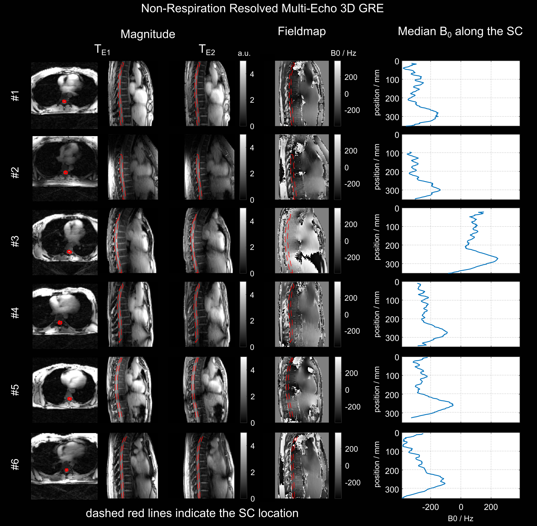
MRI scans were conducted on a 7T scanner (Magnetom 7T, Siemens, Germany) with an 32-element body array (MRI.Tools, Germany) in 8TX/32RX mode. The study included six healthy volunteers (3M/3F, 26-32years, BMI 20-35kg/m2) following IRB approval and written consent.We acquired 3D thoracic multi-echo GRE datasets with a radial phase encoding5 during free-breathing using the vendor-supplied B0 Tune-Up shim (9min10sec, FOV=250x312x312mm3, TE1/TE2/TE3/TR=1.51/2.79/4.07/6.1ms, 5° FA, 2048 radial-phase lines) . Phase errors from bipolar readout gradient polarity were corrected before iterative SENSE reconstruction. GRE data was reconstructed non-respiration resolved (NRR) and respiration resolved (RR) for six respiration states via self-navigation to achieve an isotropic voxel size of 1.4mm.5
3D NRR/RR B0 field maps were computed from the first and second echo (1.51/2.79ms) providing a dynamic B0 range of +/-390Hz. The T/L-SC volume was manually defined from the first and sixth RR datasets (exhale/inhale) and NRR data. For the sake of clarity, a single sagittal slice is displayed for each subject, though the analysis covered the entire T/L-SC volume.
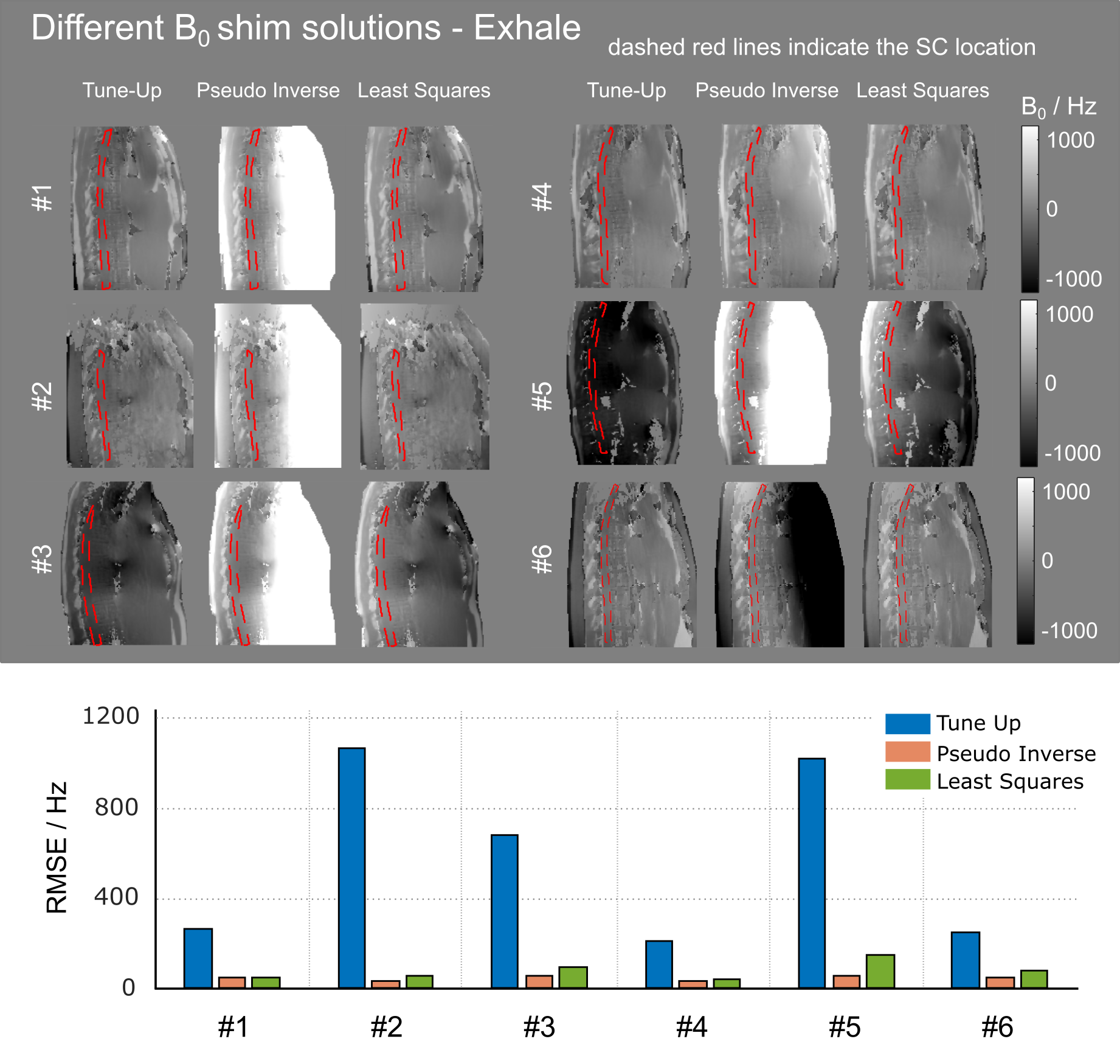
We optimized subject-tailored B0 shim solutions up to the second order using the Shimming Toolbox4, involving automated fieldmap unwrapping (scikit-image, threshold=0.01) and spherical harmonic optimization via i) a Pseudo Inverse (PI) and ii) a Least Squares (LS) approach using the mean-squared-error, dilation-kernel=5 and regularization-factor=10-6 (LS). We also simulated the effect of respiration by applying shims designed for one respiration state to another.
Results and Discussion
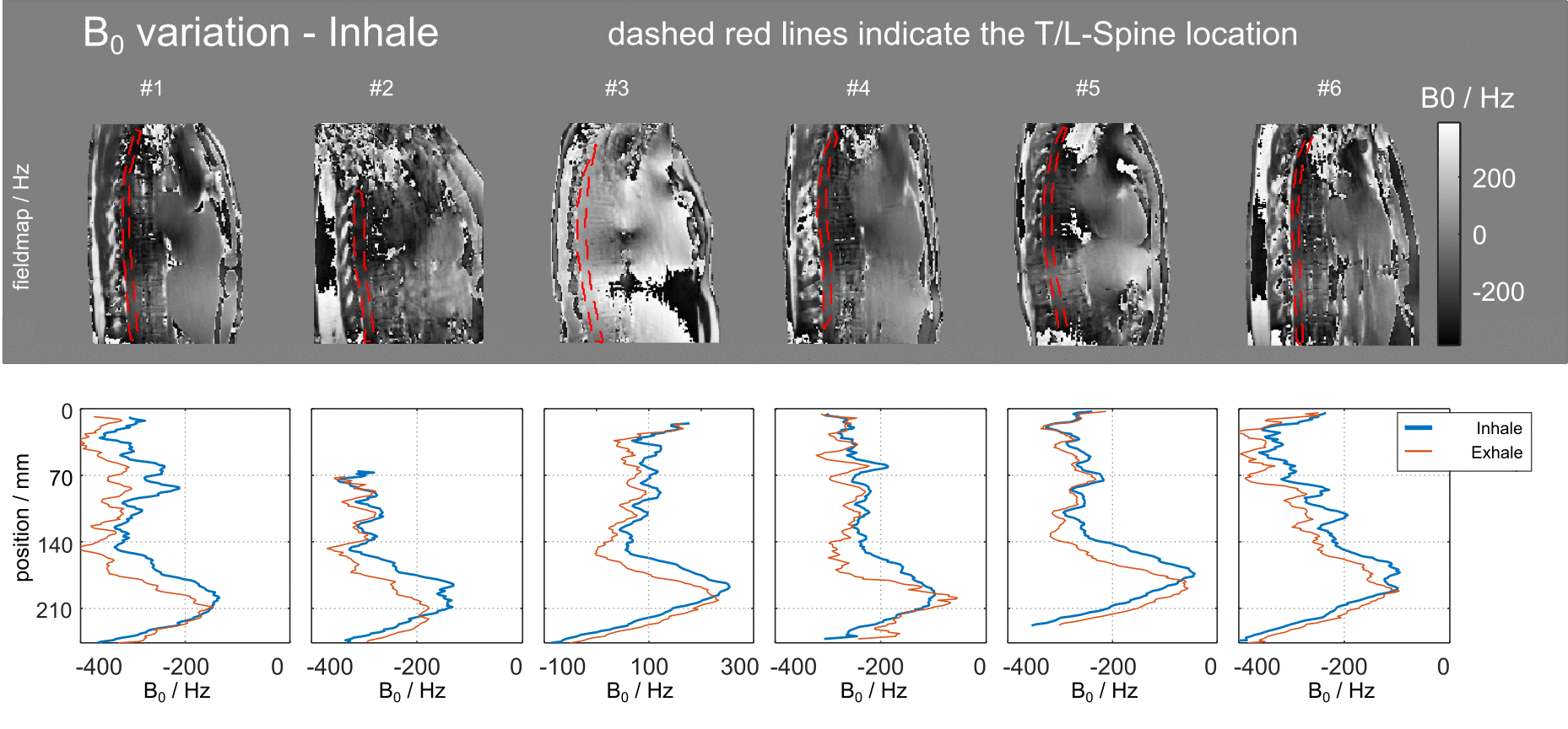
Figure 1 shows 3D NRR GRE data for six volunteers, revealing consistent B0 variations in the T/L-SC, despite notable anatomical inter-subject variations. High-frequency B0 field changes related to susceptibility changes adjacent to the SC and an increase in the lower SC region were observed, with a median B0 variation of 250Hz and high-frequency variations of 46Hz across all volunteers.Figure 2 highlights two key differences in the RR B0 field maps in the T/L-SC during exhale and inhale: Approximately 50Hz offset and a shift of the B0 variations in the lower spine, corresponding to motion of the right hemidiaphragm (averaging 14mm) during breathing. Note that these differences could be more pronounced with deep respiration, which may induce up to five times larger head-to-feet motion.6
Figure 3 illustrates the impact of three B0 shims (Tune-Up, PI, LS) on exhale B0 field maps. The two optimized shims (PI/LS) effectively reduced B0 offsets, and led to a significant reduction in the root-mean-squared-error (RMSE) of the Tune-Up shim, lowering it by approximately 90% from 472Hz (Tune-Up) to 48/68Hz (PI/LS).
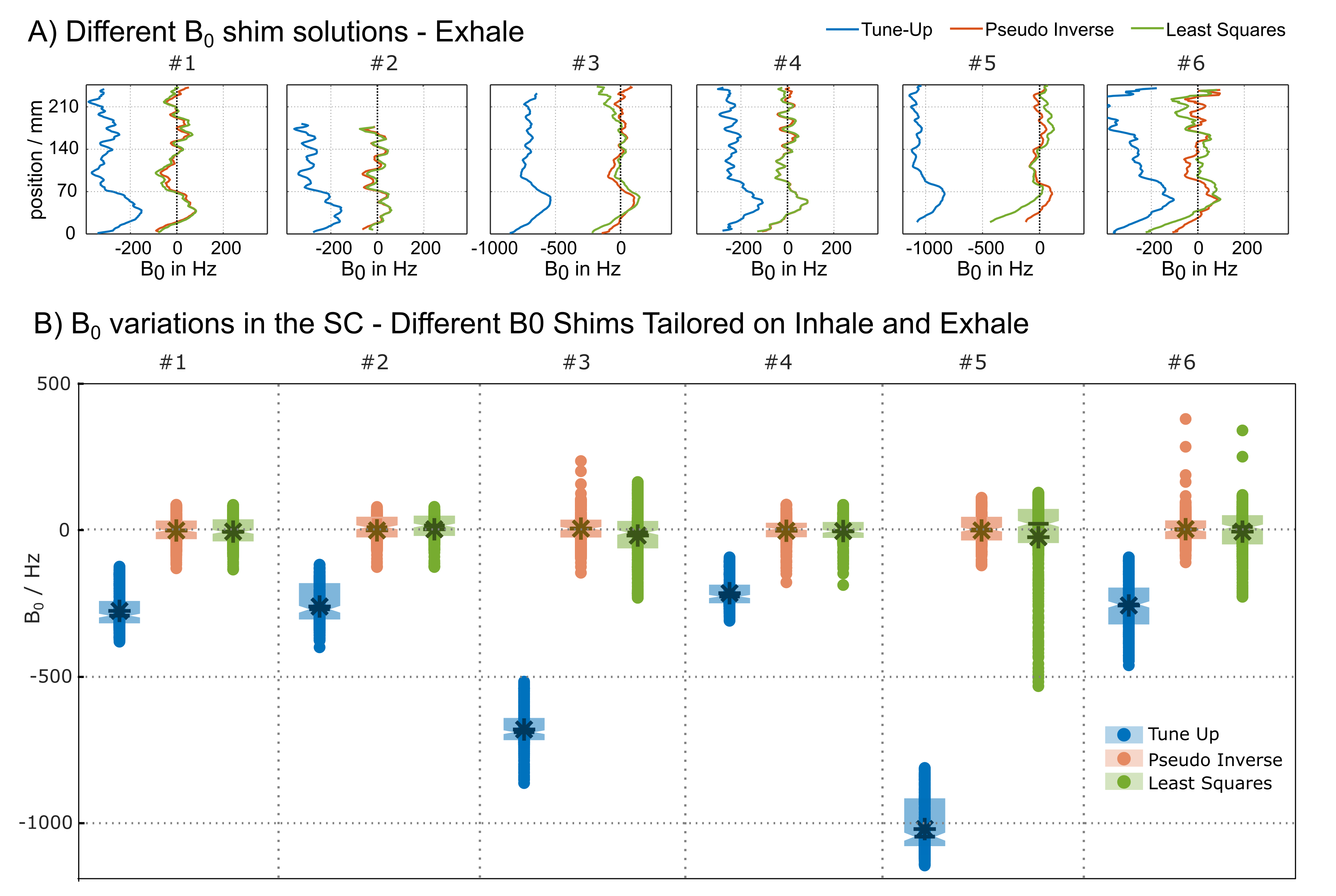
Figure 4 shows the mean B0 variation in the T/L-SC for different shim solutions, shown in Fig.3. It highlights reduced B0 deviations after tailored B0 shimming. Except for subject #3 and #5, where we observe more outliers due to a suboptimal LS solution, the average standard deviation decreased from 62Hz to 43/53Hz, representing a reduction of 31/15%. Preliminary data using up to 3rd-order B0 shims indicate an additional 23% reduction in the average standard deviation compared to 2nd-order B0 shims. These trends were consistent in the exhale dataset and confirmed by boxplots summarizing both exhale and inhale datasets, indicating a reduction in the interquartile range for all six volunteers using LS or PI B0 shims.
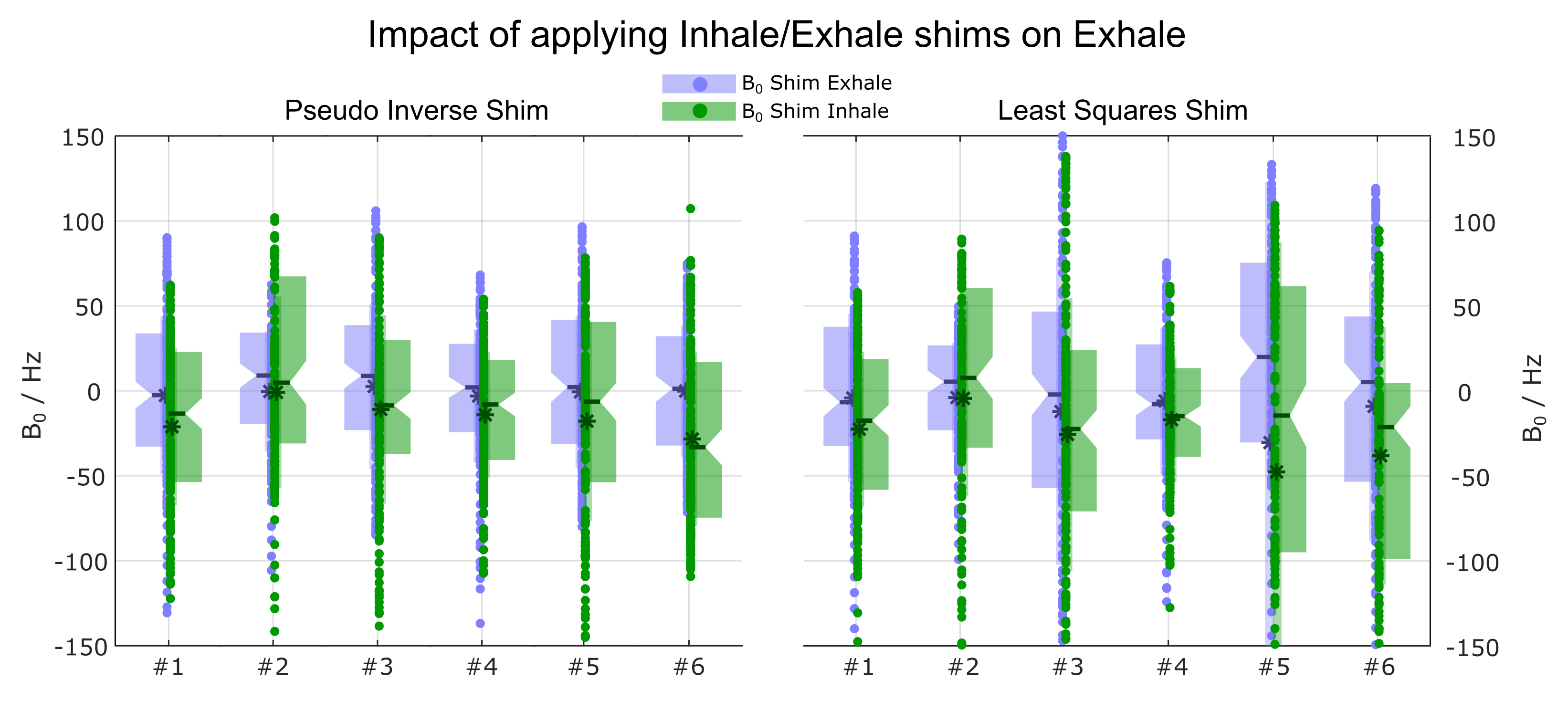
Figure 5 shows the impact of applying PI and LS B0 shims, tailored for exhale/inhale, on the exhale fieldmap of each volunteer. Incorrect shims caused an additional B0 variation of up to 40Hz and increased interquartile range. Further analysis is needed to explore the influence of deep breathing on shim performance, addressing the necessity of RR maps over NRR maps.
Conclusion
This study describes B0 variations in the T/L-SC at 7T, the impact of respiration and the performance of different tailored B0 shims. Moreover, it shows that tailored B0 shims reduce the standard deviation of B0 down to approximately 50Hz across the entire T/L-SC, thus enabling SC imaging in these mid-to-lower SC areas.Acknowledgements
We gratefully acknowledge funding from the German Research Foundation SCHM 2677/2-1, SCHM 2677/4-1 and GRK2260, BIOQIC.References
[1] Ladd, E, Bachert, P, Meyerspeer, M, et al. Pros and cons of ultra-high-field MRI/MRS for human application, Prog. Nuc. Magn. Reson. Spec. 2018; 109:1-50. doi: 10.1016/j.pnmrs.2018.06.001
[2] Barry, RL, Vannesjo, SJ, By, S, Gore, JC, Smith, SA. Spinal cord MRI at 7T. Neuroimage 168, 437–451. doi: 10.1016/j.neuroimage.2017.07.003.
[3] Padormo, F., Beqiri, A., Hajnal, J. V., and Malik, S. J. (2016), Parallel transmission for ultrahigh‐field imaging. NMR Biomed., 29: 1145– 1161. doi: 10.1002/nbm.3313
[4] D'Astous A, Cereza G, Papp D, Gilbert KM, Stockmann JP, Alonso-Ortiz E, Cohen-Adad J. Shimming toolbox: An open-source software toolbox for B0 and B1 shimming in MRI. Magn Reson Med. 2022; 1-17. doi:10.1002/mrm.29528
[5] Dietrich, S, Aigner, CS, Mayer, J, et al. Motion-compensated fat-water imaging for 3D cardiac MRI at ultra-high fields. Magn Reson Med. 2022; 87: 2621–2636. doi:10.1002/mrm.29144
[6] Aigner, CS, Dietrich, S, Schmitter, S. Respiration induced B1+ changes and their impact on universal and tailored 3D kT-point parallel transmission pulses for 7T cardiac imaging. Magn Reson Med. 2022; 87: 2862–2871. doi:10.1002/mrm.29183
Figures