1062
Correcting Susceptibility artifacts with Unified RF/Shim Coil (UNIC) in Cervical Spine MRI for Subjects with and without Orthodontic Implants1Biomedical Imaging Research Institute, Cedars-Sinai Medical Center, Los Angeles, CA, United States, 2Department of Imaging, Cedars-Sinai Medical Center, Los Angeles, CA, United States, 3Department of Bioengineering, University of California, Los Angeles, Los Angeles, CA, United States, 4Department of Radiology, Weill Cornell Medical, New York, NY, United States, 5Department of Radiation Oncology, Emory University School of Medicine, Atlanta, GA, United States
Synopsis
Keywords: Spinal Cord, Spinal Cord, B0 Shimming; Cervical spine; DWI; DTI
Motivation: Metallic orthodontic devices can introduce strong susceptibility artifacts that hinder clinical interpretation of cervical spine MRI.
Goal(s): Using novel unified RF/shim coils (UNIC) to reduce field variation caused by metal susceptibility artifact.
Approach: Total of eighteen subjects (5 with braces) were scanned with a UNIC coil research prototype.
Results: The integrated UNIC shim array significantly increased the voxel percentage from 28% with scanner-shimming to 46% in DWI, reducing distortion by 61% with braces and 15% without braces in DWI, and by 41% in DTI. It also decreased B0 field variation by 63% with braces and 31% without.
Impact: Unified RF/shim coil delivered improved quality and corrected image distortions in cervical spine scans by mitigating magnetic field variation, including those from orthodontic braces, ensuring more accurate diffusion measurements and consistently reducing susceptibility artifacts.
Introduction
Brain and cervical cord MR imaging are crucial for diagnosing various central nervous system pathologies. Diffusion-weighted imaging (DWI) and diffusion tensor imaging (DTI) offer non-invasive methods for assessing tissue microstructure and fiber tractography1–3. However, the presence of metallic dental implants or tissue-air interface can introduce artifacts, compromising magnetic field integrity and causing image distortions2. Traditional second-order spherical harmonic (SH) shim coils built into MRI scanners often struggle to correct complex field variations4. The emergence of Unified RF Coil Arrays with integrated Shimming (UNIC)5 introduces a novel approach bringing a decoupled shim array closer to the anatomy of interest6, thus maximizing the effectiveness of both RF and shim functions. UNIC has shown promise in applications such as cardiac7, liver8, and hip metal implants9. Nevertheless, their effectiveness in cervical spinal applications remains unexplored. To our best knowledge, this is the first study to evaluate the correction of metal implant-induced artifacts in cervical cord MRI using novel local shimming coils.Methods
Subjects: In this IRB-approved study, a total of 18 subjects (n=5 with braces) were recruited and scanned on 3T Siemens Biograph mMR scanner.Unified RF/shim coil: The UNIC coil prototype for B0 shimming and RF reception was used, which demonstrated uncompromised SNR by adding a decoupled 42-channel shim array to the RF arrays (12-channel) 5.
Imaging data acquisition: As shown in Figure1, protocols included Sagittal T2-weighted turbo-spin-echo (T2w-TSE), T2-Sampling Perfection with Application-Optimized Contrasts using different flip-angle Evolution (SPACE), multi-segmented (MS)-DWI, standard single-shot (SS)-DTI. Additionally, a dual-echo gradient-echo sequence to calculate shim currents for UNIC. C-spine region was shimmed using scanner 2nd order SH and UNIC shims, respectively.
Region of interest: A radiologist manually segmented the cervical spine on a single slice where it was fully visible in T2w-TSE, for T2w-TSE, apparent diffusion coefficient (ADC) and fractional anisotropy (FA) images, with and without UNIC shimming.
Statistical analysis: DWI and DTI distortion were quantified using Dice and Jaccard coefficients, voxel gain in DWI of braces, and precision of ADC and FA values by coefficient of variation (CV). B0 field maps' mean, standard deviation (SD), and root-mean-square-error (RMSE) were calculated. Differences between UNIC and standard shimming's efficacy were statistically tested using the Wilcoxon signed-rank test.
Results
Qualitative analysis of C-spine in subjects with and without orthodontal implants: Figure 2 shows that the UNIC coil improves image quality, with better delineation of the cervical spine in T2-SPACE images and less distortion in DWI and ADC images compared to traditional scanner shims. Figure 3 further confirms improved DWI and DTI image quality and B0 field homogeneity in subjects without dental braces.Quantitative analysis of C-spine in subjects with and without orthodontal implants: Figure 4 presents enhanced UNIC shimming's efficacy in image accuracy for orthodontal implant, as indicated by marked improvements in Dice (UNIC: 0.5±0.5 vs. scanner: 0.3±0.4) and Jaccard coefficients (UNIC: 0.3±0.4 vs. scanner: 0.2±0.2), with a 41% voxel increase relative to scanner shim. Moreover, UNIC achieved a significant reduction in B0 off-resonance (SD UNIC: 166.3±80.7Hz vs. scanner: 300.7±228Hz).
For subjects without implants, Figure 5 shows UNIC shimming's superior distortion correction in DWI, as reflected in higher Dice (UNIC: 0.7±0.1 vs. scanner: 0.6±0.1) and Jaccard (UNIC: 0.54±0.13 vs. scanner: 0.47±0.15) scores, alongside a 20% reduction in the coefficient of variation for ADC values. DTI also benefitted, with UNIC shimming yielding higher Dice (UNIC: 0.6±0.2 vs. scanner: 0.5±0.2) and Jaccard (UNIC: 0.4±0.2 vs. scanner: 0.3±0.2) results, a lower mean FA value, and a 12% CV reduction.
Additionally, a significant reduction in B0 off-resonance was observed in non-implant subjects using UNIC shimming (SD, UNIC: 16.6±5.9Hz vs. scanner: 38.5±15.1Hz).
Discussion
Our UNIC coil prototype demonstrates its effectiveness in enhancing the quality of DWI and DTI images for c-spine. It surpasses scanner shimming and excels in areas with/without metal artifacts. Application of the UNIC shim yielded a notably more homogeneous B0 field, significantly reducing susceptibility artifacts in patients with orthodontal braces. Qualitative assessments indicate that UNIC markedly improves the clarity of T2-SPACE sequence and corrects distortion in MRI images, regardless of the presence of dental hardware. Quantitatively, UNIC has shown high precision in both subjects with and without implants. In the future, we will further optimize the UNIC shim coil layout for spinal imaging based on this work, implement dynamic slice-optimized shimming, and extend its application to subjects with spinal metallic implants, potentially transforming spinal cord imaging.Conclusion
The UNIC coil prototype improves the quality of cervical spine MRI, reduces distortion, and surpasses standard scanner shims, and offers potential improvement in c-spine diagnostics.Acknowledgements
Hsin-Jung Yang and Hui Han contributed equally to this work. This work was supported by NIH R01NS121544; R43 NS120795; R01 HL156818.References
1. Park EH, Lee YH, Jeong EK, Roh YH, Suh JS. Diffusion tensor imaging focusing on lower cervical spinal cord using 2D reduced FOV interleaved multislice single-shot diffusion-weighted echo-planar imaging: comparison with conventional single-shot diffusion-weighted echo-planar imaging. Magnetic Resonance Imaging. 2015;33(4):401-406.
2. Koch KM, Nencka AS, Klein A, et al. Diffusion-weighted MRI of the spinal cord in cervical spondylotic myelopathy after instrumented fusion. Frontiers in Neurology. 2023;14.
3. Rutman AM, Peterson DJ, Cohen WA, Mossa-Basha M. Diffusion Tensor Imaging of the Spinal Cord: Clinical Value, Investigational Applications, and Technical Limitations. Current Problems in Diagnostic Radiology. 2018;47(4):257-269.
4. Topfer R, Starewicz P, Lo KM, et al. A 24-channel shim array for the human spinal cord: Design, evaluation, and application. Magnetic Resonance in Medicine. 2016;76(5):1604-1611.
5. Han, H. Multi-Coil B0 Shimming in the Body. Proceedings of the 28th Annual Meeting of the ISMRM 2020, Virtual Meeting; E0912.
6. Yang, H.-J.; Serry, F.; Hu, P.; Fan, Z.; Shim, H.; Christodoulou, A.; Wang, N.; Kwan, A.; Xie, Y.; Huang, Y.; et al. Ultra-Homogeneous B0 Field for High-Field Body Magnetic Resonance Imaging with Unified Shim-RF Coil. Nature Biomedical Engineering (under revision).
7. Yang, H.-J.; Stager, J.; Azab, L.; Liu, W.; Lu M.; Huang, Y. et al. Whole Heart High-Order B0 Shimming at 3T Using a UNIfied Coil (UNIC) for RF Receive and Shimming. Proceedings of the 28th Annual Meeting of the ISMRM 2020, Virtual Meeting; p2183.
8. Wang, N.; Serry, FM.; Ocasio, M.; Xie Y.; Huang, Y.; Li, X.; et al. Integrated high-order B0 shimming for multiparametric quantitative liver imagingat 3T using a UNIfied Coil (UNIC). Proceedings of the 31st Annual Meeting of the ISMRM-ESMRM 2022, London; p0028.
9. Serry, FM.; Chen, J.; Christodoulou, AG.; Huang Y.; Han, F, et al. Improving MRI Near Metal with Local B0 Shimming using a Unified Shim-RF Coil (UNIC): First Case Study, Hip Prosthesis in Phantom. Proceedings of the 28th Annual Meeting of the ISMRM 2020, Virtual Meeting; p3110.
Figures
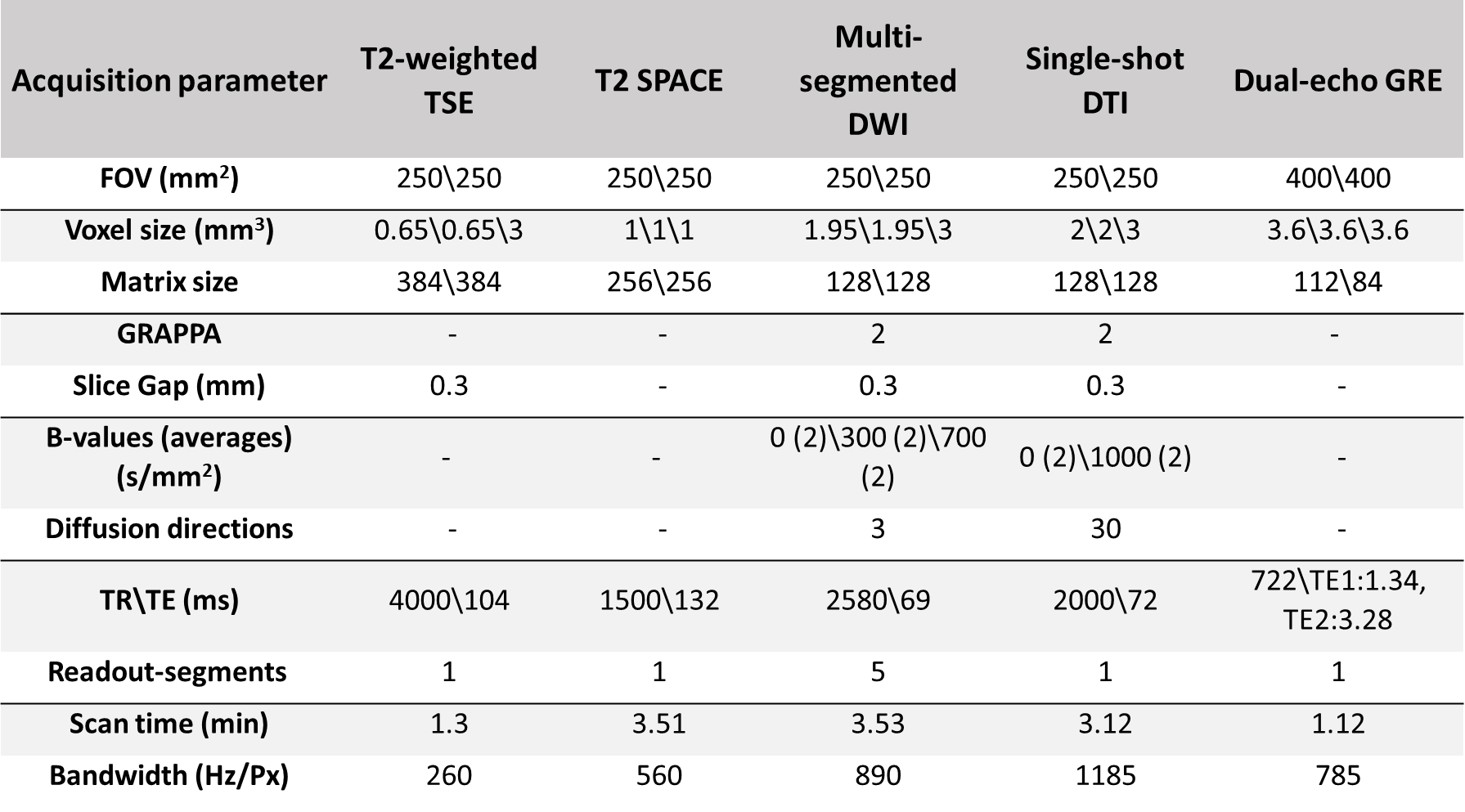
Figure1: Study protocol including T2w-TSE, T2-SPACE, multi-segmented DWI, single-shot DTI, and dual-echo gradient-echo (GRE) sequence.
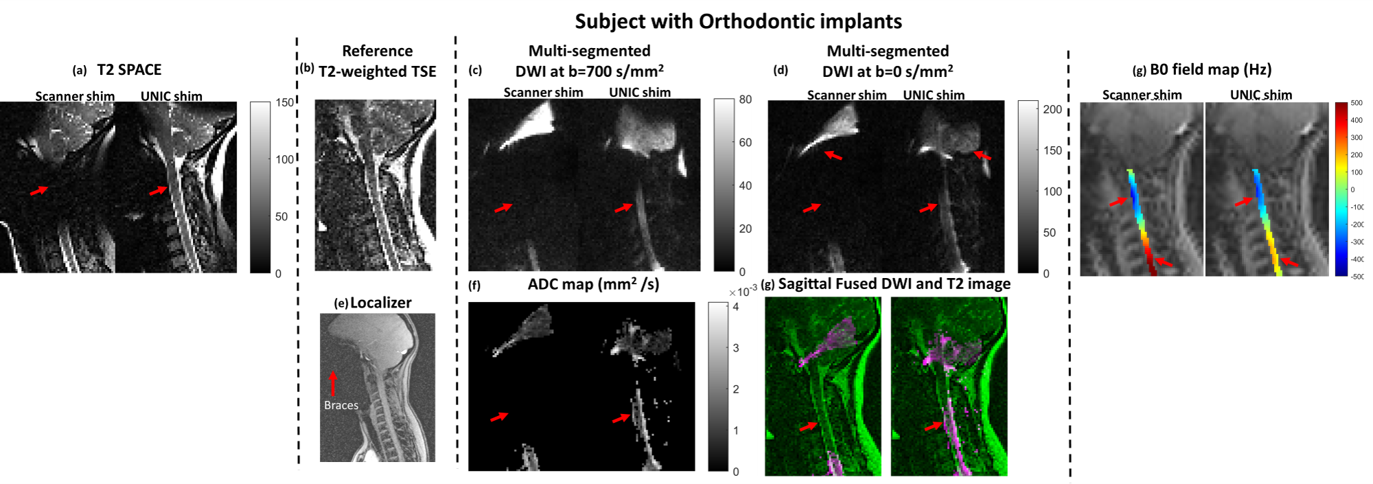
Figure2: Comparison between UNIC and standard scanner shim for different acquisition sequences in Orthodontal implant subjects. (a) T2 SPACE, (b) T2-w TSE, (c,d,f) DWI and ADC image with distortion correction by UNIC, (e) Localizer showing braces location, (g) fused DWI and T2 images shows voxel gained after UNIC shimming and (h) B0 fieldmap in c-spine region. Red arrow mark shows improvement in image quality or field correction with UNIC shimming.
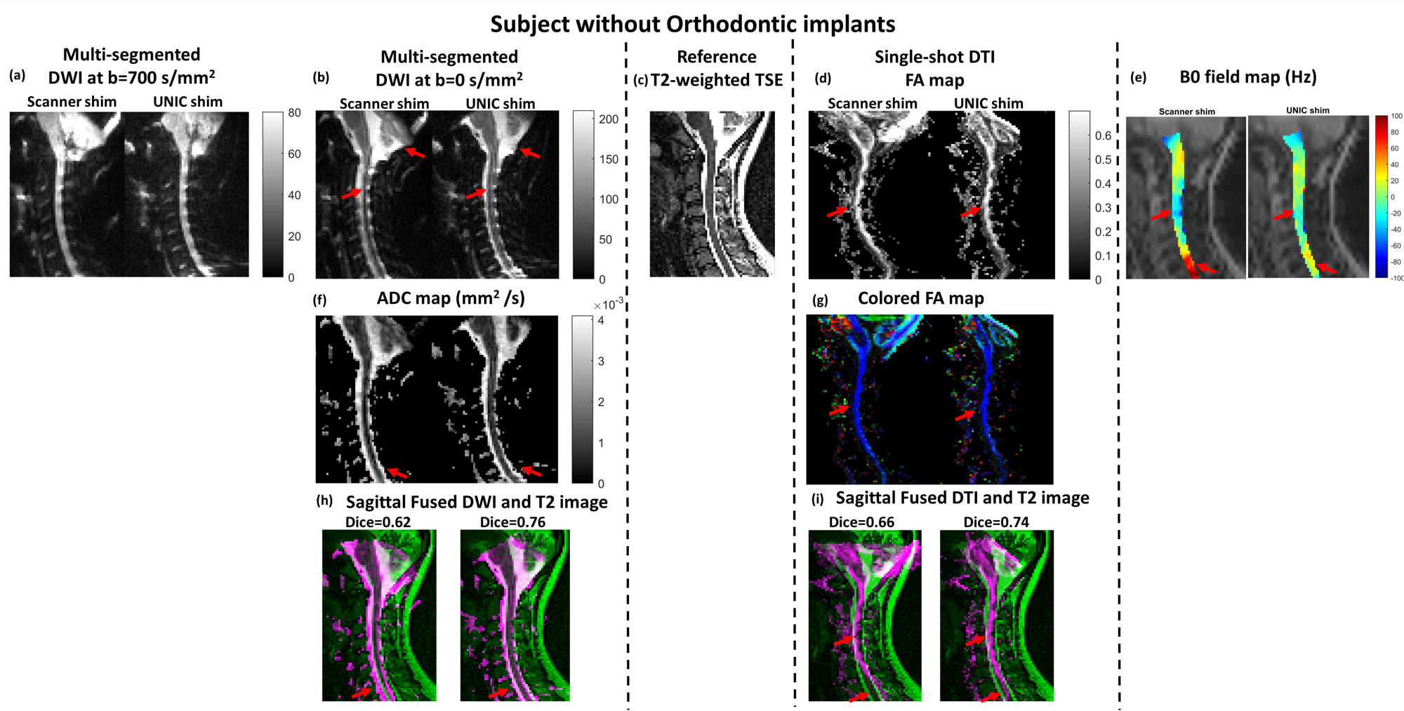
Figure3: Comparison between UNIC and standard scanner shim for different acquisition sequences in non-Orthodontal implant subjects. (a,b,f) DWI and ADC image with distortion correction by UNIC, (h) fused DWI and T2 images shows voxel gained after UNIC shimming, (c) T2-w TSE (d,g) DTI FA map and colored FA image where blue color on c-spine region indicate fiber tract oriented head to foot, (h) fused DTI and T2 images shows voxel gained after UNIC shimming, B0 fieldmap in c-spine region. Red arrow mark shows improvement in image quality or field correction with UNIC shimming.
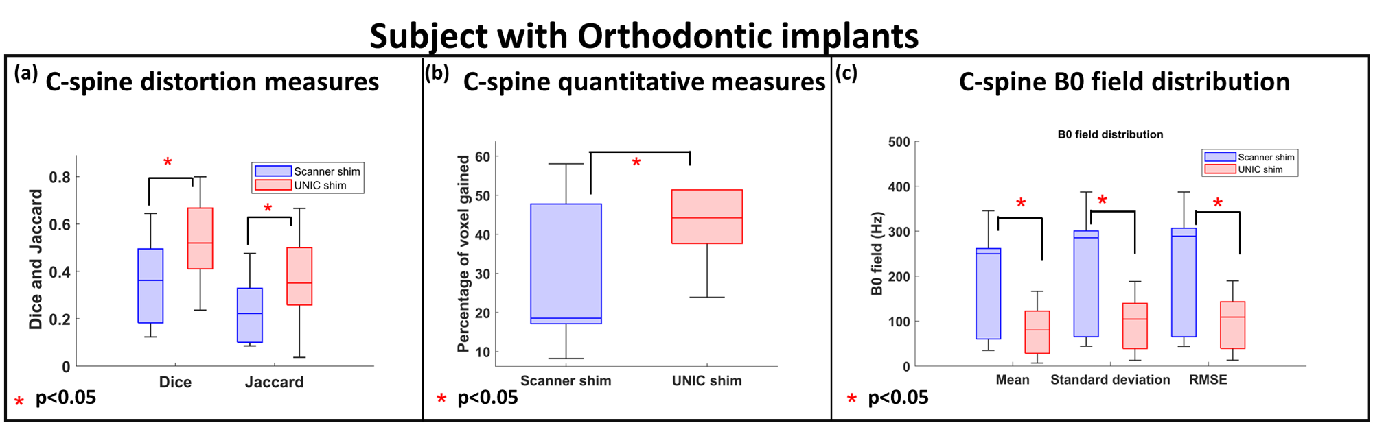
Figure4: Quantitative analysis of DWI distortion and percentage voxel gain with B0 field correction by UNIC in subjects with Orthodontal implant subjects.
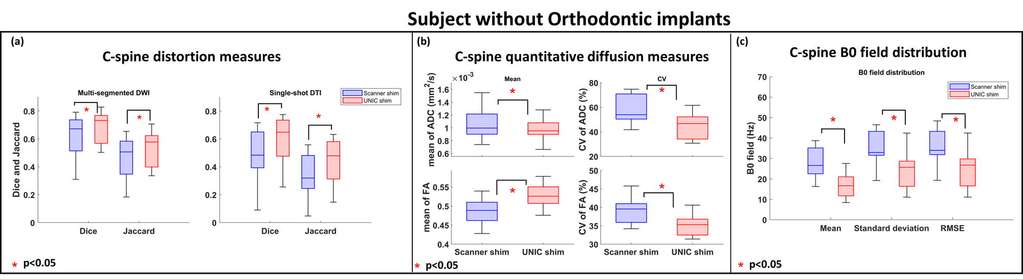
Figure5: Quantitative analysis of DWI and DTI distortion and percentage voxel gain with B0 field correction by UNIC in subjects with non-Orthodontal implant subjects.