1059
Personalized Lumbosacral Spinal Nerve Roots 3D Reconstruction and Computer Simulation for Spinal Cord Stimulation1Institute of Science and Technology for Brain-Inspired Intelligence, Fudan University, Shanghai, China, 2School of Information Science and Technology, Fudan University, Shanghai, China, 3MR Research Collaboration Team, Siemens Healthineers Ltd., Shanghai, China
Synopsis
Keywords: Spinal Cord, Spinal Cord, Spinal Cord Stimulation, Nerve Roots, 3D reconstrcution
Motivation: Spinal cord stimulation requires personalized 3D reconstructed model to address challenges arising from individual variability.
Goal(s): This study aims to image the entire spinal cord with lumbosacral nerve roots and propose a method for reconstructing a 3D model for simulation.
Approach: Three MRI sequences were collected for reconstruction and nerve roots annotation. Then the 3D spinal cord model was reconstructed using a custom interpolating algorithm guided by anatomical rules.
Results: A tailored imaging protocol and reconstruction method designed for the spinal cord and its nerve roots have been proposed, resulting in a 3D personalized spinal cord model.
Impact: The protocol offers an effective guide for researchers and clinicians conducting spinal cord and nerve roots imaging. Additionally, it enables the creation of personalized computational spine models with nerve roots and rootlets from MRI images, facilitating simulation studies.
Introduction
Spinal cord stimulation is a rapidly advancing medical technology with significant potential for treating conditions like pain, spasticity, and paralysis1. However, the invasive nature of stimulators poses challenges in clinical trials and parameter optimization. Therefore, there is a critical need for convenient and effective simulation tools to aid in designing implantation strategies and stimulation parameters2. Additionally, generic spinal cord models are insufficient due to high inter-subject variability3, emphasizing the importance of developing imaging-based personalized modeling approaches. Such approaches are crucial for applications like electric field simulation, neural modeling, and surgical planning.Method
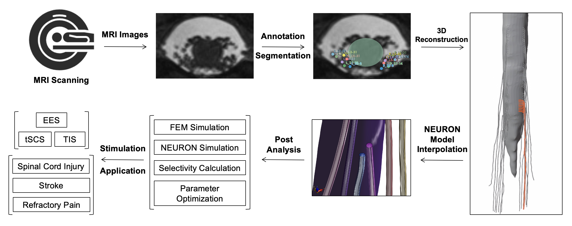
Acquisition: The spinal cords of six healthy volunteers were scanned using a 3T whole-body MRI system(MAGNETOM Prisma, Siemens Healthineers, Erlangen, Germany) quipped with a 20-channel head coil, an 18-channel body coil, and a spine coil. Three MRI sequences were employed to locate various spinal structures. Whole spine T2-weighted TSE images were obtained to outline the spine (TR/TE=4320/104ms, voxel size=0.6*0.6*3.0mm). Double echo steady state sequence (DESS) was used to identify ganglions of each vertebral segment, enabling the determination of nerve root exit points from the spinal canal (TR/TE=11.59/4.24ms, voxel size=0.6*0.6*0.6mm). 3D high-resolution CISS (Constructive Interference in Steady State) images were acquired to identify each spinal nerve root in transverse view (TR/TE=9.80/4.46ms, voxel size=0.4*0.4*1.8mm).Data annotation: The data annotation process has been meticulously designed by an expert spine surgeon, adhering closely to physiological principles. Each MRI sequence was independently annotated by two skilled annotators. In cases where there were notable discrepancies in the annotation results, thorough discussions were conducted to reach a consensus. The final annotation results were obtained by averaging the annotations from the two annotators.
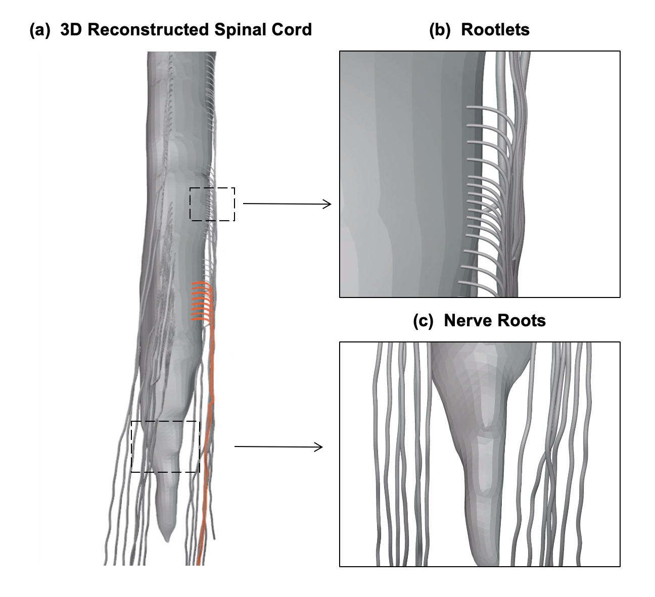
3D reconstruction: The spinal cord segmentation was processed using Spinal Cord Toolbox (SCT)4. The segments of the spinal cord were determined after identifying the entry and exit points of the nerve roots into the cord. The number and interval of root filaments within the segments were determined to generate attachment points in accordance with anatomical conventions5–9.These points were sequentially linked as rootlets to the main root via a uniform interpolation method based on our custom algorithm.
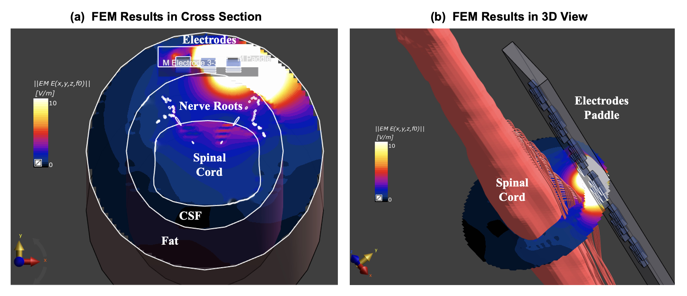
Post-analysis: The reconstructed 3D model can be imported into finite element simulation platforms or coupled with computational neural models for physical field and neural recruitment analysis. For instance, throughout our study, we undertook a finite element electric field simulation for epidural spinal cord stimulation. The electric field propagated by the electrode was computed with the quasi-static solver in Sim4Life (Zurich Med Tech, Zurich, Switzerland) at 1 kHz with an automatically sized grid.
Results
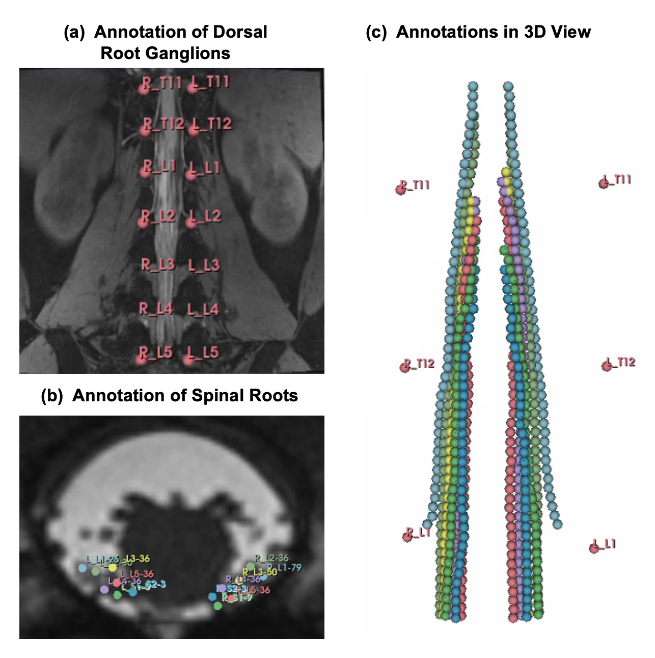
Imaging and protocol: Figure 1a presents the composited T2 TSE image, providing a comprehensive view of the overall spinal cord structure. The DESS sequence enhances nerve root imaging, enabling clear visualization of their trajectories within the vertebral canal(Figure 1b). The CISS sequence, optimized for both signal-to-noise ratio and experimental purposes, offers detailed cross-sectional views (Figure 1c).Annotation and construction results: Figure 2 provides detailed annotations for exit points, root trajectories, and rootlet entry points (Figures 2a, 2b, and 2c, respectively). The reconstructed model, generated using a custom algorithm, is presented in Figure 3. The complete method pipeline is illustrated in Figure 4.
FEM results: Figure 4a depicts a slice view of the simulated electric field distribution in Sim4Life, targeting region where rootlets enter the spinal cord. In addition, Figure 4b presents a 3D visualization of the distribution.
Discussions
The lower activation threshold of the dorsal root10 compared to the spinal cord's white matter has led to innovative electrical spinal cord stimulation methods. Personalized spinal cord models, including nerve root structures, enhance precision in established techniques like epidural11 and transcutaneous spinal cord stimulation12, as well as emerging methods. These advancements hold promise for treating spinal cord injuries, refractory pain, and stroke, improving stimulation selectivity and treatment outcomes.This study presents a method for reconstructing a 3D model of spinal cord with lumbosacral nerve roots from a customized imaging protocol, supporting precise and personalized neurostimulation therapies. Despite previous challenges in nerve root imaging, our openly shared acquisition protocol, validated on six participants, ensures reproducible results.Conclusion
We proposed a tailored MR imaging protocol for the reconstruction of a 3D spinal cord model and its nerve roots. The acquired spinal cord MRI images clearly depict the spinal cord structure and the trajectories of nerve roots within the vertebral canal. Utilizing our custom reconstruction method, the resulting spinal cord model accurately represents the spatial distribution of the nerve roots, enabling its applicability in simulation studies.Acknowledgements
Thanks for the volunteers and the support of Zhangjiang Brain Image Center.References
- Krames, E. S., Hunter Peckham, P., Rezai, A. & Aboelsaad, F. Chapter 1 - What Is Neuromodulation? in Neuromodulation (eds. Krames, E. S., Peckham, P. H. & Rezai, A. R.) 3–8 (Academic Press, 2009). doi:10.1016/B978-0-12-374248-3.00002-1.
- Liang, L. et al. A systematic review of computational models for the design of spinal cord stimulation therapies: from neural circuits to patient‐specific simulations. The Journal of Physiology JP282884 (2022) doi:10.1113/JP282884.
- Canbay, S. et al. Anatomical relationship and positions of the lumbar and sacral segments of the spinal cord according to the vertebral bodies and the spinal roots: Anatomical Relations of the Lumbosacral Spine. Clin. Anat. 27, 227–233 (2014).
- De Leener, B. et al. SCT: Spinal Cord Toolbox, an open-source software for processing spinal cord MRI data. NeuroImage 145, 24–43 (2017).
- Mendez, A. et al. Segment-Specific Orientation of the Dorsal and Ventral Roots for Precise Therapeutic Targeting of Human Spinal Cord. Mayo Clinic Proceedings 96, 1426–1437 (2021).
- ERIC J.WALL. Cauda Equina Anatomy. SPINE 15, (1990).
- Hauck, E. F., Wittkowski, W. & Bothe, H. W. Intradural microanatomy of the nerve roots S1–S5 at their origin from the conus medullaris: Laboratory investigation. SPI 9, 207–212 (2008).
- Arslan, M. et al. Lumbosacral intrathecal nerve roots: an anatomical study. Acta Neurochir 153, 1435–1442 (2011).
- Zhou, M.-W. et al. Microsurgical Anatomy of Lumbosacral Nerve Rootlets for Highly Selective Rhizotomy in Chronic Spinal Cord Injury. Anat Rec 293, 2123–2128 (2010).
- Capogrosso, M. et al. A Computational Model for Epidural Electrical Stimulation of Spinal Sensorimotor Circuits. Journal of Neuroscience 33, 19326–19340 (2013).
- Rowald, A. et al. Activity-dependent spinal cord neuromodulation rapidly restores trunk and leg motor functions after complete paralysis. Nat Med 28, 260–271 (2022).
- Nagel, S. J., Lempka, S. F. & Machado, A. G. Percutaneous Spinal Cord Stimulation for Chronic Pain: Indications and Patient Selection. Neurosurgery Clinics of North America 25, 723–733 (2014).
Figures
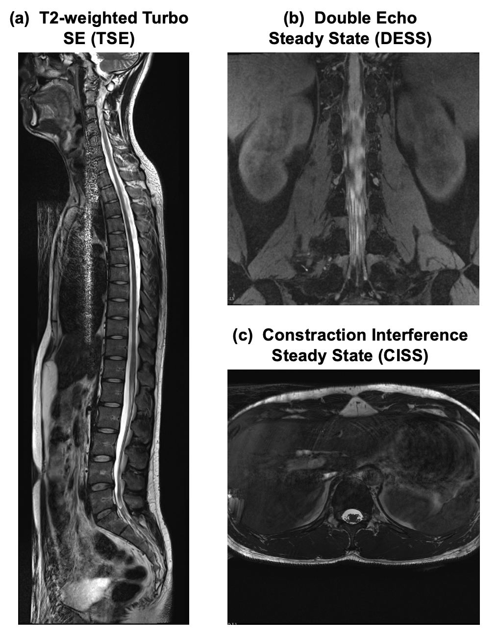
Figure 1: Imaging results of three MRI sequence base on the proposed protocol, taking one of the volunteers as an example. (1a) The composited T2 tse image, which provides view of the overall spinal cord structure. (1b) The DESS sequence, which enhances the imaging of nerve roots, allowing for a visualization of the nerve root trajectories in the peripheral vertebral canal, particularly at where the nerve roots exits(intervertebral foramen). (1c) The CISS sequence offers a relatively clear depiction of the position information of different nerve roots within a single cross-section.