1054
COVID-19-related anosmia is driven by inflammation and myelin alterations as shown by Voxel Based Analysis1Department of Brain & Behavioral Sciences, University of Pavia, Pavia, Italy, 2Digital Neuroscience Centre, IRCCS Mondino Foundation, Pavia, Italy, 3NMR Research Unit, Queen Square MS Centre, Department of Neuroinflammation, UCL Queen Square Institute of Neurology, Faculty of Brain Sciences, University College London, London, United Kingdom, 4Radiomics Group, Vall d’Hebron Institute of Oncology, Vall d’Hebron Barcelona Hospital Campus, Barcelona, Spain, 5Department of Medical Physics and Biomedical Engineering, Centre for Medical Image Computing (CMIC), University College London, London, United Kingdom, 6E-Health Center, Universitat Oberta de Catalunya, Barcelona, Spain, 7Centre for Obesity Research, Department of Medicine, University College London, London, United Kingdom, 8National Institute of Health Research, UCLH Biomedical Research Centre, London, United Kingdom
Synopsis
Keywords: White Matter, COVID-19, Neuroinflammation, Voxel Based Analysis, g-ratio
Motivation: Voxel Based Analysis (VBA) can be a powerful tool to detect localized alterations.
Goal(s): Here VBA was used to understand the underpinnings of COVID-19-related anosmia.
Approach: Quantitative magnetization transfer and diffusion-weighted imaging derived maps were used to detect pathological changes affecting white matter structures.
Results: Microstructural differences were detected between healthy controls and subjects experiencing anosmia or those who recovered from it. Results highlighted the presence of widespread inflammation in persistent anosmia subjects, with myelin damage and possible repair in those who recovered. Myelin alterations involved the olfactory circuit, as well as other brain regions, providing insights into possible mechanisms of COVID-19-related anosmia.
Impact: Voxel Based Analysis is a powerful tool to highlight local tissue disruption linked to neuroinflammatory processes. Here VBA provided an insight into microstructure and myelin changes associated to COVID-19-related persistent or recovered anosmia symptoms.
Introduction
In the wake of the global COVID-19 pandemic, the loss of smell, known as anosmia, has emerged as a common symptom of SARS-CoV-2 infection1. Nonetheless, anosmia related brain alterations are still unknown.Neurite Orientation Dispersion and Density Imaging (NODDI) model is widely used to obtain information about microstructure alterations2 while quantitative magnetization transfer (qMT) imaging3 is one of the most accurate approaches for myelin damage as shown by the high correlation between qMT metrics and myelin histological staining4. Myelin integrity can also be evaluated using the g-ratio, representing the ratio between inner and outer axon diameter5.
Here, qMT, NODDI and g-ratio maps were analyzed with a voxel wise approach to detect white matter (WM) inflammation, microstructure and myelin alterations in people who experienced COVID-19-related anosmia.
Methods
Subjects and MRI acquisitionHealthy controls (HC) and four groups of people who recovered from SARS-COV-2 infection were recruited: people who had COVID-19 (COVID), people with persistent anosmia (COVIDP), people who recovered from anosmia (COVIDR) and young adults who also experienced and recovered from anosmia (COVIDY). All subjects underwent a MRI protocol using a Philips Ingenia 3T scanner. Demographic and MRI details are summarized in Figure1.
Quantitative maps and g-ratio calculation
Diffusion weighted (DW) and qMT images were preprocessed to reduce noise and correct for distortions and Gibb’s artefacts. qMT scans were analyzed to obtain bound pool fraction (BPF) and T2b maps6 while DW data were fitted with NODDI to obtain the isotropic volume fraction (viso) and intra-cellular volume fraction (vintra) maps7. Then, Myelin Volume Fraction (MVF) and Fiber Volume Fraction (FVF)5 were calculated as follows: $$MVF=k\times{BPF}$$ where k = 3.4 calculated according to8, and $$$FVF= MVF+(1-MVF)\times{(1-v_{iso} )}\times{v_{intra}}$$$.
These MRI-derived metrics were finally used to calculate g-ratio maps in vivo. $$g-ratio = \sqrt{1-\frac{MVF}{FVF}}$$
Voxel based analysis (VBA)
The pipeline is shown in Figure2. Previously obtained quantitative maps were registered to MNI152 standard space, and smoothed with a 3mm Gaussian kernel. For statistical analysis, FSL randomise was used9. A two tailed t-test (covariates: age; gender) was performed between groups, using threshold-free cluster enhancement (TFCE)10 and multiple comparison correction. Statistical threshold was set at p-value=0.05 with a 10 voxels cluster extent.
Then, the extent of alterations per WM tract was quantified by calculating the percentage of statistically significant voxels in WM tracts extracted from the JHU-atlas11.
Results
WM quantitative maps for a randomly chosen HC are shown in Figure3, while metrics significantly altered between groups are reported in Figure4. BPF was increased in COVIDR with respect to HC, and g-ratio was reduced in recovered patients (COVIDR + COVIDY) compared to HC. Moreover, comparing COVIDP with COVIDR, g-ratio, viso and T2b were increased, while BPF was reduced. The percentage of significant voxels within each tract demonstrated widespread alterations beyond the olfactory circuit (Table1).Discussion and Conclusion
The main finding of the present work is that VBA identifies specific changes, spreading beyond the olfactory circuit, in people who experienced COVID-19-related anosmia identifying the presence of different mechanisms12,13. For simplicity of discussion, only tracts presenting alterations greater than 20% of their volume were considered and categorized into circuits based on their involvement in possible functions as shown in Table1.Compared to HC, patients that recovered from anosmia (either COVIDR and COVIDY) present increased BPF and reduced g-ratio suggesting possible ongoing remyelination and increased myelin thickness given that there are no differences in vintra, implying preserved axonal integrity.
Instead, COVIDP presents increased viso , g-ratio and T2b, and decreased BPF with respect to COVIDR suggesting inflammation and generalized WM damage with potential decreased myelin volume14,15. In contrast, changes in the brain of people who recovered from anosmia highlight that neuroinflammation may be promoting remyelination15.
Interestingly, alterations within WM tracts extend beyond the olfactory circuit: the noticeable involvement of the limbic circuit may depend on the strong correlation between odors and emotional processes, including memory, attention, and emotional regulation16. Variations in cerebellar tracts align with the cerebellar role in olfactory function, particularly in relation to sniff adjustment 17,while the alteration of motor tract metrics could imply the involvement of the voluntary motor system in respiratory control - questionably a compensatory response to anosmia - which is further supported by the inclusion of the brainstem18,19. The involvement of visual and somatosensory circuit suggests possible compensatory mechanisms after sensory loss20.
In conclusion, this study demonstrates that VBA highlights myelin related alterations persistent beyond neuroinflammation after SARS-COV2 infection, which should be studied longitudinally to unveil potential links between changes21.
Acknowledgements
EL is a PhD student enrolled in the National PhD in Artificial Intelligence, XXXVIII cycle, course on Health and life sciences, organized by Università Campus Bio-Medico di Roma. EG receives funding from TDC Technology Dedicated to Care. FG receives the support of a fellowship from ”la Caixa” Foundation (ID 100010434). The fellowship code is “LCF/BQ/PR22/11920010”. FP received a Guarantors of Brain fellowship 2017–2020. FP is supported by the National Institute for Health Research (NIHR), the Biomedical Research Centre initiative at University College London Hospitals (UCLH). RS receives funding from the BRC (BRC1130/HEI/RS/11041). H2020 Research and Innovation Action Grants Human Brain Project 785907 and 945539 (SGA2 and SGA3) to ED'A and FP. Moreover, the project was supported by the MNL Project “Local Neuronal Microcircuits” of the Centro Fermi (Rome, Italy) to ED'A. This work was also supported by #NEXTGENERATIONEU (NGEU) and funded by the Ministry of University and Research (MUR), National Recovery and Resilience Plan (NRRP), project MNESYS (PE0000006) - A Multiscale integrated approach to the study of the nervous system in health and disease (DN. 1553 11.10.2022). CGWK receives funding from Horizon2020 (Research and Innovation Action Grants Human Brain Project 945539 (SGA3)), BRC (#BRC704/CAP/CGW), MRC (#MR/S026088/1), Ataxia UK, Rosetrees Trust (#PGL22/100041 and #PGL21/10079). CGWK is a shareholder in Queen Square Analytics Ltd.
References
1. Lechien, J. R. et al. Olfactory and gustatory dysfunctions as a clinical presentation of mild-to-moderate forms of the coronavirus disease (COVID-19): a multicenter European study. European Archives of Oto-Rhino-Laryngology 277, 2251–2261 (2020).
2. Zhang, H., Schneider, T., Wheeler-Kingshott, C. A. M. & Alexander, D. C. NODDI: practical in vivo neurite orientation dispersion and density imaging of the human brain. Neuroimage 61, 1000–16 (2012).
3. Wolff, S. D. & S, B. R. "Magnetization transfer contrast (MTC) and tissue water proton relaxation in vivo. Magn Reson Med 10, 135–144 (1989).
4. Mancini, M. et al. An interactive meta-analysis of MRI biomarkers of Myelin. Elife 9, 1–23 (2020).
5. Stikov, N. et al. In vivo histology of the myelin g-ratio with magnetic resonance imaging. Neuroimage 118, 397–405 (2015).
6. Battiston, M. et al. Fast bound pool fraction mapping via steady-state magnetization transfer saturation using single-shot EPI. Magn Reson Med 82, 1025–1040 (2019).
7. Fick, R. H. J., Wassermann, D. & Deriche, R. The Dmipy Toolbox: Diffusion MRI Multi-Compartment Modeling and Microstructure Recovery Made Easy. Front Neuroinform 13, (2019).
8. Cercignani, M. et al. Characterizing axonal myelination within the healthy population: a tract-by-tract mapping of effects of age and gender on the fiber g-ratio. Neurobiol Aging 49, 109–118 (2017).
9. Winkler, A. M., Ridgway, G. R., Webster, M. A., Smith, S. M. & Nichols, T. E. Permutation inference for the general linear model. Neuroimage 92, 381–397 (2014).
10. Smith, S. M. & Nichols, T. E. Threshold-free cluster enhancement: Addressing problems of smoothing, threshold dependence and localisation in cluster inference. Neuroimage 44, 83–98 (2009).
11. Mori, S., Wakana, S., Nagae-Poetscher, L. M. & van Zijl, P. C. M. MRI Atlas of Human White Matter. AJNR Am J Neuroradiol 26, 1384–1385 (2006).
12. HCHM Philippens, I. et al. SARS-CoV-2 causes brain inflammation and induces Lewy body formation in macaques 1 2. doi:10.1101/2021.02.23.432474.
13. Yang, A. C. et al. Dysregulation of brain and choroid plexus cell types in severe COVID-19. Nature 595, 565–571 (2021).
14. Glass, C. K., Saijo, K., Winner, B., Marchetto, M. C. & Gage, F. H. Mechanisms Underlying Inflammation in Neurodegeneration. Cell vol. 140 918–934 Preprint at https://doi.org/10.1016/j.cell.2010.02.016 (2010).
15. Yong, H. Y. F., Rawji, K. S., Ghorbani, S., Xue, M. & Yong, V. W. The benefits of neuroinflammation for the repair of the injured central nervous system. Cellular and Molecular Immunology vol. 16 540–546 Preprint at https://doi.org/10.1038/s41423-019-0223-3 (2019).
16. Poellinger, A. et al. Activation and habituation in olfaction - An fMRI study. Neuroimage 13, 547–560 (2001).
17. Zhang, Z. hao et al. Cerebellar involvement in olfaction: An fMRI Study. Journal of Neuroimaging 31, 517–523 (2021).
18. Ciumas, C., Rheims, S. & Ryvlin, P. fMRI studies evaluating central respiratory control in humans. Frontiers in Neural Circuits vol. 16 Preprint at https://doi.org/10.3389/fncir.2022.982963 (2022).
19. Mazzatenta, A., Maffei, M., Di Giulio, C. & Neri, G. COVID-19 Smell Impairment and Crosstalk with Hypoxia Physiology. Life 12, (2022).
20. Iravani, B. et al. Acquired olfactory loss alters functional connectivity and morphology. Sci Rep 11, (2021).
21. Butowt, R. & von Bartheld, C. S. Anosmia in COVID-19: Underlying Mechanisms and Assessment of an Olfactory Route to Brain Infection. Neuroscientist 27, 582–603 (2021).
Figures
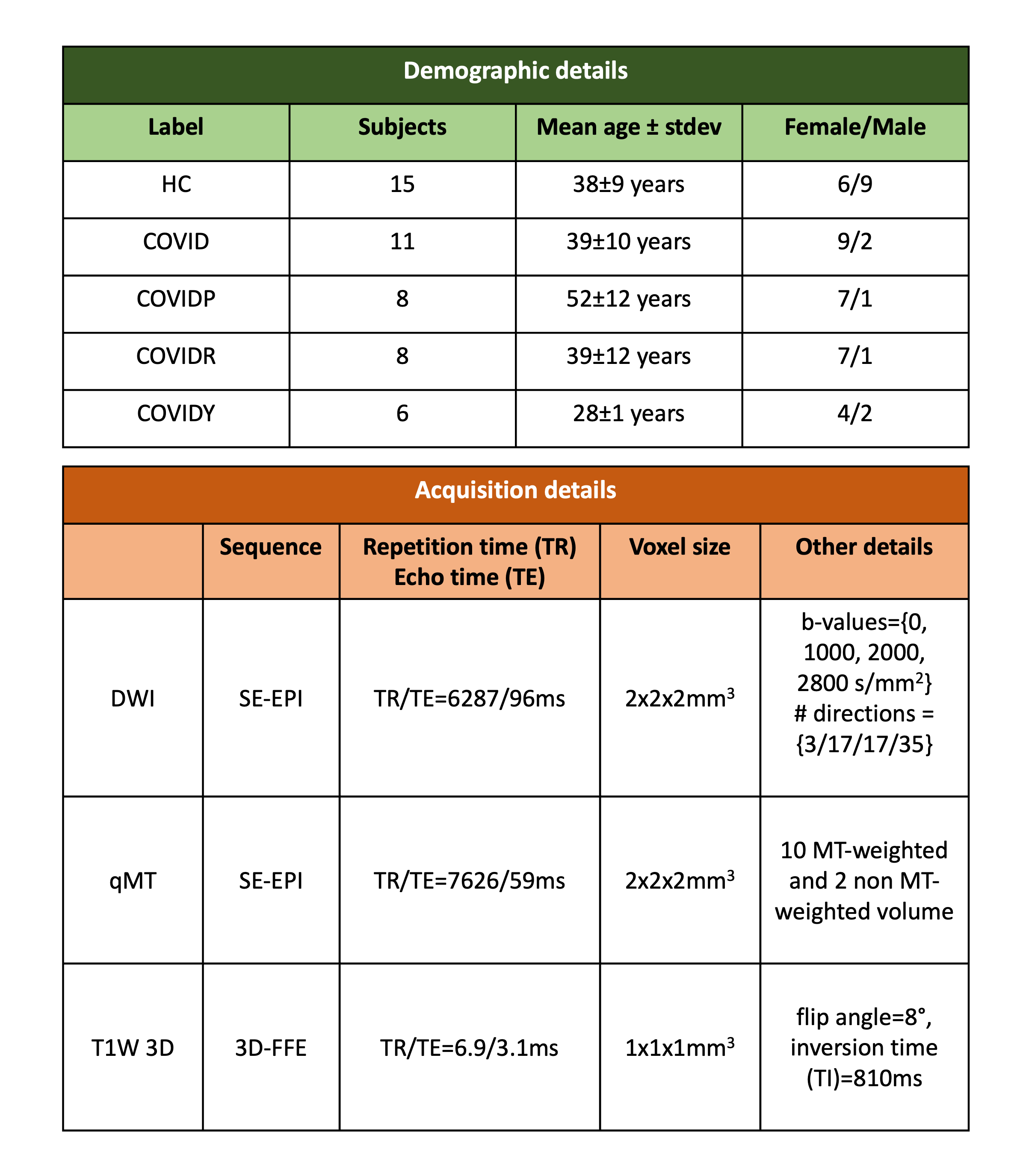
Figure1:
The first table shows the number of subjects in each group, with their age and gender distribution. HC = healthy controls, COVID = people who recovered from COVID-19, COVIDP = people with COVID-19-related persistent anosmia, COVIDR = people who recovered from COVID-19-related anosmia, COVIDY = young adults who recovered from anosmia.
The second table shows the MRI acquisition protocol. DWI = diffusion-weighted imaging; qMT = quantitative magnetization transfer; T1W = T1 weighted.
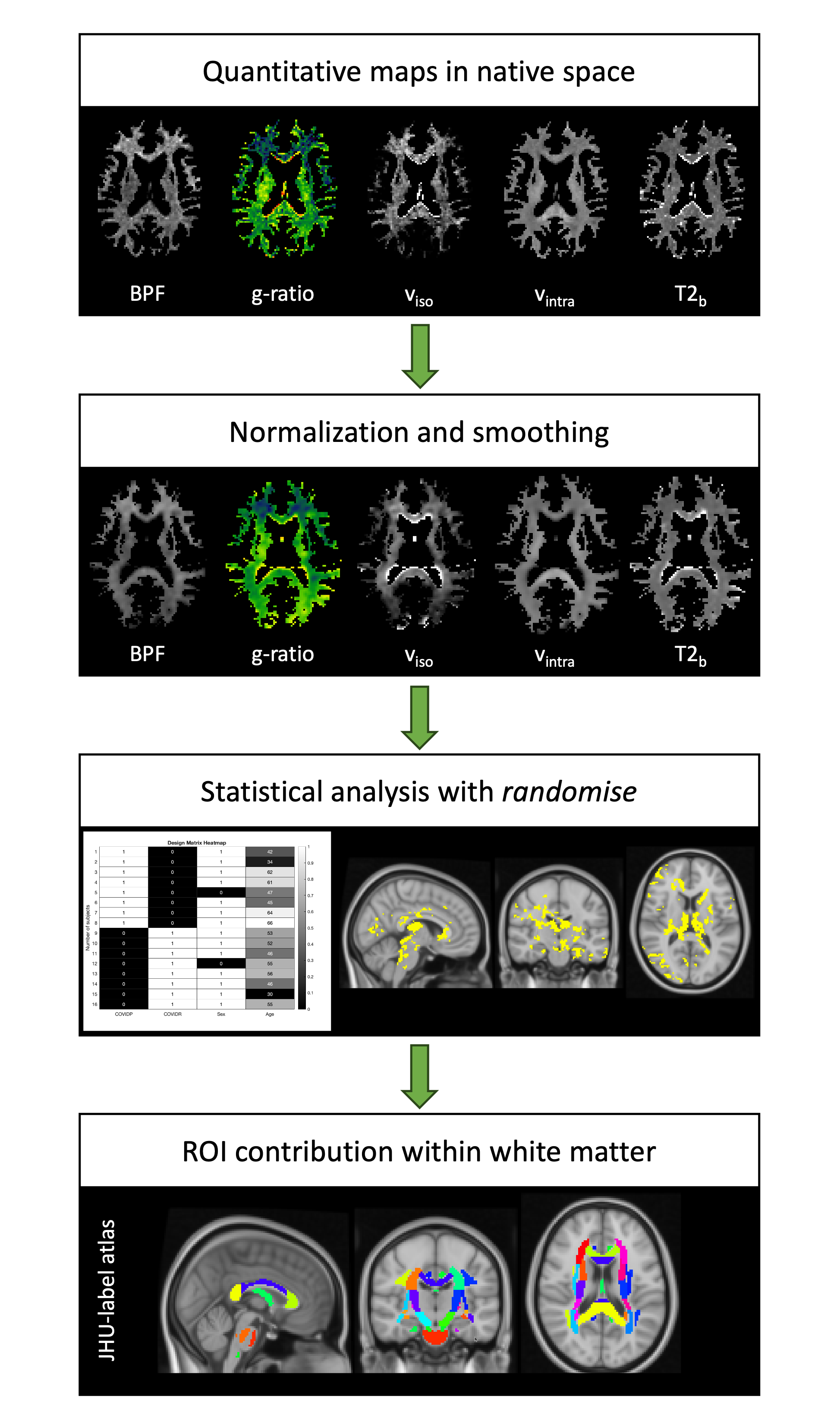
Figure2: Voxel based analysis (VBA) pipeline. From top to bottom: quantitative maps in white matter (WM) for a randomly chosen healthy control (HC); normalized maps in MNI space; design matrix to compare pair of groups with age and gender as covariates (left), and the resulting statistical maps at p-value<0.05 (right); tracts definition using the JHU-atlas. ROI = Regions Of Interest.
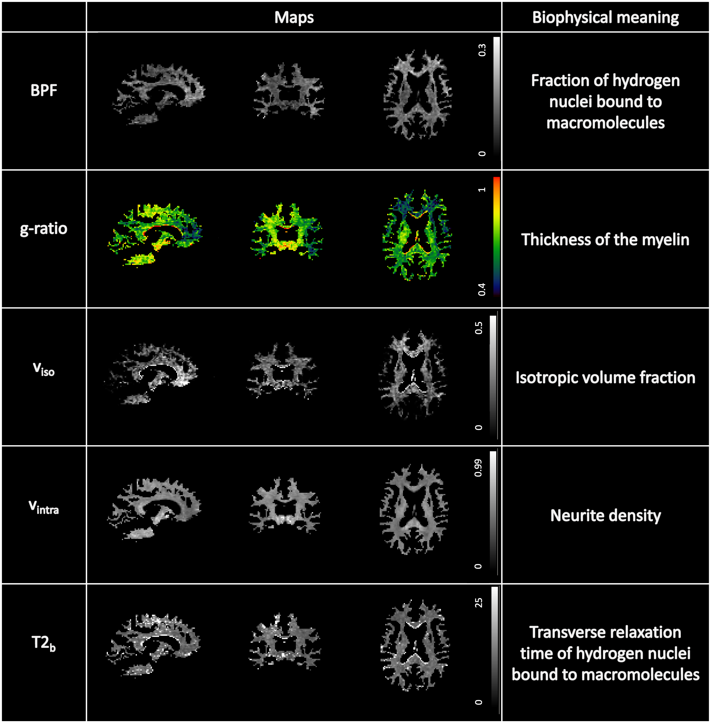
Figure3: White matter quantitative maps for a randomly chosen subject and their biophysical meaning. Metrics considered are: Bound Pool Fraction (BPF), g-ratio, isotropic volume fraction (viso), intra-cellular volume fraction (vintra) and T2b.
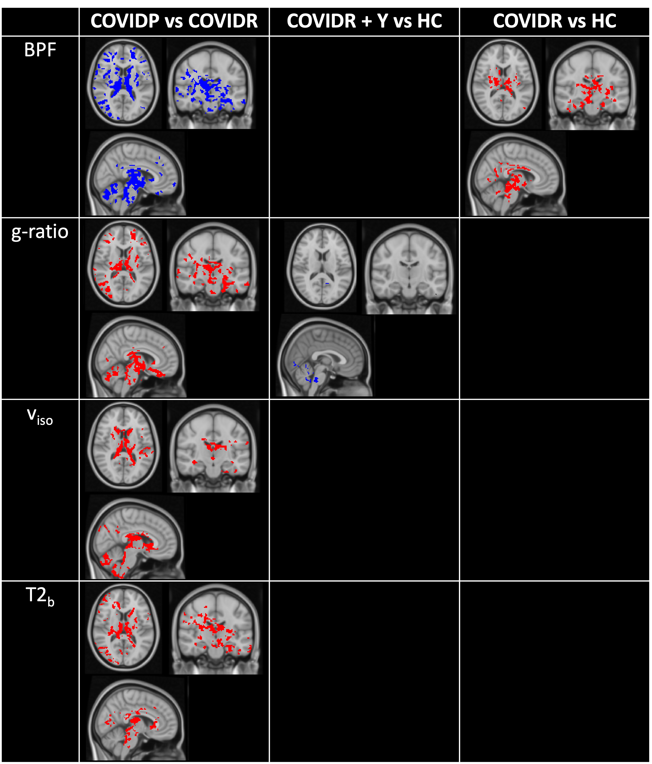
Figure4: Voxel wise significant alterations emerging from some group comparisons, overlaid on the MNI template. BPF = Bound Pool Fraction, viso = isotropic volume fraction. Red represents higher values in the first group compared to the second, while blue represents lower values in the first group compared to the second. HC = healthy controls, COVID = people who had recovered from COVID-19, COVIDP = people with COVID-19-related persistent anosmia, COVIDR = people recovered from COVID-19-related anosmia, COVIDY = young adults who also recovered from anosmia.
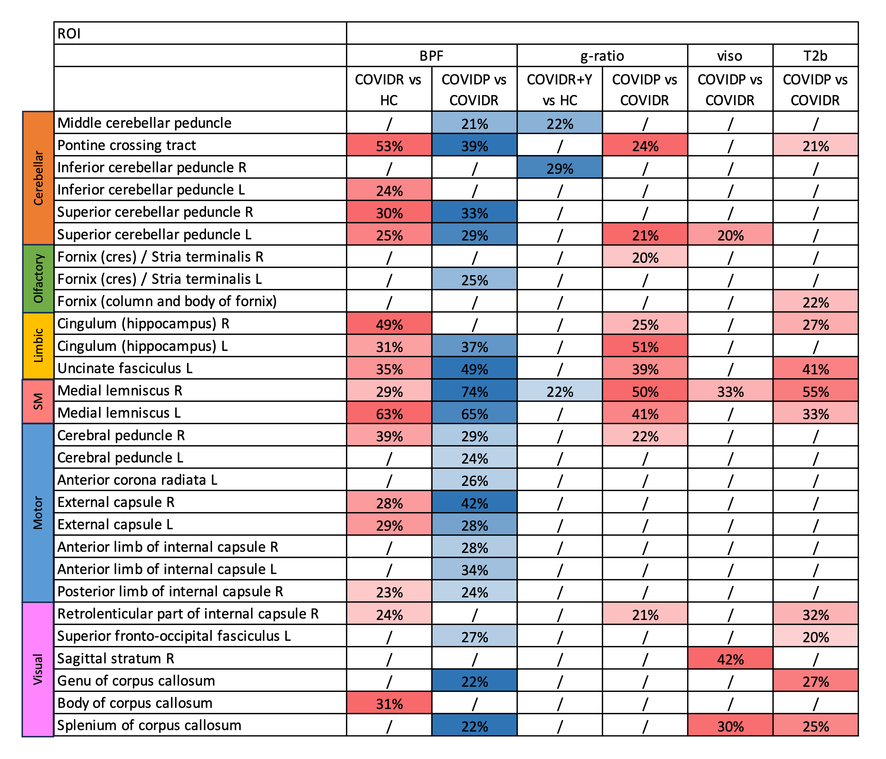
Table1: White matter tract showing a percentage of significant altered voxels, emerging from voxel based group comparisons, divided into circuits. SM = somatosensory. BPF = Bound Pool Fraction, viso = isotropic volume fraction. Red represents higher values in the first group compared to the second, while blue represents the opposite. HC = healthy controls, COVID = people who recovered from COVID-19, COVIDP = people with COVID-19-related persistent anosmia, COVIDR = people who recovered from COVID-19-related anosmia, COVIDY = young adults who recovered from anosmia.