1049
Association of Iron Deposition in Early-Stage Multiple Sclerosis Lesion with Remyelination Capacity: chi-separation imaging study1Radiology, Johns Hopkins University, Baltimore, MD, United States, 2Kennedy Krieger Institute, Baltimore, MD, United States, 3Neurology, Seoul St. Mary's Hospital, Seoul, Korea, Republic of, 4Siemens Healthineers, Seoul, Korea, Republic of, 5Mathematics and Statistics, University of South Florida, Tampa, FL, United States, 6Biomedical Engineering, Hankuk University of Foreign Studies, Yongin, Korea, Republic of, 7Electrical and Computer Engineering, Seoul National University, Seoul, Korea, Republic of, 8Radiology, Seoul St. Mary's Hospital, Seoul, Korea, Republic of
Synopsis
Keywords: Multiple Sclerosis, Multiple Sclerosis, 𝜒-separation
Motivation: In multiple sclerosis (MS) lesion, factors influencing myelin dynamics and future remyelination are under active investigation.
Goal(s): To assess dynamic changes of MS lesions from their early stage and explore the factors related to their future remyelination outcomes.
Approach: Longitudinal changes of MRI phenotypes in MS lesions were assessed from their early stage, particularly focusing on longitudinal alternations in diamagnetic myelin and paramagnetic iron signals using susceptibility source-separation (chi[𝜒]-separation).
.
Results: 26 lesions show remyelination and 36 did not. The hyperintensity in paramagnetic iron signal (hyper-paramagnetic sign, HPS) at early stage of lesion development was significantly associated to future remyelination.
Impact: Iron deposition sign in early-stage MS lesion, which detected by added sensitivity of 𝜒-separation to iron and myelin, can offer potential imaging marker for the impaired remyelination capability in MS pathology.
Introduction
Multiple sclerosis (MS) is one of the most prevalent neuroinflammatory diseases of the CNS.1 The characteristic of MS is focal demyelination in the CNS white matter, which can be followed by subsequent remyelination. Hence, the alterations in myelin, encompassing demyelination and remyelination, have been recognized as a key histological feature in MS lesions correlated with the clinical course of the disease.2-4 Meanwhile, impaired iron homeostasis is another important histopathological aspect of MS lesions. Iron deposition in microglia and macrophage is associated with oxidative stress on neurons5-7 and polarization toward a pro-inflammatory profile.5-7 Therefore, studying brain iron and its clinical aspects is a promising approach to elucidate iron-related pathology in MS.5,7Recently, magnetic susceptibility source separation (𝜒-separation or chi-separation) imaging8 has been proposed, which is a novel susceptibility-based MRI method that allows separate quantification of diamagnetic (𝜒dia) and paramagnetic (𝜒para) susceptibility. As the susceptibility in the brain is predominantly determined by iron and myelin,9 𝜒-separation imaging technique has provided valuable information on alternations in the two substrates in MS lesions.8,10 In this study, we aimed to investigate longitudinal changes of myelin and iron in MS lesions by leveraging the increased specificity of 𝜒-separation imaging to both substrates and evaluate the clinical and radiological markers that influence future remyelination. In particular, we hypothesized that early-stage iron deposition in MS lesions could be correlated with their impaired remyelination capacity.
Methods
To explore MS lesion dynamics of myelin and iron from their early stage of lesion development, we identified and included lesions satisfying the following criteria for the analysis: 38 newly-noted lesion during follow-ups and 24 contrast-enhancing lesions11,12 from 23 RRMS patients (33 ± 10 y/o). We used two MRI protocols at different 3T MRI systems (MAGNETOM Vida and Ingenia), including T2 FLAIR, Contrast-enhanced T1-weighted images, and multi-echo gradient echo (MEGE) and T2-mapping sequences for 𝜒-separation imaging.For 𝜒-separation, MEGE data was preprocessed to calculate R2* (= 1/T2*) and local frequency maps, and T2-weighted contrasts were used for estimating R2 (= 1/T2) map. After preprocessing of MEGE and other images, estimation of absolute values of paramagnetic susceptibility (𝜒para) and diamagnetic susceptibility (𝜒dia), relative to water susceptibility was jointly estimated using a deep learning reconstruction algorithm trained to estimate the multi-orientation 𝜒-separation maps.13
For the included lesions, the following clinical and radiologic characteristics were assessed from their detection and over the repeated MR assessment, focused on myelin and iron signal changes found on 𝜒dia and 𝜒para maps. From these observations, we investigated the clinic-radiological characteristics related to the future remyelination of MS lesions.
Results
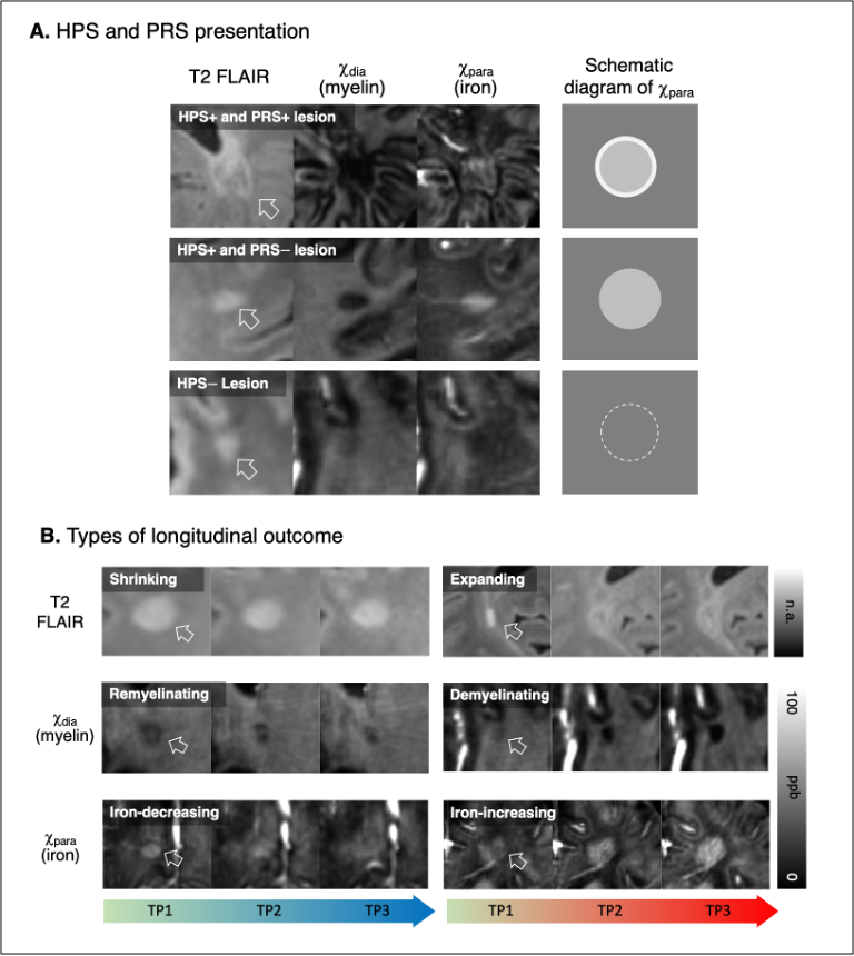
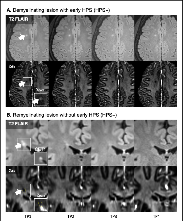
Most early-stage lesions (58/62) were myelin loss (hypointensity on 𝜒dia) at lesion detection, and less than half of them (27/62) showed hyperintense on 𝜒para (hyper-paramagnetic sign, HPS, Figure 1). Paramagnetic rim (PRS) was observed in about 17 % (11/62) of them.From 226 observations of 𝜒-separation in 62 early-stage lesions, 26 lesions showed remyelination, 14 showed stable myelin signal and 22 showed progressed myelin signal loss, suggesting progressive demyelination. HPS was much frequently observed in lesions with failed remyelination (25/36, 69%) as compared to remyelinated lesions (2/26, 8%, P<0.01, Figure 2), while the paramagnetic rim sign at lesion detection was not (2/26 vs 9/36, P=0.10). Lesion with future remyelination was significantly smaller in its detection as compared to other groups (175± 157 mm3 vs 163±110 or 425±523 mm3, P=0.02). Lesion location, contrast enhancement, disease duration was not significantly associated to the future remyelination.
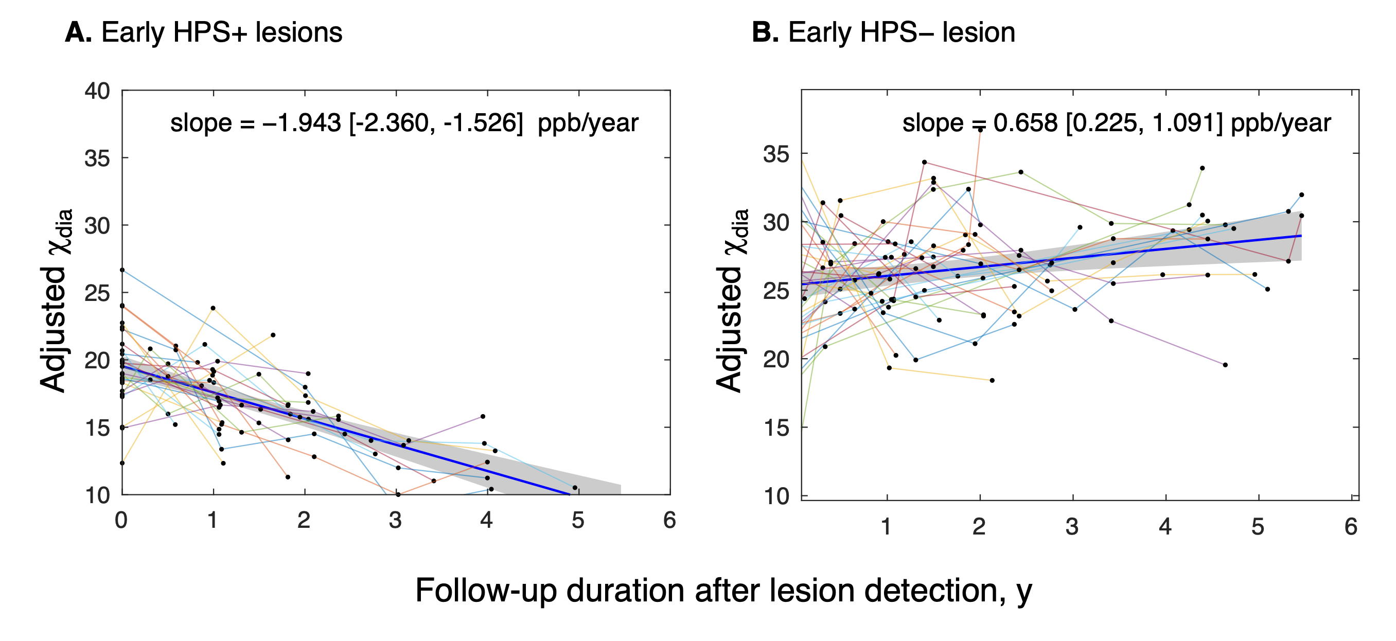
Notably, on GLMM analysis, absence of early-stage HPS (P<0.001) and EDSS at lesion detection (P=0.017) was significantly associated to future remyelination. Quantitatively, myelin (𝜒dia) signals was longitudinally decreasing (-1.953 ppb/yr, demyelinating) in HPS+ lesions, but increasing (0.668 ppb/yr, remyelinating) in HPS- lesions (Figure 3). In addition, significant interaction between presence of HPS and lesion age was observed (P<0.001).
Discussion and conclusion.
In this study, we employed an 𝜒-separation to investigate longitudinal changes of myelin and iron signals in MS lesions and their interplay, with a specific focus on the effects of early-stage iron deposition on the trajectory of lesion evolution. We successfully identified various types of longitudinal outcomes of MS lesions in myelin signal and their association with early-stage lesional iron deposition. Our results suggested that early-stage iron deposition in MS lesions could be closely associated with their impaired remyelination capacity as well as clinical relapse of MS.Therefore, early HPS may serve as a potential marker of remyelination failure and potentially indicate clinical worsening. 𝜒-separation imaging can be a promising non-invasive in-vivo imaging tool for monitoring key histopathological substrates in MS lesions, myelin and iron.
Acknowledgements
This study was supported by the National Research Foundation of Korea funded by the Korean government (MSIT) (RS-2023-00208409 and NRF-2021R1A2B5B03002783)References
1. Reich DS, Lucchinetti CF, Calabresi PA. Multiple Sclerosis. N Engl J Med 2018;378:169-180.
2. Rahmanzadeh R, Galbusera R, Lu PJ, et al. A New Advanced MRI Biomarker for Remyelinated Lesions in Multiple Sclerosis. Ann Neurol 2022;92:486-502.
3. Klotz L, Antel J, Kuhlmann T. Inflammation in multiple sclerosis: consequences for remyelination and disease progression. Nat Rev Neurol 2023;19:305-320.
4. Franklin RJM, Simons M. CNS remyelination and inflammation: From basic mechanisms to therapeutic opportunities. Neuron 2022;110:3549-3565.
5. Absinta M, Maric D, Gharagozloo M, et al. A lymphocyte-microglia-astrocyte axis in chronic active multiple sclerosis. Nature 2021;597:709-714.
6. Hametner S, Wimmer I, Haider L, et al. Iron and neurodegeneration in the multiple sclerosis brain. Ann Neurol 2013;74:848-861.
7. Absinta M, Sati P, Masuzzo F, et al. Association of Chronic Active Multiple Sclerosis Lesions With Disability In Vivo. JAMA Neurol 2019;76:1474-1483.
8. Shin HG, Lee J, Yun YH, et al. chi-separation: Magnetic susceptibility source separation toward iron and myelin mapping in the brain. Neuroimage 2021;240:118371.
9. Duyn JH, Schenck J. Contributions to magnetic susceptibility of brain tissue. NMR Biomed 2017;30.
10. Kim W, Shin HG, Lee H, et al. chi-Separation Imaging for Diagnosis of Multiple Sclerosis versus Neuromyelitis Optica Spectrum Disorder. Radiology 2023;307:e220941.
11. Chen W, Gauthier SA, Gupta A, et al. Quantitative susceptibility mapping of multiple sclerosis lesions at various ages. Radiology 2014;271:183-192.
12. Absinta M, Sati P, Schindler M, et al. Persistent 7-tesla phase rim predicts poor outcome in new multiple sclerosis patient lesions. J Clin Invest 2016;126:2597-2609.
13. Kim M, Shin, H-G., Oh, C., Jeong, H., Ji, S., An, H., Kim, J., Jang, J., Bilgic, B., Lee, J. Chi-sepnet: Susceptibility source separation using deep neural networks. Joint Annual Meeting ISMRM-ESMRMB & ISMRT 31st Annual Meeting 2022:2464.
Figures