1048
In vivo assessment of astrocyte reactivity in patients with progressive supranuclear palsy1National Institutes for Quantum Science and Technology, Chiba, Japan, 2Tokyo Medical and Dental University, Tokyo, Japan, 3Sunnybrook Research Institute, Tronto, ON, Canada, 4University of Toronto, Tronto, ON, Canada, 5Niigata University, Niigata, Japan
Synopsis
Keywords: Dementia, Neuro, magnetic resonance spectroscopy, progressive supranuclear palsy, astrocyte reactivity
Motivation: Although astrocytic pathology is a pathological hallmark of progressive supranuclear palsy (PSP), the role of astrocytes in the pathophysiology of PSP is not fully understood.
Goal(s): This study aimed to evaluate astrocyte reactivity in vivo in patients with PSP.
Approach: Astrocyte reactivity was assessed by magnetic resonance spectroscopy and plasma biomarkers, which were verified via tau-PET and histopathological analysis.
Results: Our results suggest that, in the anterior cingulate cortex, astrocyte reactivity precedes pronounced tau deposition and neurodegenerative processes and modulates brain function in PSP. Elevated myo-inositol was associated with high lactate levels, suggesting a link between reactive astrocytes and brain energy metabolism changes.
Impact: This study assessed astrocyte reactivity in vivo using magnetic resonance spectroscopy and plasma biomarkers, providing insights into the involvement of astrocytes in the pathogenesis of progressive supranuclear palsy.
INTRODUCTION
Emerging evidence highlights the association of reactive astrocytes with neurodegenerative diseases1,2. While astrocytic pathology, characterised by the presence of tufted astrocytes, is a pathological hallmark of progressive supranuclear palsy (PSP)3, the role of astrocytes in the pathophysiology of PSP is not fully understood. In this study, we aimed to assess astrocyte reactivity in vivo in patients with PSP using magnetic resonance spectroscopy (MRS) and plasma biomarkers. Furthermore, given the central role of astrocytes in brain energy metabolism and their glycolytic profile, which implies a preference for lactate production4, this study investigated alterations in brain energy metabolism by measuring brain lactate levels and examined their relationship with astrocyte reactivity.METHODS
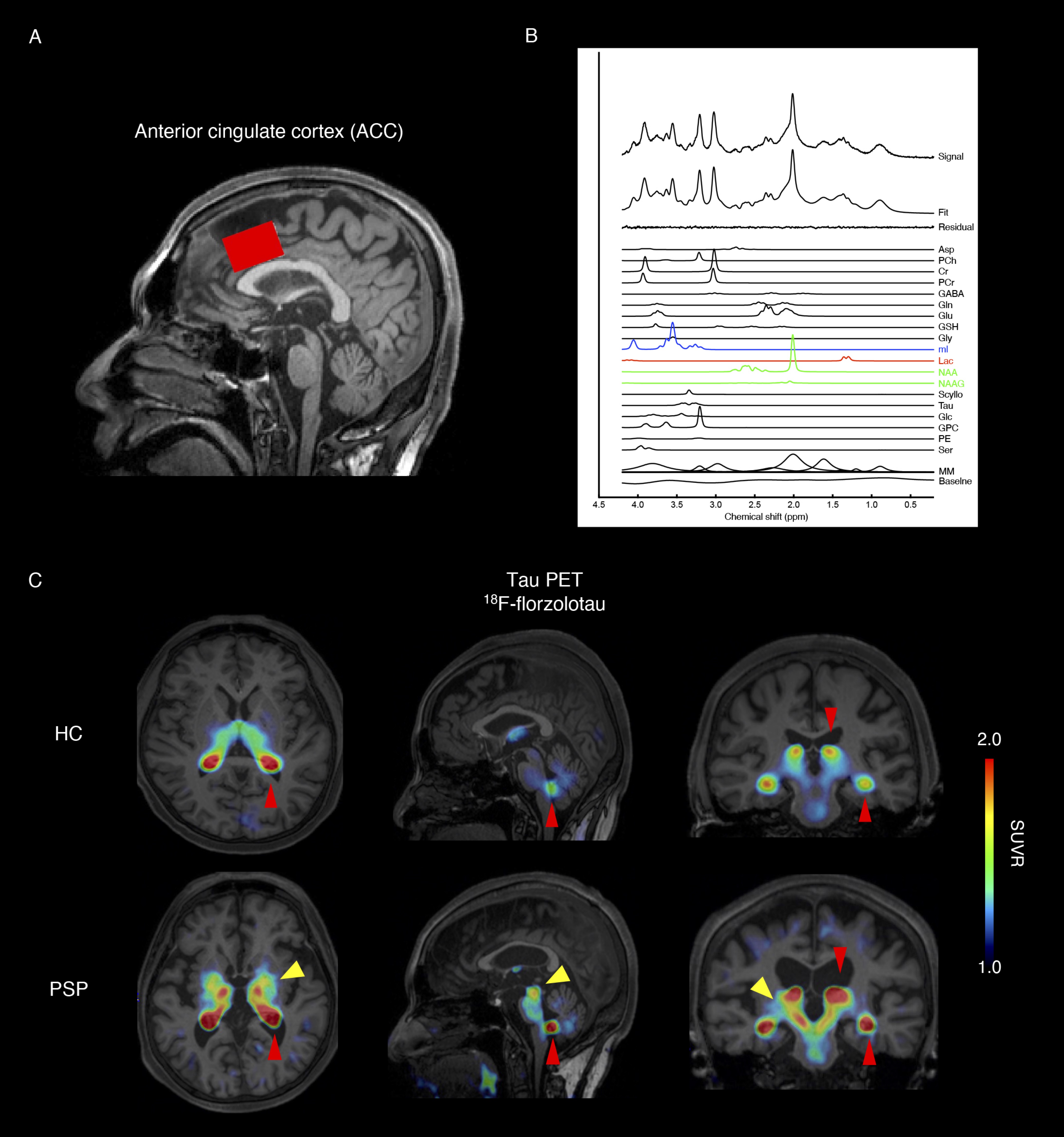
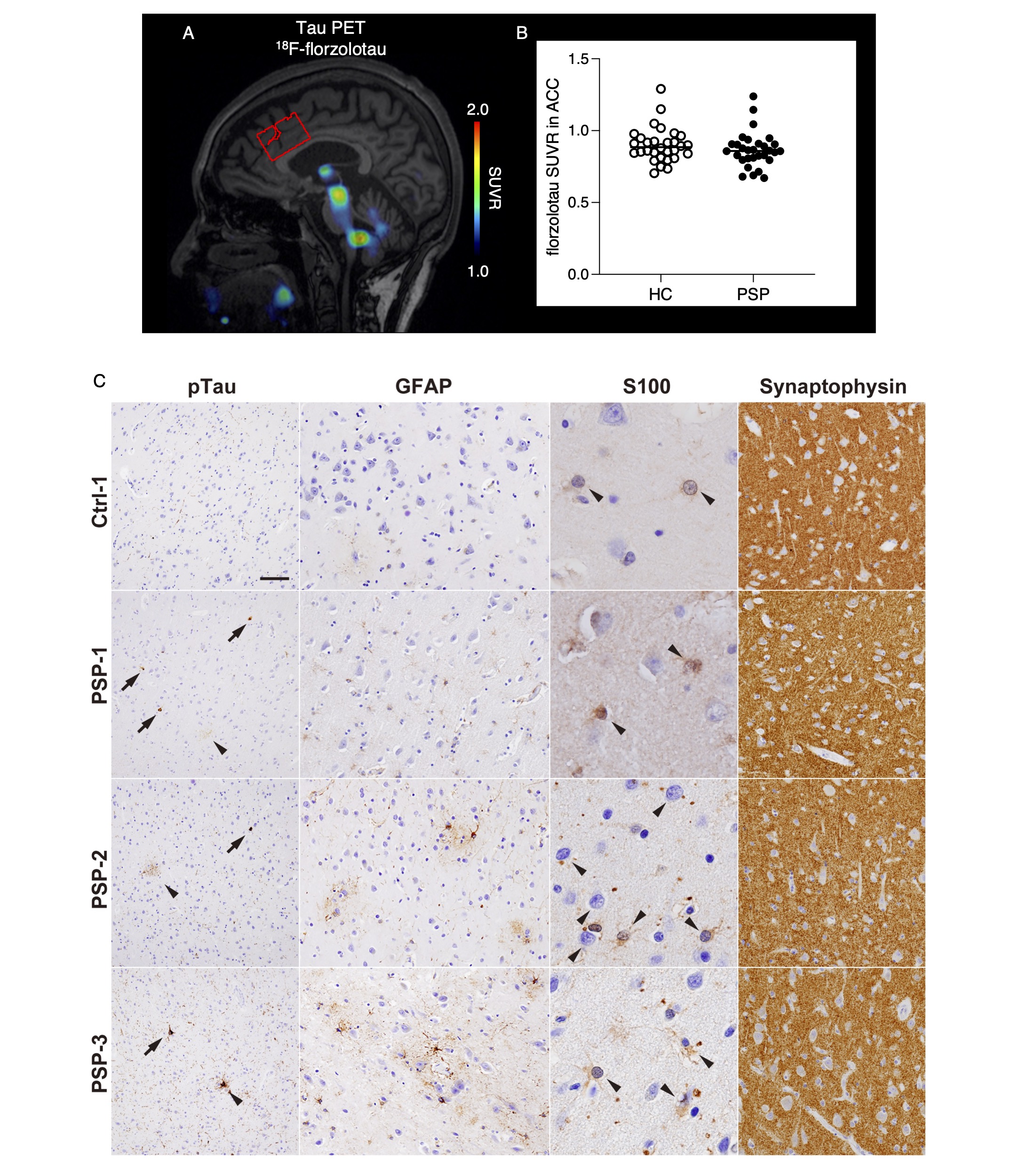
The study included 30 patients with PSP-Richardson’s syndrome and 30 healthy controls (HC); in patients with PSP, tau deposition was confirmed by tau-PET using 18F-florzolotau (Figure 1). Myo-inositol, an astroglial marker, lactate, and total N-acetyl-aspartate (tNAA: NAA + N-acetyl–aspartyl–glutamate [NAAG]) as an neuronal biomarker were quantified in the anterior cingulate cortex (ACC) via MRS (Figure 1). The ACC was studied because previous functional imaging studies have shown its involvement as a distinctive feature of PSP. The ACC is involved in cognitive processes, such as executive function, which often exhibit deficits in patients with PSP. MRI and MRS were performed using a 3.0-T scanner (MAGNETOM Verio; Siemens Healthcare) with a 32-channel receiving head coil, using a short TE spin-echo full-intensity acquired localized single voxel spectroscopy (SPECIAL) sequence with TE of 8.5 ms, TR of 3000 ms, and 128 averages5. MRS data were processed using LCModel software for a linear combination of model spectra with simulated basis functions6. We used a neurochemical basis set that incorporated nine macromolecular basis functions, each sourced from the individual peaks in a macromolecular spectrum that was obtained by the summation of experimentally acquired metabolite-nulled spectra from six healthy, adult volunteers. A representative spectrum is illustrated in Figure. 1. Partial volume effects were corrected by extracting the grey matter, white matter and cerebrospinal fluid fractions within the volume of interest (VOIs) derived from segmentation of T1-weighted images using Gannet 3.0 software7. Plasma levels of glial fibrillary acidic protein (GFAP) as another astrocytic marker, neurofilament light chain (NfL), and tau phosphorylated at threonine 181 were measured. Additionally, reactive astrocytes, tau deposition, and synaptic loss in the ACC were assessed in post-mortem brain samples from patients with PSP with comparable disease durations to those of participants.RESULTS
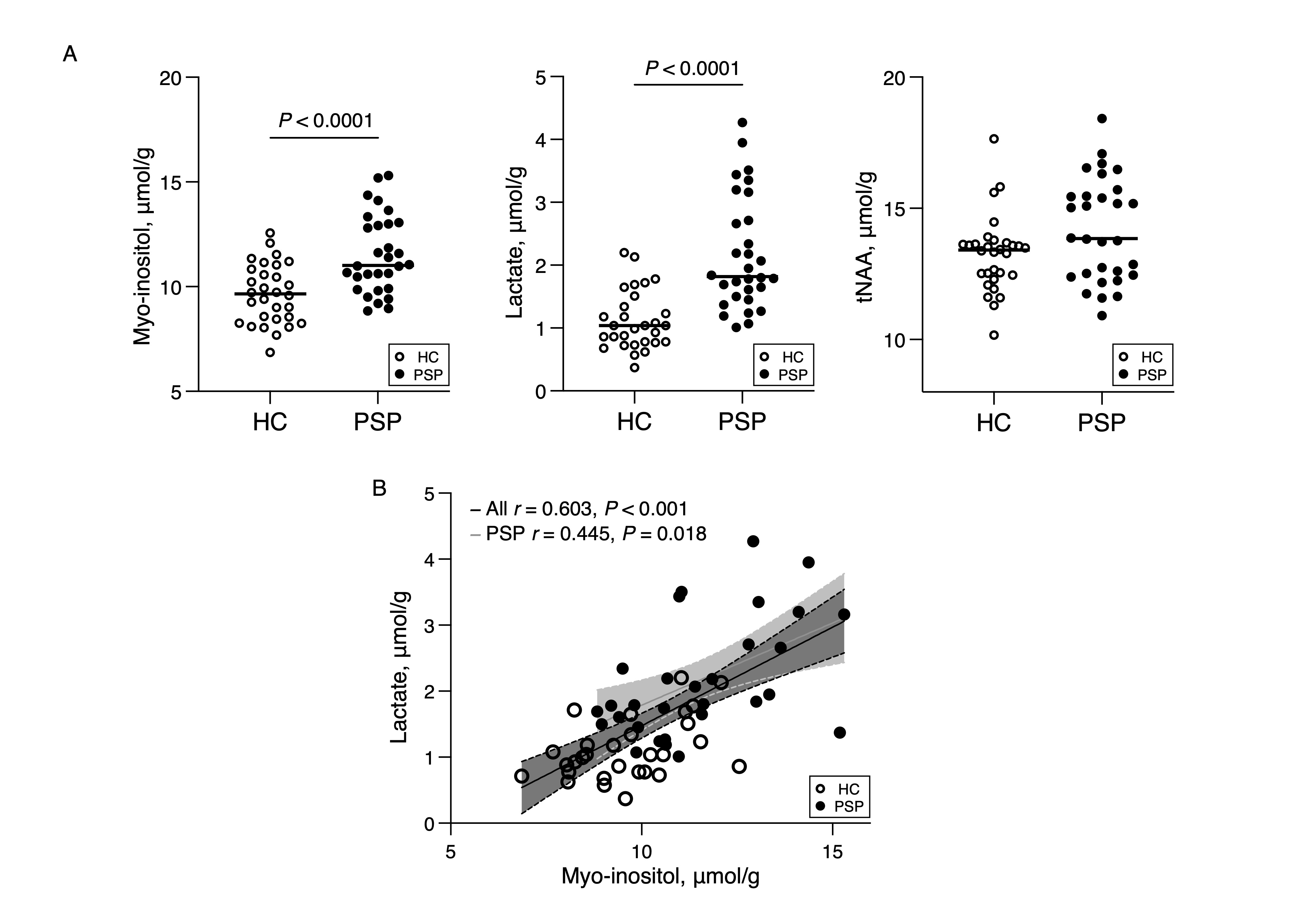
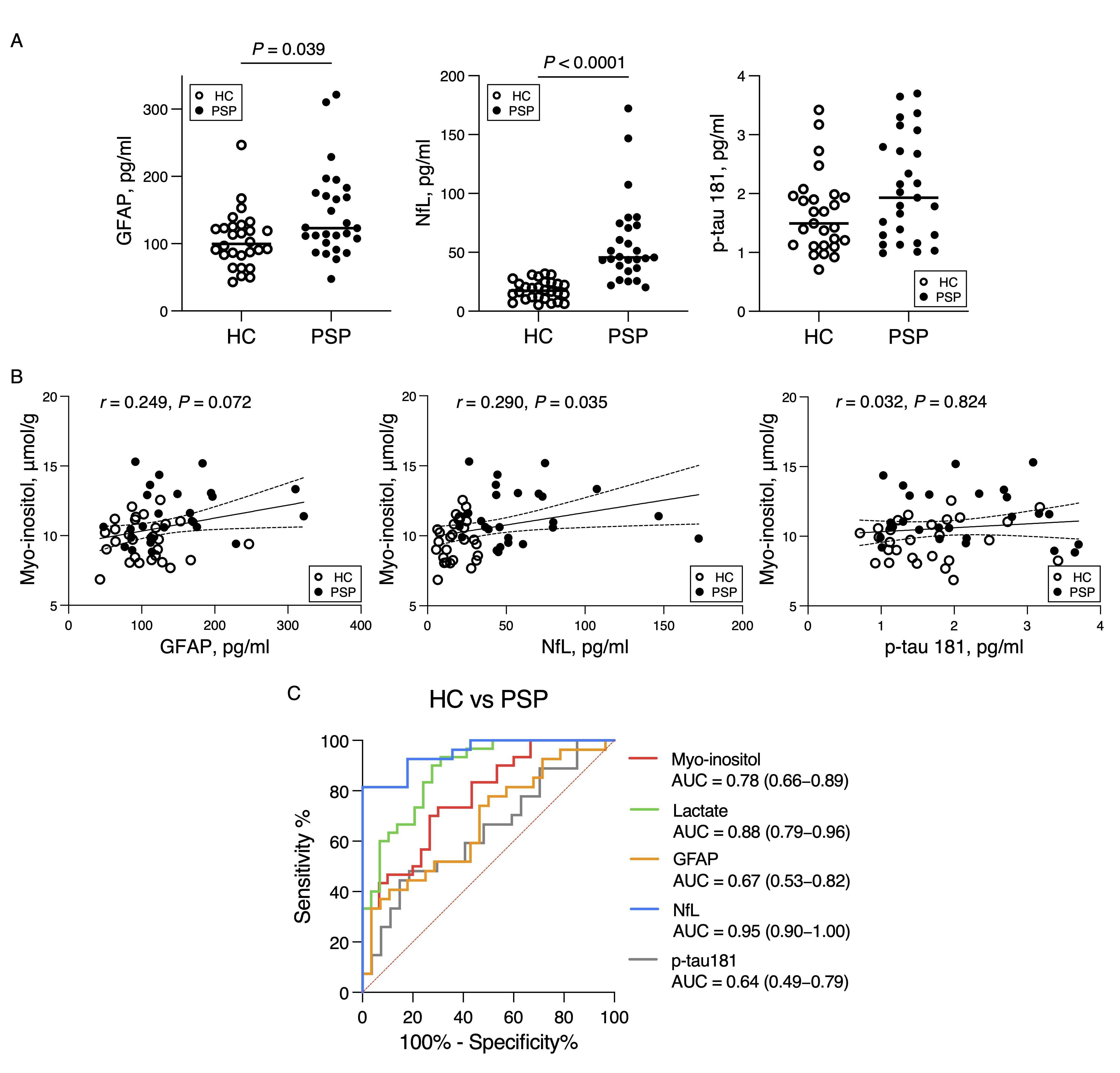
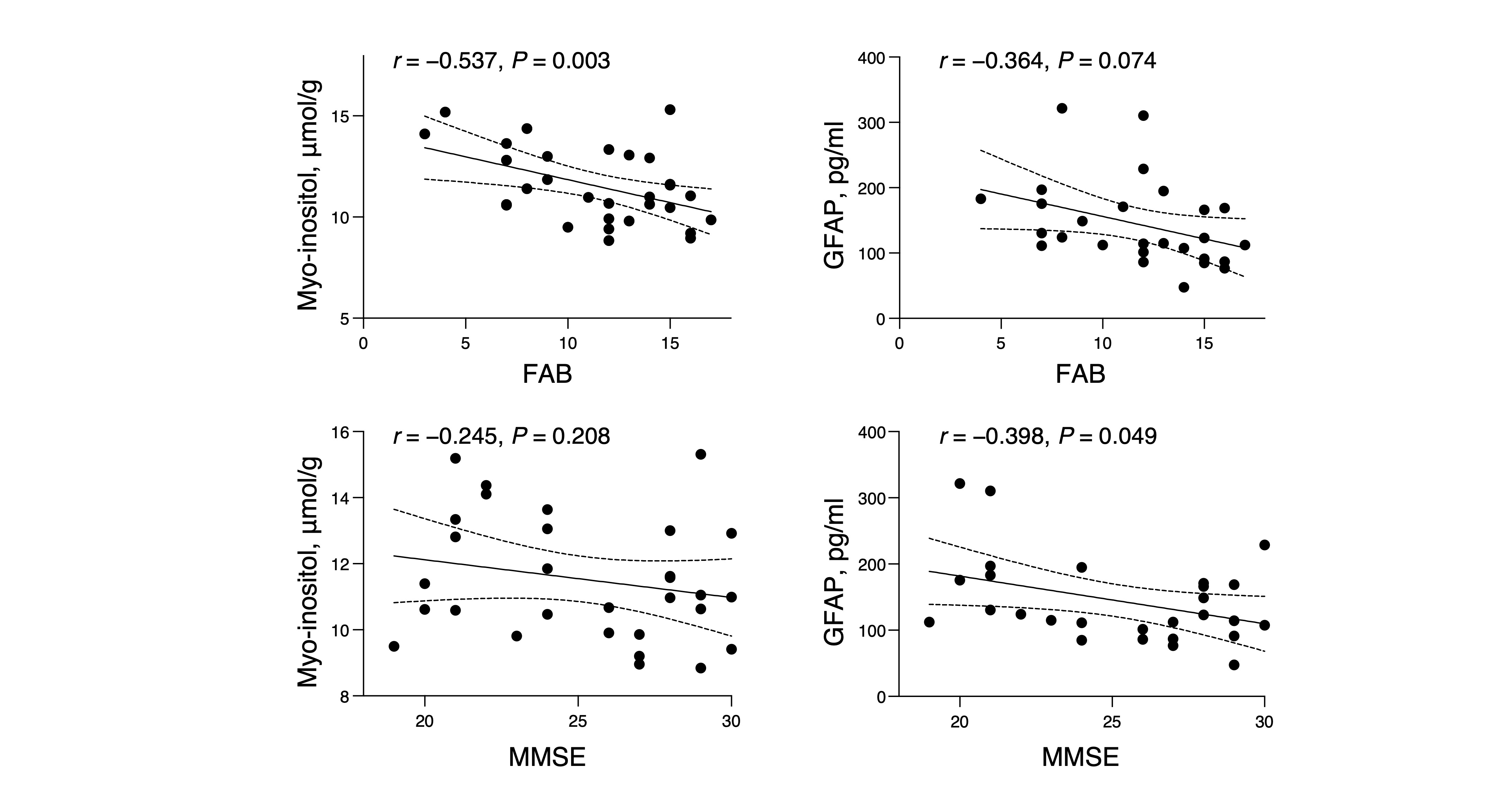
The average ± standard deviation of the Cramer-Rao lower bound for myo-inositol, lactate, and tNAA were 3.7% ± 1.0%, 15.9% ± 6.5%, and 1.8% ± 0.7%, respectively. The level of myo-inositol in the ACC were significantly higher in patients with PSP than those in healthy controls (Figure 2). The lactate level in the ACC was high in the PSP group and correlated significantly with high myo-inositol levels (Figure 2). The plasma concentrations of GFAP and NfL were significantly higher in the PSP group than those in the HC group (Figure 3). Plasma NfL outperformed other biomarkers in discriminating PSP from HC (area under the curve [AUC] = 0.95), followed by lactate and myo-inositol (AUC = 0.88 and 0.78, respectively) (Figure 3). Increased myo-inositol and plasma GFAP levels were significantly associated with scores on the Frontal Assessment Battery and Mini-Mental State Examination, respectively (Figure 4). Although the voxel-based analysis showed atrophy in the midbrain, cerebellum, thalamus, globus pallidus of participants with PSP, no atrophy was evident in the ACC. Tau-PET retention was assessed in the same ACC VOI as the MRS evaluation, revealing no difference in tau-PET retention in the ACC between the PSP group and the HC group (Figure 5). Histological analysis of the ACC in patients with PSP showed reactive astrocytes, but no marked tau deposition or synaptic loss (Figure 5).DISCUSSION and CONCLUSION
We found high levels of astrocyte biomarkers (myo-inositol in the ACC and plasma GFAP) in patients with PSP, suggesting astrocyte reactivity, and these biomarkers correlated with cognitive decline. Elevated myo-inositol levels were associated with high lactate levels, suggesting a link between reactive astrocytes and brain energy metabolism changes. Our results indicate that astrocyte reactivity in the ACC occurs before the progression of tau pathology and neurodegenerative processes in the region and affects brain function in PSP.Acknowledgements
The authors thank all patients and their caregivers for their participation in this study, and clinical research coordinators, PET and MRI operators, radiochemists, and research ethics advisers at QST for their assistance with the current projects. We thank APRINOIA Therapeutics for kindly sharing the precursor of 18F-florzolotau. This work was supported by AMED under grant no. JP22dk0207063, JP19dm0207072, JP22dk0207055, and JP21zf0127004, and by JSPS KAKENHI grant no. JP19H01041 and JP21K18268.References
1. Brandebura AN, Paumier A, Onur TS, Allen NJ. Astrocyte contribution to dysfunction, risk and progression in neurodegenerative disorders. Nat Rev Neurosci. 2023;24(1):23-39.
2. Lawrence JM, Schardien K, Wigdahl B, Nonnemacher MR. Roles of neuropathology-associated reactive astrocytes: a systematic review. Acta Neuropathol Commun. 2023;11(1):42.
3. Dickson DW, Rademakers R, Hutton ML. Progressive supranuclear palsy: pathology and genetics. Brain Pathol. 2007;17(1):74-82.
4. Beard E, Lengacher S, Dias S, Magistretti PJ, Finsterwald C. Astrocytes as Key Regulators of Brain Energy Metabolism: New Therapeutic Perspectives. Front Physiol. 2021;12:825816.
5. Mekle R, Mlynárik V, Gambarota G, Hergt M, Krueger G, Gruetter R. MR spectroscopy of the human brain with enhanced signal intensity at ultrashort echo times on a clinical platform at 3T and 7T. Magn Reson Med. 2009;61(6):1279-85.
6. Provencher SW. Estimation of metabolite concentrations from localized in vivo proton NMR spectra. Magn Reson Med. 1993;30(6):672-9.
7. Harris AD, Puts NA, Edden RA. Tissue correction for GABA-edited MRS: Considerations of voxel composition, tissue segmentation, and tissue relaxations. J Magn Reson Imaging. 2015;42(5):1431-40.
Figures