1042
T1 relaxometry or EPR signal intensity – Which is best for quantifying iron oxide nanoparticles in tissues non-destructively?1University of Minnesota, Minneapolis, MN, United States
Synopsis
Keywords: Electron Paramagnetic Resonance, Electron Paramagnetic Resonance, iron oxide nanoparticles, T1 relaxometry
Motivation: Currently there is no low-cost method to nondestructively quantify iron oxide nanoparticles (IONPs) in tissue across a wide concentration range (0.05-100 mg Fe/mL).
Goal(s): Our lab has developed a low-cost, LOngitudinally Detected Electron Paramagnetic Resonance (LOD-EPR) system. This work aims to evaluate LOD-EPR IONP quantification accuracy.
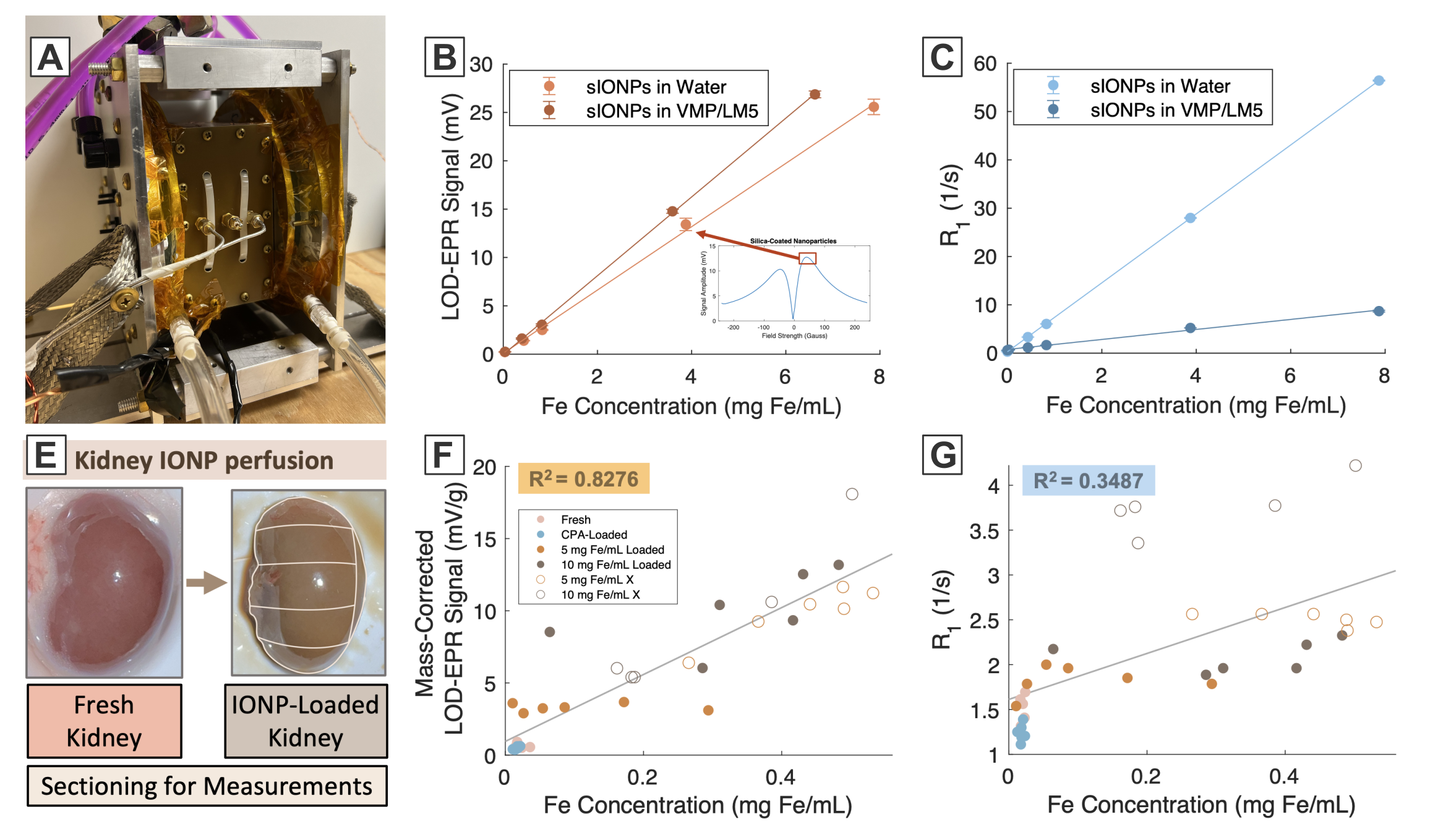
Approach: We compare IONP Fe quantification accuracy of R1 (=1/T1) from MR relaxometry versus LOD-EPR signal in solution and IONP-perfused rat kidney sections used in cryopreservation.
Results: LOD-EPR signal vs Fe concentration is linear in 0.05-10 mg Fe/mL IONP solutions and in IONP-perfused tissue, whereas R1 vs Fe concentration is linear in solution but not in tissue.
Impact: Accurate quantification of IONPs in tissues at room temperature can be done using low-cost, benchtop LOD-EPR. Our primary application is in IONP rewarming of cryopreserved organs, however other applications such as dosimetry and oxygen sensing should also be possible.
Purpose
Iron oxide nanoparticles (IONP) have a multitude of applications including drug delivery[1], neuromodulation[2], and IONP-based rewarming (nanowarming) of cryopreserved organs[3]. However, IONPs demonstrate complex behaviors in tissue (uneven distribution, aggregation, etc.). Translation of IONPs to clinical applications can be accelerated with non-invasive quantification of IONPs in tissue. Magnetic resonance imaging (MRI) and nuclear magnetic resonance (NMR) have this capability, however, have limited detection range and high cost. Increased IONP concentrations correlate with faster water T1, T2, or T2* due to increasing local magnetic field gradients[4][5]. MRI and NMR have low limits of detection, but also low upper limits of detection due to exceedingly short T1, T2, or T2* at high IONP concentrations. Even with ultra-short or zero-TE sequences such as SWeep Imaging with Fourier Transform, the detection range is ~0-3 mg Fe/mL[6]. This does not meet the needs of the above applications, for example nanowarming requires measurement between 0.05-100 mg Fe/mL.Electron paramagnetic resonance (EPR) is a direct method for quantifying IONPs, as signal is proportional to unpaired electrons in Fe, however conventional EPR performs at GHz frequencies, limiting penetration depth in large aqueous samples, and typically requiring frozen samples due to ns-µs electron relaxation. We have developed a low-cost, benchtop LOngitudinally-Detected EPR (LOD-EPR) system that transmits in the MHz range with simultaneous transmit and receive (STAR). This allows for even sample excitation at large volumes at room temperature. Here, we compare benchtop NMR T1 relaxometry to LOD-EPR in quantifying IONPs in solution and in IONP-perfused tissues. Our target application is in cryopreservation and nanowarming of organs for transplant, however this system should also enable quantitative EPR at a low-cost relative to conventional EPR in areas such as radiation dosimetry and oxygen sensing.
Methods
LOD-EPR applies a 11.5 MHz continuous-wave carrier frequency modulated by a 47.8 kHz sine wave to the transverse plane of the sample in quadrature. A pair of Helmholtz coils are used to sweep the B0 field during acquisition (-250-250 Gauss). The signal is longitudinally detected at the modulation frequency, enabling STAR by geometric and frequency isolation.A 10-point average over the LOD-EPR spectral peak and R1 (=1/T1) for a solution or tissue are obtained and compared to the sample Fe concentration validated by inductively-coupled mass spectroscopy (ICP-MS), the gold standard for Fe quantification. Solutions of commercially available IONP EMG308 are coated in silica (sIONP) to minimize aggregation and measured at varying concentrations (0.05-10 mg Fe/mL) in water and cryoprotective agent VMP/LM5 using both systems. Lastly, sIONPs are perfused into rat kidneys alongside cryoprotective agent VMP/LM5 in a typical protocol for kidney cryopreservation and rewarming at varying IONP concentrations (fresh kidneys, CPA-perfused kidneys, 5 mg Fe/mL, and 10 mg Fe/mL). The kidneys are then sectioned into n=5-6 sections, measured on each system, and compared in terms of Fe quantification accuracy.
Results and Discussion
Linearity of LOD-EPR signal and R1 vs Fe concentration is shown for sIONPs in water and VMP/LM5 (R2>0.99). R1 vs Fe concentration for sIONPs drastically changes depending on whether the surrounding solution is water or VMP/LM5, whereas LOD-EPR signal vs Fe concentration changes slightly with surrounding solution composition. The large change in R1 may be related to both changes in baseline R1 of the surrounding solution and solution viscosity affecting IONP easy axis alignment with the external field. The latter may be the primary mechanism affecting LOD-EPR signal. Most notably, R1 does not correlate well with Fe concentration in sIONP- and cryoprotectant-perfused rat kidney sections (R2 = 0.3487), while LOD-EPR does (R2 = 0.8276). This may be a result of inherent tissue R1 varying between kidneys, particle aggregation, and variable IONP and cryoprotective agent distribution among kidney sections.Overall, LOD-EPR shows promise as a low-cost, benchtop method of IONP quantification in tissues for cryopreservation and other applications. To better address issues of uneven distribution of IONPs during IONP perfusion into tissues, we aim to develop an LOD-EPR imaging system. The utility of LOD-EPR for other applications is also being investigated in alanine radiation dosimetry and oxygen sensing.
Acknowledgements
This material is based upon work supported by the National Science Foundation under Grant No. EEC 1941543, P41 EB027061, and The Malcolm B. Hanson Endowed Chair in Radiology. We also thank Dr. Zonghu Han for his guidance in kidney perfusion.References
[1] Estelrich, J.; Escribano, E.; Queralt, J.; Busquets, M. A. Iron Oxide Nanoparticles for Magnetically-Guided and Magnetically-Responsive Drug Delivery. International Journal of Molecular Sciences 2015, 16 (4), 8070–8101. https://doi.org/10.3390/ijms16048070.
[2] Chen, R.; Romero, G.; Christiansen, M. G.; Mohr, A.; Anikeeva, P. Wireless Magnetothermal Deep Brain Stimulation. Science 2015, 347 (6229), 1477–1480. https://doi.org/10.1126/science.1261821.
[3] Sharma, A.; Rao, J. S.; Han, Z.; Gangwar, L.; Namsrai, B.; Gao, Z.; Ring, H. L.; Magnuson, E.; Etheridge, M.; Wowk, B.; Fahy, G. M.; Garwood, M.; Finger, E. B.; Bischof, J. C. Vitrification and Nanowarming of Kidneys. Adv Sci (Weinh) 2021, 8 (19), e2101691. https://doi.org/10.1002/advs.202101691.
[4] Chambon, C.; Clement, O.; Le Blanche, A.; Schouman-Claeys, E.; Frija, G. Superparamagnetic Iron Oxides as Positive MR Contrast Agents: In Vitro and in Vivo Evidence. Magnetic Resonance Imaging 1993, 11 (4), 509–519. https://doi.org/10.1016/0730-725X(93)90470-X.
[5] Hardy, P. A.; Henkelman, R. M. Transverse Relaxation Rate Enhancement Caused by Magnetic Particulates. Magnetic Resonance Imaging 1989, 7 (3), 265–275. https://doi.org/10.1016/0730-725X(89)90549-3.
[6] Etheridge, M.; Hurley, K.; Zhang, J.; Jeon, S.; Ring, H.; Hogan, C.; Haynes, C.; Garwood, M.; Bischof, J. Accounting for Biological Aggregation in Heating and Imaging of Magnetic Nanoparticles. Technology 2014, 2, 214–228. https://doi.org/10.1142/S2339547814500198.
Figures