1041
Molecular MRI of neuroinflammation using a redox-active iron complex1Athinoula A. Martinos Center for Biomedical Imaging, Department of Radiology, Massachusetts General Hospital/Harvard Medical School, Charlestown, MA, United States, 2Department of Radiology, The Third Affiliated Hospital of Zhengzhou University, Zhengzhou, China, 3Genetics and Aging Research Unit, McCance Center for Brain Health, Mass General Institute for Neurodegenerative Diseases (MIND), Department of Neurology, Massachusetts General Hospital/Harvard Medical School, Charlestown, MA, United States
Synopsis
Keywords: Contrast Agents, Contrast Agent, Neuroinflammation
Motivation: Neuroinflammation is a critical pathophysiological process implicated in the development of neurodegenerative disorders.
Goal(s): Imaging to detect, monitor, and surveil neuroinflammatory processes could profoundly improve how patients suffering neurodegenerative diseases are diagnosed and managed.
Approach: We evaluated brain imaging with the oxidatively activated contrast agent, Fe-PyC3A, as a proxy for inflammatory microglial activity in a mouse model of lipopolysaccharide(LPS)-induced neuroinflammation.
Results: Fe-PyC3A generated significantly greater enhancement in LPS-treated mice than in saline-treated controls, correlating with immunohistochemical quantification of microglial activation. Imaging using Gd-DOTA as negative control probe and NOX-2 deficient mice as loss of function control links Fe-PyC3A enhancement with reactive microglial activity.
Impact: MRI using oxidatively activated probes as a potential marker to detect and quantify neuroinflammatory processes could profoundly improve how patients suffering neurodegenerative diseases are diagnosed and managed.
Introduction
Neuroinflammation is a key factor driving neurodegeneration [1]. During active neuroinflammation, resident brain macrophages termed microglia adopt a ‘reactive’ phenotype that generates a neurotoxic microenvironment in part due to the secretion of reactive oxygen species. We hypothesize that extracellular ROS released by reactive microglia during neuroinflammation will mediate the oxidation of Fe-PyC3A from the low-relaxivity Fe2+ state to the high-relaxivity Fe3+ state [2], generating strong MR signal enhancement. Here, we performed brain imaging with the oxidatively activated ROS-sensing MRI contrast agent, Fe-PyC3A, in a mouse model of lipopolysaccharide (LPS)-induced neuroinflammation for non-invasive detection of microglial changes.Methods
A total of 24 C57BL/6 male mice and 4 gp91phox knockout (NOX-2 KO) mice (8-10 weeks old) received i.p. injection of either LPS (3 mg/kg) as a model of neuroinflammation or saline (3 mg/kg) as controls. After 24h, animals were anesthetized with isoflurane (1.5%) and imaged with a 4.7 Tesla Bruker MRI scanner equipped with a custom-built volume coil. Prior to i.v. injection of 0.3 mmol/kg Fe-PyC3A (N = 8 LPS, N = 6 saline) or 0.1 mmol/kg Gd-DOTA (N = 5 LPS, N = 5 saline), the blood-brain barrier was transiently disrupted via i.v. injection of 25% wt/wt mannitol. 2D multi-slice T1-weighted gradient echo images were acquired dynamically prior to and up to 20 minutes post-injection of Fe-PyC3A or Gd-DOTA. We selected hippocampus and cortex as regions of interest (ROIs) and analyzed the (post-pre) injection percentage change in signal intensity (SI%) from these ROIs and (post-pre) injection change in brain ROIs vs. muscle contrast to noise ratio (ΔCNR) from 2 -10 min after Fe-PyC3A or Gd-DOTA injection. Mice were euthanized 1h post-injection, and brain tissue was harvested for immunohistochemical evaluation of microglial activation (Iba1). The proportional area of Iba-1 expression (microglia area %) in the hippocampus and cortex was measured in ImageJ.Results and Discussion
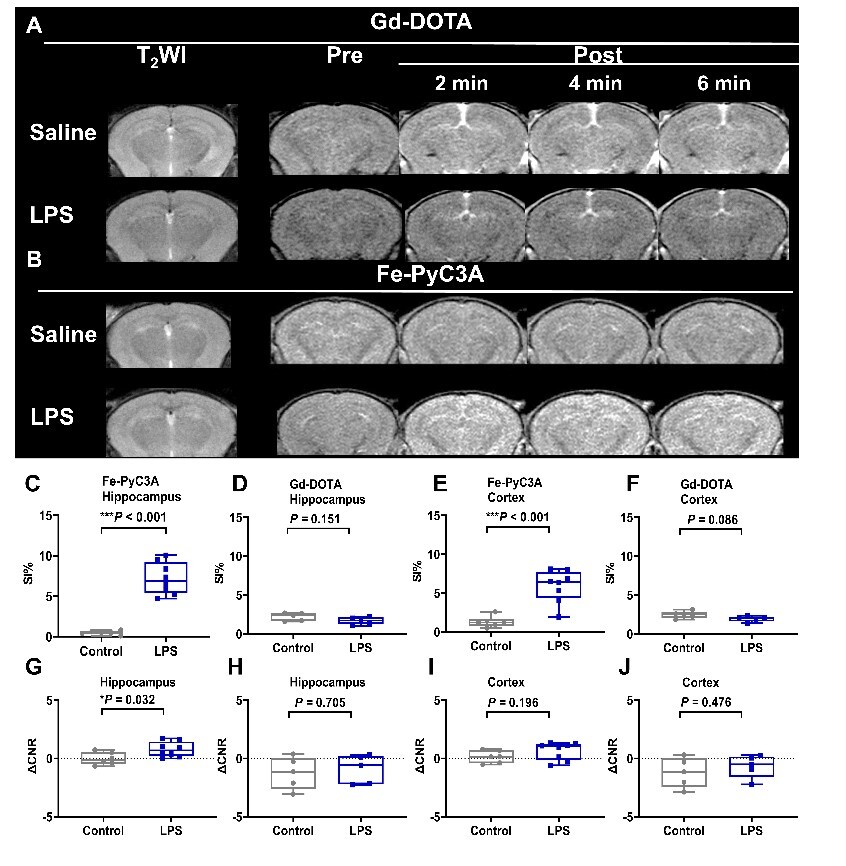
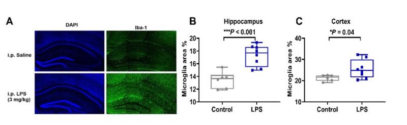
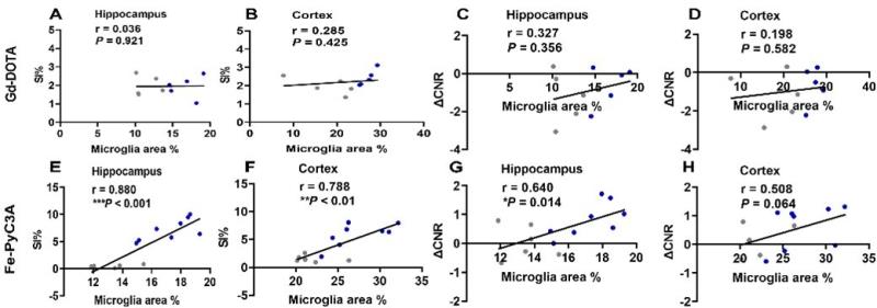
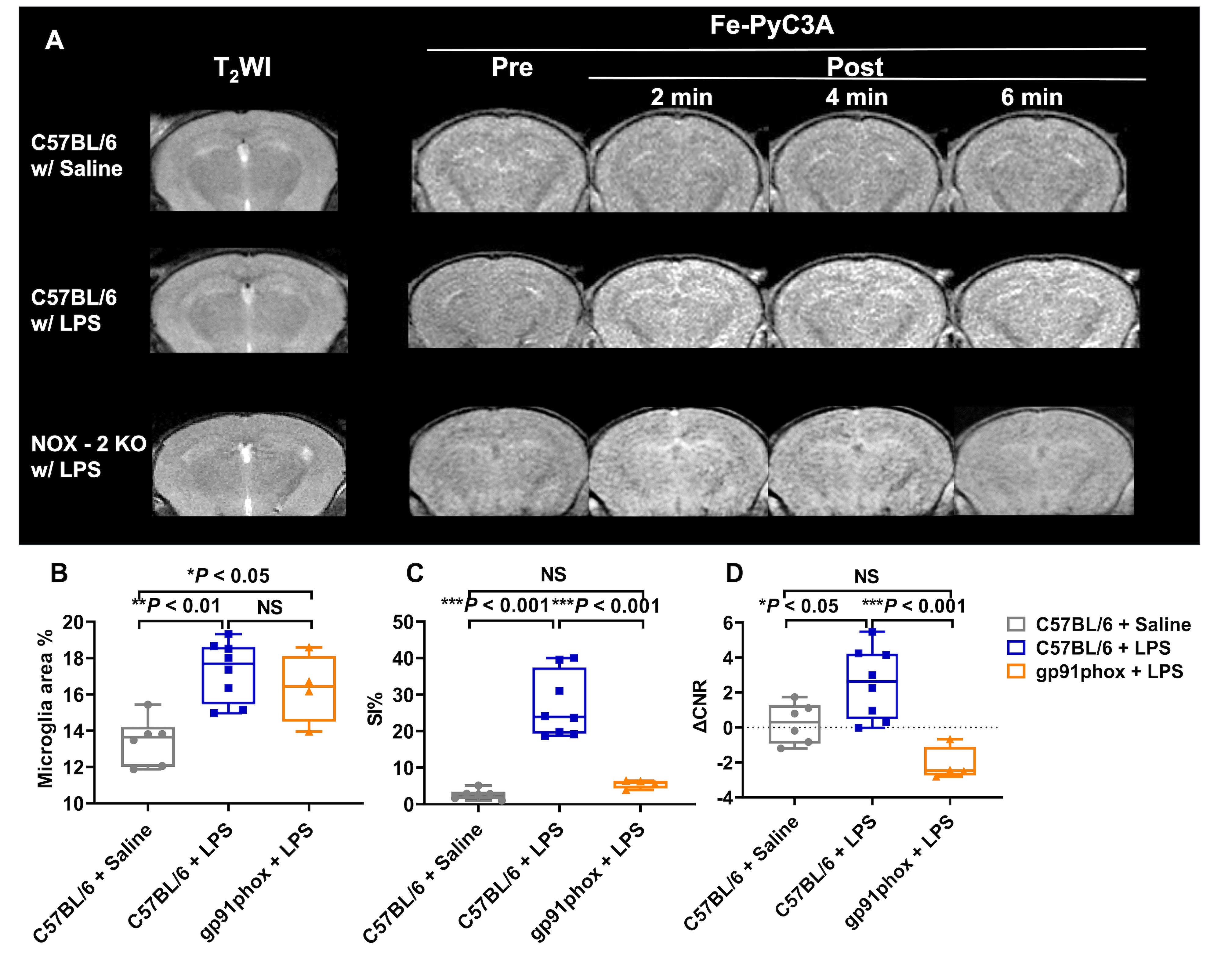
Dynamic images recorded prior to and after Fe-PyC3A or Gd-DOTA injection into saline and LPS-treated mice demonstrate differential brain signal enhancement in the LPS-treated mice as shown in Figure 1A, B. We found that both SI% and ΔCNR in the hippocampus and cortex at 2 min post-injection of Fe-PyC3A are significantly higher in LPS-treated mice than in saline-treated controls (Figure 1G - J). No significant difference in the SI% and ΔCNR was observed in the hippocampus and cortex between LPS-treated mice and controls using Gd-DOTA (Figure 1C - F). IBA-1 immunostaining demonstrates significantly greater microglial activation in LPS-treated mice vs. controls with 17.3% ± 1.64% vs. 13.4% ± 1.31% (P < 0.001), for the hippocampus, and 25.5% ± 4.34% vs. 21.2% ± 1.40% (P = 0.040) for the cortex (Figure 2A - C). The proportional area of Iba-1 expression significantly and positively correlated with hippocampal SI% (r = 0.880, P < 0.001) and cortex (r = 0.788, P < 0.01) (Figure 3E, F), and ΔCNR (r = 0.640, P = 0.014, Figure 3G), for mice imaged with Fe-PyC3A. In contrast, for mice imaged with Gd-DOTA, no significant correlations between SI% or ΔCNR and Iba-1 positive area were observed in the hippocampus and cortex (Figure 3A-D, H). To further evaluate the specificity of Fe-PyC3A to ROS secreted during neuroinflammation, we compared Iba-1 positive area, Fe-PyC3A induced SI% and ΔCNR in gp91phox knockout (NOX-2 KO) mice. NOX-2 KO mice are deficient in NADPH oxidase II and therefore exhibit severely diminished capabcity to generate ROS. We found that both NOX-2 KO mice and C57BL/6 mice with LPS had greater Iba-1 positive area than C57BL/6 mice with saline (Figure 4). We also found no significant differences between NOX-2 KO mice with LPS and C57BL/6 mice with saline in SI% (P = 0.748) and ΔCNR (P = 0.090), demonstrating the specificity of Fe-PyC3A contrast enhancement for ROS generated through microglial NOX-2 activity.Conclusion
Fe-PyC3A provided strong, selective contrast enhancement in the hippocampus and cortex of LPS-treated mice. MR imaging using oxidatively activated MR imaging probes such as Fe-PyC3A merits further evaluation as a marker to diagnose and quantify neuroinflammation in neurodegenerative disorders.Acknowledgements
EMG and IYZ co-corresponding authors. This work was supported by the National Institute on Aging via an Administrative Supplement to R01DK120663. EMG has equity and receivesconsulting income from reveal pharmaceutical.References
[1] Mi Y, Qi G, Vitali F, et al. Loss of fatty acid degradation by astrocytic mitochondria triggers neuroinflammation and neurodegeneration. Nat Metab. 2023;5(3):445-465.[2] Wang H, Jordan VC, Ramsay IA, et al. Molecular Magnetic Resonance Imaging Using a Redox-Active Iron Complex. J Am Chem Soc. 2019;141(14):5916-5925.Figures