1040
Nanodiamonds as a novel T1-contrast agent for MRI1Intelligent Systems, Max Planck Institute, Stuttgart, Germany
Synopsis
Keywords: Novel Contrast Mechanisms, Contrast Agent, nanodiamonds, cell labeling
Motivation: The structural defects in diamond particles, are known for their paramagnetic properties. Here we aim to determine if the presence of paramagnetic centers in detonation nanodiamonds particles can be exploited to enhance longitudinal relaxation time (T1).
Goal(s): Introduce nanodiamonds as a novel T1-contrast agent and contrast differences with gadolinium chelates.
Approach: Using high-field, 7 T MRI, longitudinal and transverse relaxation rates were measured and compared between detonation and air-oxidized detonation nanodiamonds. In-vivo demonstration was carried out using chicken embryos.
Results: Air-oxidized detonation nanodiamonds have superior longitudinal and transverse relaxivity compared to detonation nanodiamonds. We demonstrate their potential as an alternative, gadolinium-free T1-contrast agent.
Impact: Nanodiamonds hold great promise for biomedical applications mostly due to their biocompatibility, non-toxicity and versatile functionalization. A possibility for direct visualization by means of T1-weighted MRI is opening new venues for tracking over time without a concern for Gd3+-toxicity.
Introduction
Nanodiamonds (NDs) are already envisioned for multifunctional integration involving imaging, biosensing, drug-carrying and cell tracking. Most of in vivo imaging is conducted using fluorescent NDs, or nanodiamonds conjugated to Gd3+ ions 1-3. Here we demonstrate T1-relaxation in the presence of detonation nanodiamonds and air-oxidized detonation nanodiamonds as alternative, gadolinium-free contrast agents.Methods
Detonation nanodiamonds (>98.3% pure, size range 3-10 nm) were purchased from US Research Nanomaterials, Inc. By heating the nanodiamond powder in air at 520oC for 65 min we produced air-oxidized detonation nanodiamonds (DND). To calculate longitudinal (T1) and transverse (T2) relaxation times, six different concentrations of DNDs and air-oxidized DNDs were prepared (0.025, 0.0625, 0.125, 0.25, 0.5, 0.75 mM) in double-distilled water. Using 7 T Bruker (Bruker, Germany) preclinical scanner and 40 mm quadrature coil, T1 and T2 were quantified. T1 was measured using a saturation recovery method with a variable repetition time (RAREVTR); TR = (60,70, 80, 90, 100, 200, 300, 400, 500, 600, 700, 800, 900, 1000, 2000, 3000, 4000, 5000, 6000 ms), and fixed echo time (TE) = 6.68 ms, RARE factor = 2, 117x117 μm2 in-plane resolution, 1 mm slice thickness, NEX = 1. T2 was measured using single slice multi-echo spin echo sequence with fixed repetition time TR=3000 ms and 30 echo times (TE=5.4–162 ms), 150x150 μm2 in-plane resolution, 1 mm slice thickness, NEX = 2. Image processing and the parameter fitting (r1,2, R1,2 and T1,2) was done using ImageJ and Origin. HRTEM was performed on advanced TEM (JEOL ARM200F, JEOL Co Ltd.) using an accelerating voltage of 200 kV.To demonstrate in vivo potential of DND, as a T1-contrast agent, embryonic development day (EDD) 14, 15 and 16 chicken embryos were used (Valo Biomedia GmbH, Germany). Prior to DND intravenous (IV) injections, to prevent rapid agglomeration of DND in the blood, air-oxidized DND were mixed with sodium citrate solution (pH 7.0). We imaged, N=5 control embryos (no injection), N=5 IV injected with 50 µl of 0.625 mM air-oxidized DND in citrate solution, and N=5 IV injected with gadobutrol (Gadavist, 50 µl of 30 mM). MRI was performed immediately after IV injections and repeated at 24 and 48 h.
Results
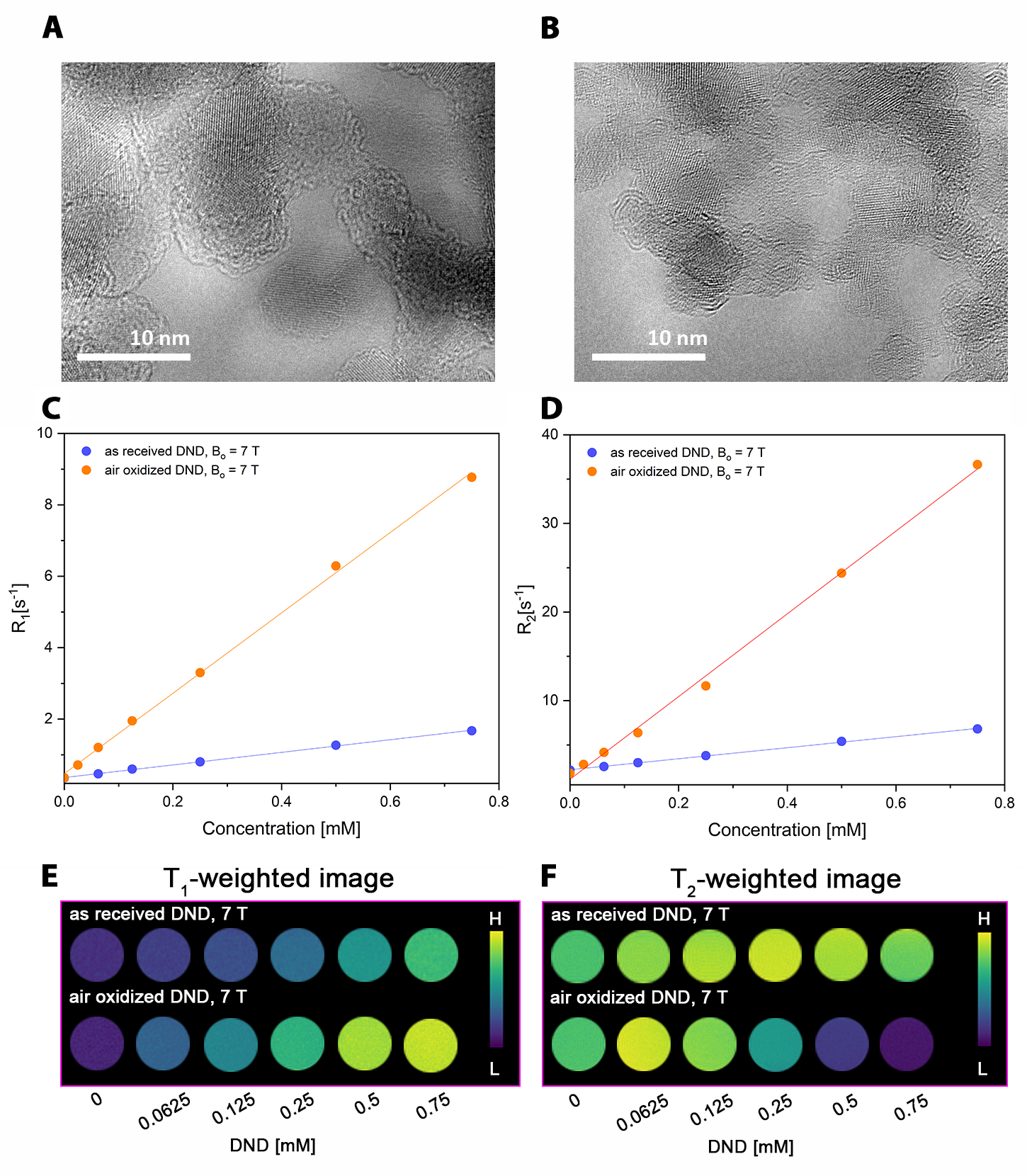
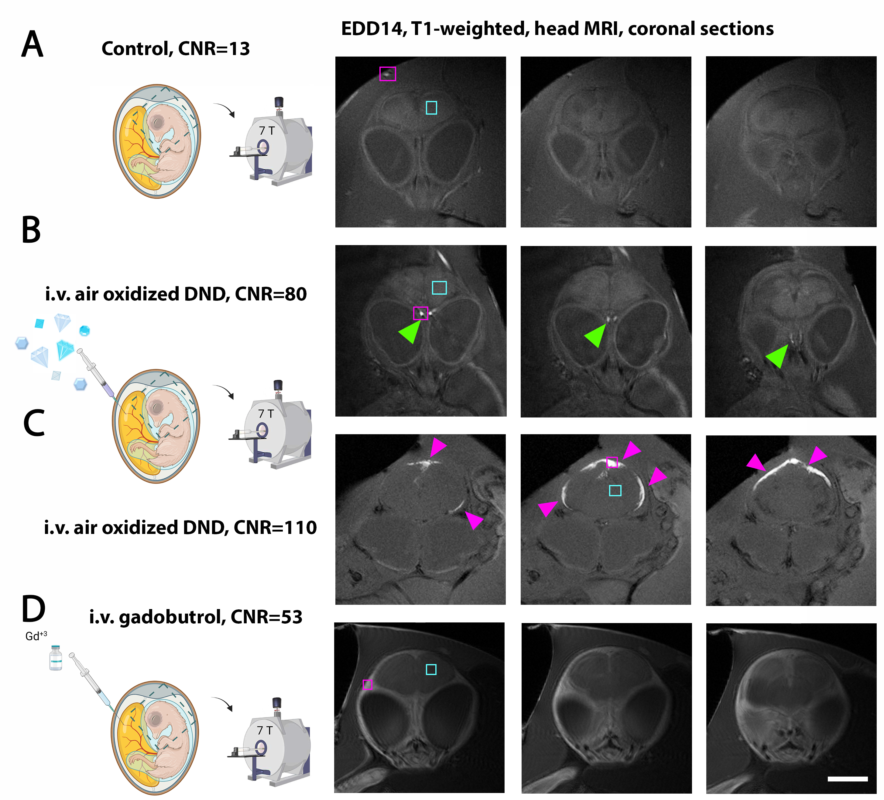
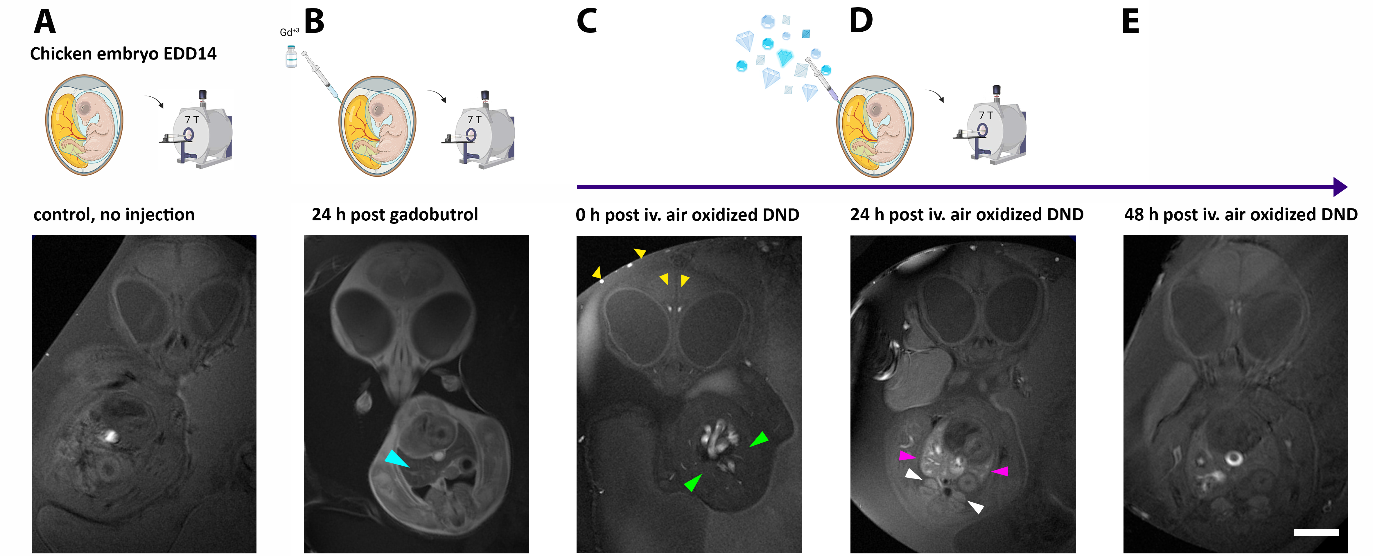
Detonation and air-oxidized detonation nanodiamonds were characterized for their potential to reduce T1 and T2. Both DND particles have a crystalline structure, as demonstrated using high resolution transmission electron microscopy (HRTEM) (Fig. 1A, B), with air-oxidized DND being only slightly smaller. Longitudinal relaxivity rate was r1 = (1.77±0.03) [mM-1s-1] for detonation nanodiamonds (DND) and six times higher for air-oxidized DNDs r1 = (11.26±0.18) [mM-1s-1] (Fig. 1C). Transverse relaxivity was r2 = (6.23 ± 0.09) [mM-1s-1] for DNDs and seven times higher for air-oxidized DNDs r2 = (46.68±1.05) [mM-1s-1] (Fig. 1D). A strong enhancement of the vasculature on T1-weighted MRI with high CNR is prominent for embryos injected with air-oxidized DND particles (Fig. 2B, green arrowheads) and especially in second embryo with enlarged cranial veins (Fig. 2C, magenta arrowheads), compared to control (Fig. 2A) or gadobutrol injected embryos (Fig. 2C). At 24 h post intravascular (IV) injection, both liver (Fig. 3D, magenta arrowheads) and kidney (Fig. 3D, white arrowheads) became prominent and strong signal enhancement was evident. In comparison, no liver or kidney enhancement were present immediately after DND particle IV injection (Fig. 3C, green arrowhead) or in control embryo (Fig. 3A). This is in contrast to gadobutrol-injected embryos, where after 24 h the liver was markedly darker compared to other tissues (Fig. 3B, cyan arrowhead). The most likely explanation for the enhanced liver signal is the phagocytosis of blood-circulating DND particles by the reticuloendothelial system, including Kupffer cells in the liver. A similar mechanism has been already established for SPIONs4.Discussion
Presented MR images point the main differences in the performance between air-oxidized DND particles and gadobutrol as the contrast agent. The small gadobutrol molecule is an extravascular contrast agent, and provides good tissue enhancement (except the brain tissue, to some extent, due to the intact blood brain barrier), while DND particles are intravascular (blood pool) agents leading to strong vascular enhancement and high CNR of the vasculature.The enhanced regions of the liver at 24 h post air-oxidized DND injection point to the capacity of air-oxidized DND for cell labeling and tracking and their visualization as bright signal.
Conclusion
The presence of paramagnetic centers within nanodiamond particles enables their efficient detection on T1-weighted MRI images at biologically-relevant concentrations without the need for conjugation with gadolinium or use of hyperpolarization techniques, simplifying both detection and manufacturing processes.Acknowledgements
We would like to acknowledge Dr. Elke Weiler for her kind help with chicken embryos.References
1. Manus, L. M. et al. Gd(III)-Nanodiamond Conjugates for MRI Contrast Enhancement. Nano Letters 10, 484-489, doi:10.1021/nl903264h (2010).
2. Rammohan, N. et al. Nanodiamond-Gadolinium(III) Aggregates for Tracking Cancer Growth In Vivo at High Field. Nano Lett 16, 7551-7564, doi:10.1021/acs.nanolett.6b03378 (2016).
3. Panich, A. M. et al. PVP-coated Gd-grafted nanodiamonds as a novel and potentially safer contrast agent for in vivo MRI. Magn Reson Med 86, 935-942, doi:10.1002/mrm.28762 (2021).
4. Wahajuddin & Arora, S. Superparamagnetic iron oxide nanoparticles: magnetic nanoplatforms as drug carriers. Int J Nanomedicine 7, 3445-3471, doi:10.2147/ijn.S30320 (2012).
Figures