1038
A Novel PET/MRI Hybrid Probe enables Non-invasive Imaging of Perfusion and Excretion in vivo1Werner Siemens Imaging Center, University Hospital Tuebingen, Tuebingen, Germany, 2Cluster of Excellence iFIT (EXC 2180) “Image-Guided and Functionally Instructed Tumor Therapies”, University of Tuebingen, Tuebingen, Germany, 3Institute of Organic Chemistry and Biochemistry of the CAS, Prague, Czech Republic, 4German Cancer Consortium (DKTK), partner site Tübingen, German Cancer Research Center (DKFZ), Heidelberg, Germany
Synopsis
Keywords: Contrast Agents, PET/MR, Perfusion Kidney Gadolinium
Motivation: Our work is driven by the conviction that the integration of PET and MRI in molecular imaging has the potential to revolutionize diagnostic precision and patient care.
Goal(s): Enhance precision molecular imaging through the development of a unique hybrid PET/MRI perfusion probe, [18F][Gd(FL1)].
Approach: Develop a PET/MRI probe, [18F][Gd(FL1)], that is exceptionally stable, rapidly synthesized, efficiently radiolabeled, and allows simultaneous PET/MR imaging, focusing on tissue perfusion and renal filtration in preclinical models.
Results: After achieving high-yield radiosynthesis with a unique approach, our dual-modality probe showed consistent signals in both imaging modalities and was reliably quantified thanks to the combination of PET and MRI.
Impact: The first-of-its-kind hybrid PET/MR probe [18F][Gd(FL1)] can be readily and efficiently radio-synthetized and allows simultaneous PET/MR quantitative measurement of perfusion and excretion. This probe opens the path for quantifying molecular imaging probes in research and clinical practice.
Introduction
PET/MRI systems are rapidly growing in numbers in clinical infrastructures and practice in oncology, cardiology, and neurology1. Combined PET/MRI scanners match PET outstanding sensitivity and quantitative signal detection with MRI excellent functional and anatomical detail. While PET/MRI holds immense potential in molecular imaging, most simultaneous PET/MRI applications have concentrated on reducing the burden of examinations and durations associated with sequential PET and MRI scans rather than exploring the full potential of hybrid applications for precise molecular imaging2. Mainly, this is due to the lack of hybrid molecular probes that can be detected with PET and MRI simultaneously. Here we present a novel hybrid PET/MRI perfusion probe, [18F][Gd(FL1)], that is quickly synthesized, readily radiolabelled, shows excellent stability in vivo and suitable pharmacokinetic properties allowing for monitoring tissue perfusion with simultaneous PET/MR imaging. Thanks to its swift renal clearance, [18F][Gd(FL1)] can also assess renal filtration in vivo.Methods
[18F][Gd(FL1)] was synthesized by direct radiolabelling of a macrocyclic Gd(III) precursor chelate with 18F- by fluorine isotopic exchange. The synthesis was optimized to yield tracer batches relevant for clinical translation (1.3 GBq). After HPLC purification, in order to enhance MR contrast, a "hot/cold" cocktail was prepared by doping the radiolabelled product with non-radioactive [19F][Gd(FL1)]. Phantom and in vivo imaging experiments with healthy mice (n = 6) were performed on a 7T preclinical scanner (Bruker Clinscan) with an in-house built small animal PET insert. DCE FLASH images, T1 maps (VFA method), and PET scans were acquired simultaneously over 62 minutes after a bolus injection of the probe (0.09 ± 0.03 mmol/kg of Gd; 0.91 ± 0.16 MBq). Ex vivo biodistribution was assessed by gamma counting and autoradiography (18F), ICP-OES and LA-ICP-MS (Gd content). Glomerular filtration rates (GFRs) were estimated from the natural logarithm of the kidney's time-activity curves3. The ex vivo analysis also included histological validation (PAS staining) for mice whose PET/MR scans suggested an atypical renal filtration.Results and Discussion
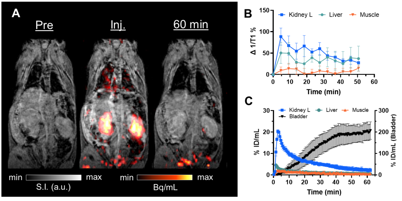
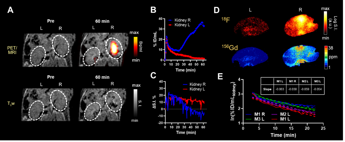
Simultaneous PET/MRI of phantoms containing titrations of [18F][Gd(FL1)] (Fig. 1A) revealed a perfect linear correlation between T1 relaxation rates and PET signal (Fig.1B). Concentrations determined via gamma counting and ICP-MS showed perfect linear correlations (R2 = 0.98; Fig.1C). The physicochemical characterization of the probe showed that [18F][Gd(FL1)] has similar kinetic inertness (half-life in 0.1 M HCl, t1/2 = 45h), number of coordinated water molecules (q = 1), but slower water exchange rate (kex = 1.7 × 10⁶) compared to Gd-DOTA (t1/2 = 44h, q = 1, kex = 4.1 × 10⁶), commercially available as Dotarem. In healthy mice, [18F][Gd(FL1)] showed consistent signal in both imaging modalities (Fig. 2A). The probe showed quick renal clearance into the bladder, making it a suitable tool for perfusion imaging. Dynamic contrast enhancement and time-activity curves confirmed fast clearance with no 18F radioactivity or gadolinium retention in healthy tissue (Fig. 2B-C), which was also confirmed by all the ex vivo biodistribution analyses. Negligible uptake in bone and HPLC analysis of urine indicated no de-fluorination or de-complexation. In vivo, PET/MR imaging provided accurate quantification of the probe and the tissue contrast (T1 maps and T1-w) with high spatial/temporal resolution. The images obtained from both MR and PET scans of two mice kidneys showed unexpected observations, which were quite surprising (as depicted in Fig. 3A). In these mice, one kidney showed a substantial abnormal accumulation of [18F][Gd(FL1)], resulting in a significant T2 effect and hypointense MR signal in the kidney pelvis and cortex starting from 25 minutes after injection. The simultaneously obtained PET images showed spots of high uptake in the same regions, confirming the presence of the probe and allowing a better quantification thanks to the linearity of the PET signal (Fig.3B-C). The estimated GFR of these kidneys was substantially different from those without probe accumulation (Fig.3E table). Combining PET and MRI signals from a single probe has proven instrumental in distinguishing normal and abnormal renal clearance. Ex vivo autoradiography and LA-ICP-MS images of these kidneys showed the same distribution patterns (Fig.3D). Histological examination of both kidneys of these mice assessed relevant findings in the glomeruli of the kidneys with unexpected retention of the probe, explaining the observations made during imaging.Conclusions
We designed, developed, and characterized a first-of-its-kind PET/MRI perfusion probe [18F][Gd(FL1)]. Preclinical in vivo PET/MR imaging in mice revealed the advantages of combining the properties of the two modalities in a single probe to obtain complementary quantitative functional and anatomical information on healthy and diseased tissues. Notably, the probe has demonstrated significant potential in the quantitative imaging of kidney function, marking a significant advance in the molecular imaging field.Acknowledgements
The authors thank Jan Blahut, Jan Ráliš and Martin Dračínský from the Charles University in Prague (Czech Republic), Ulrike Seeling, Astrid Küppers and Stephan Küppers from the Forschungszentrum Jülich (Germany), Irene Gonzalez-Menendez and Leticia Quintanilla de Fend from the Institute for Pathology, Eberhard-Karls University of Tübingen, for performing ICP-OES, LA-ICP-MS and histology analyses.
References
[1] Fendler WP, et al. J Nucl Med. 2016 Dec;57(12):2016-2021.
[2] Lois C, et al. Eur J Nucl Med Mol Imaging. 2012 Nov;39(11):1756-66.
[3] Calvert ND, et al. Nat Commun. 2023 Jul 5;14(1):3965.
Figures
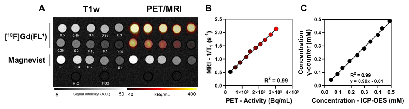
A PET-MRI imaging of a phantom containing titrations of [18F][Gd(FL1)]. Concentrations are reported as mM. Magnevist titration, H2O, and PBS were included as references for MR images and negative controls for PET images. B Correlation plot between radioactivity measured with PET and relaxation rates (1/T1). C Linear regression plot of the concentration of [18F][Gd(FL1)] measured by ICP-OES or determined via γ-counter.