1031
A 32-Channel Cap for Temporal Lobes Exploration at 11.7 T1Université Paris-Saclay, CEA, Joliot, NeuroSpin, Gif-Sur-Yvette, France, 2Multiwave Imaging SAS, Marseille, France, 3Université Paris-Saclay, CEA, IRFU, Gif-sur-Yvette, France, 4Imaging Centre of Excellence, University of Glasgow, Glasgow, United Kingdom
Synopsis
Keywords: RF Arrays & Systems, RF Arrays & Systems
Motivation: Our newly operational 11.7 T machine provides an improved signal-to-noise ratio (SNR) in the human brain. This gain in SNR can be even enhanced by designing region-focused receive arrays.
Goal(s): Our main goal was to maximize the SNR in the temporal lobes and provide high-acceleration capabilities for fMRI studies.
Approach: We developed a modular 32-channel receive array made of non-overlapped hexagonal loops placed on a flexible cap, using high-impedance coils (HIC). We compared our coil to a 32-channel whole-brain receive array at 11.7 T.
Results: The cap receive array provides a significant SNR boost in the targeted region.
Impact: Our cap receive array should ease sub-millimeter resolution fMRI with high SNR in the temporal lobes. Moreover, the detachable hexagonal modules could easily be re-arranged to target any other brain region, with no need for retuning.
Introduction
The unique 11.7 T scanner installed at NeuroSpin (CEA, France) offers unprecedented opportunities for human fMRI studies. It was previously shown that the SNR follows a close to quadratic increase with B01,2. This gain can be even pushed further locally using high-density receive arrays at ultra-high-field MRI3,4. However, when pushing towards the mesoscopic submillimeter scale, the amount of data collected from such arrays becomes too significant for whole-brain fMRI studies. Therefore, it motivates the need for region-focused receive arrays. In particular, the temporal lobes and Broca’s region are of primary interest to neuroscientists, as they are associated with language and speech functions. The high-impedance coils (HIC) were recently introduced in the MRI community and provide new vistas for conformable receive array designs5,6. Their intrinsic robustness to the loading and improved decoupling ease the implementation of dense receive arrays.In this abstract, we introduce a 32-channel receive array whose loops are placed on a flexible cap closely fitting the patient’s head and mainly dedicated to the temporal lobes. The experimental results are compared with a previously described7 32-channel whole-brain receive array which produced the first in-vivo images at 11.7 T8.
Methods
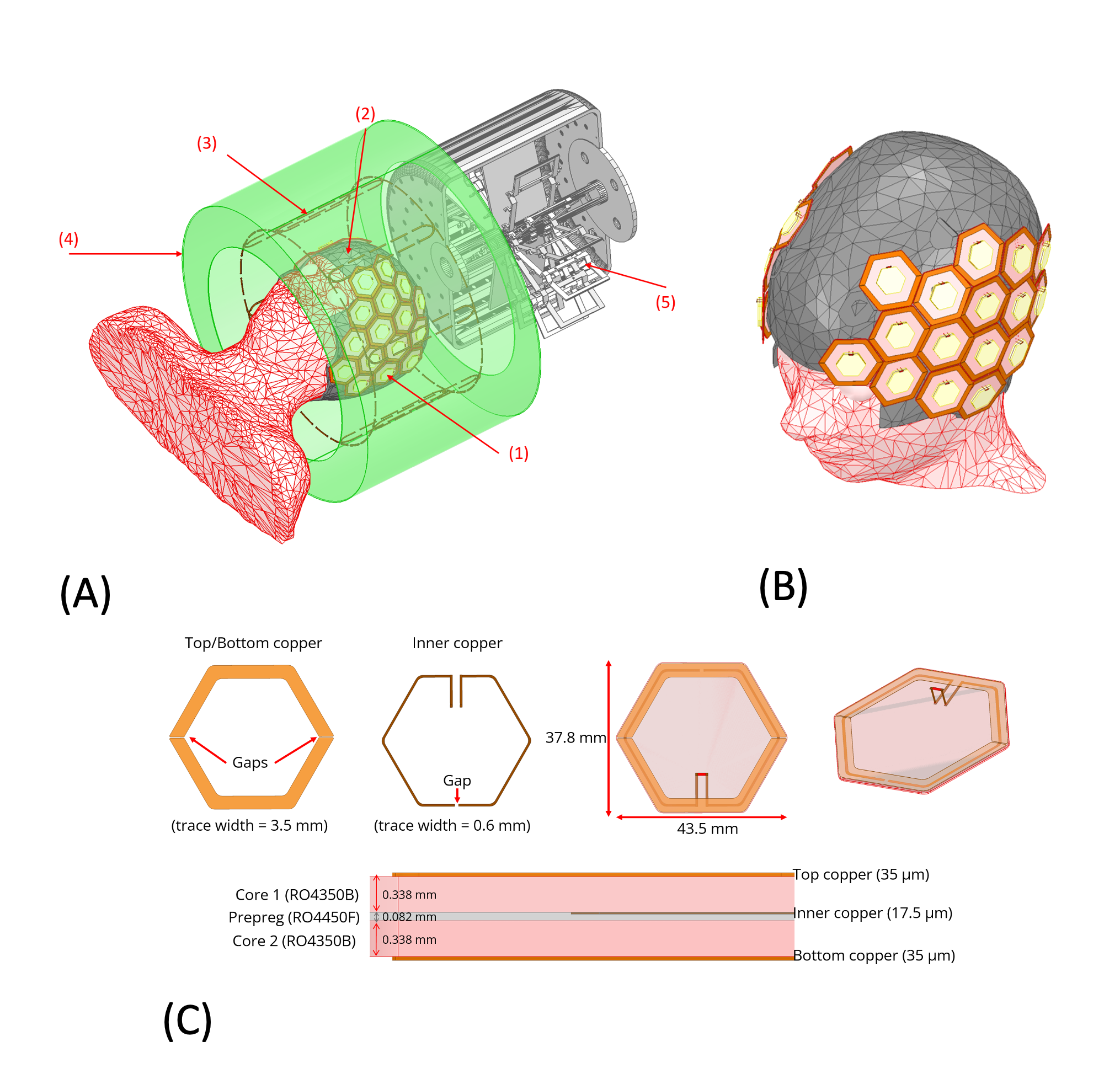
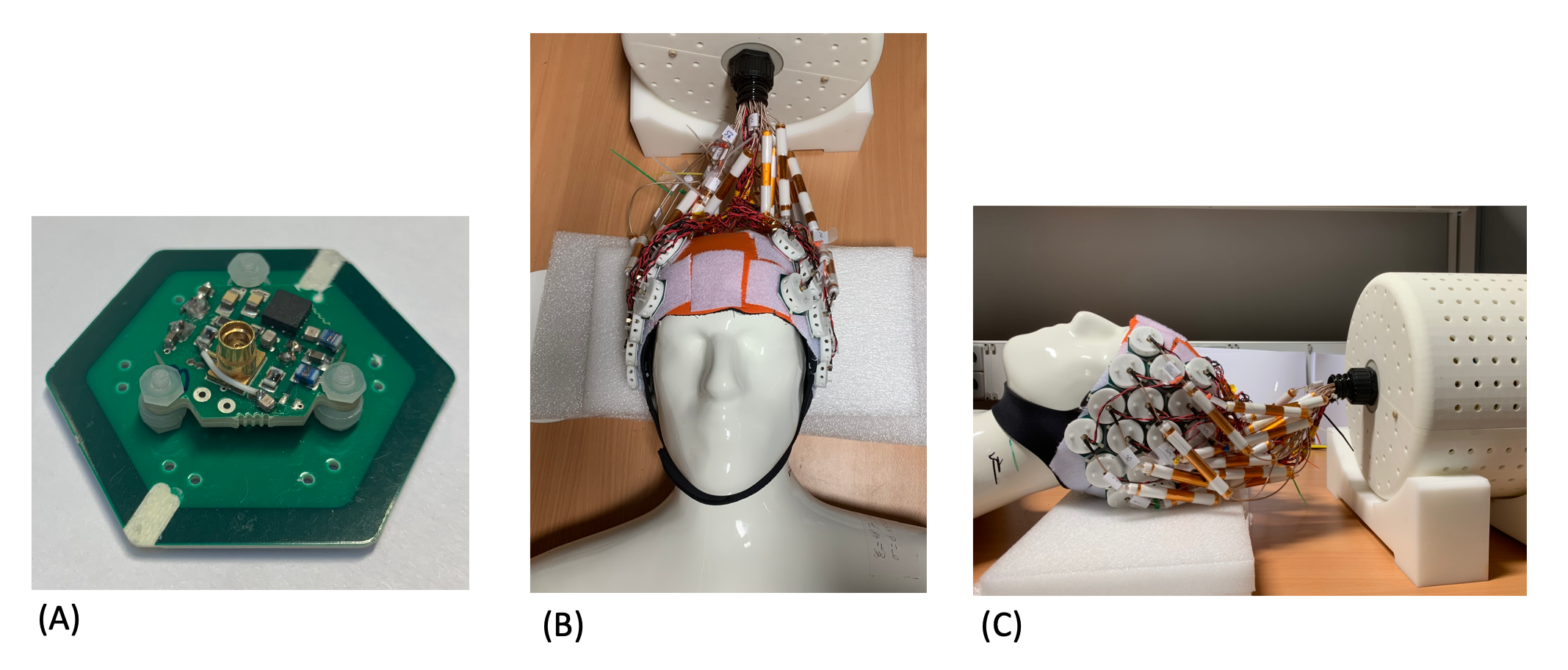
The 32-channel receive array is composed of high-impedance hexagonal loops with a 21.75 mm circumradius, mainly covering the temporal lobes following a honeycomb pattern (Figures 1A and 1B). Velcro-loop straps are sewed on a flexible cap made of neoprene and nylon fabric, and adhesive Velcro-hook is placed below each loop PCB to place the loop freely on the cap (Figure 2). The loops are self-resonating at the proton Larmor frequency (499.4 MHz), without any lumped component. Tuning is made with two gaps in the outer conductors and one gap in the inner conductor6 of a stripline. To fine-tune the loops, the gaps on the outer conductors are widened using a milling machine. The loops are printed using a rigid Rogers 4350B dielectric (Rogers Corporation, AZ, USA) with 0.8 mm thickness (Figure 1C). Miniaturized preamplifiers (noise figure = 0.5 dB, gain = 22 dB) are built in-house and placed directly on the loops PCBs, providing a low impedance (about 50 Ω) to the loop input for preamplifier decoupling5 and a noise-matching to a high resistance given by the loop (about 1500 Ω). These preamplifiers are encapsulated in a 3D-printed housing to protect the components. Floating cable traps9 are 3D-printed using Polycarbonate material and are placed at approximately 7 cm from the preamplifiers. No tuning capacitors are required since the cable trap length directly equals λ/4 (89 mm). The transmit coil is made of 8 large Tx-only loops10, detuned in the reception mode.The experimental SNR is obtained with a head and shoulders phantom (εr = 48.7; σ = 0.65 S/m; T1 = 600 ms) using a GRE sequence (2 mm isotropic resolution, TR = 30 ms, TE = 3 ms, 10°-flip angle pTx pulse based on 7 kT-points11, acquisition matrix = 128x112x88), and a 0-V acquisition for noise measurement. The SNR is reconstructed with a noise-covariance weighted root sum-of-squares12 and corrected for transmit inhomogeneities based on the simulated flip angle maps2. The g-maps are computed in post-processing with the SENSE formula13. The results are compared using the same protocol with the 32-channel whole-brain receive array7.
Results
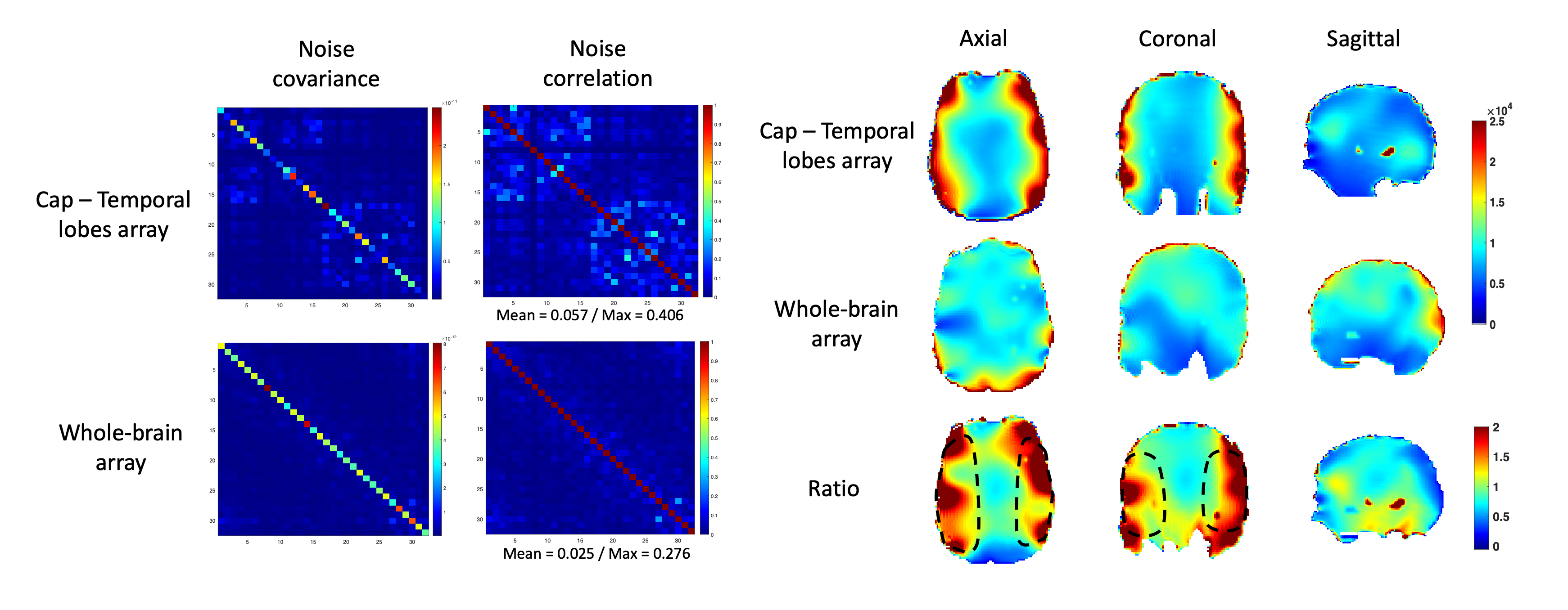
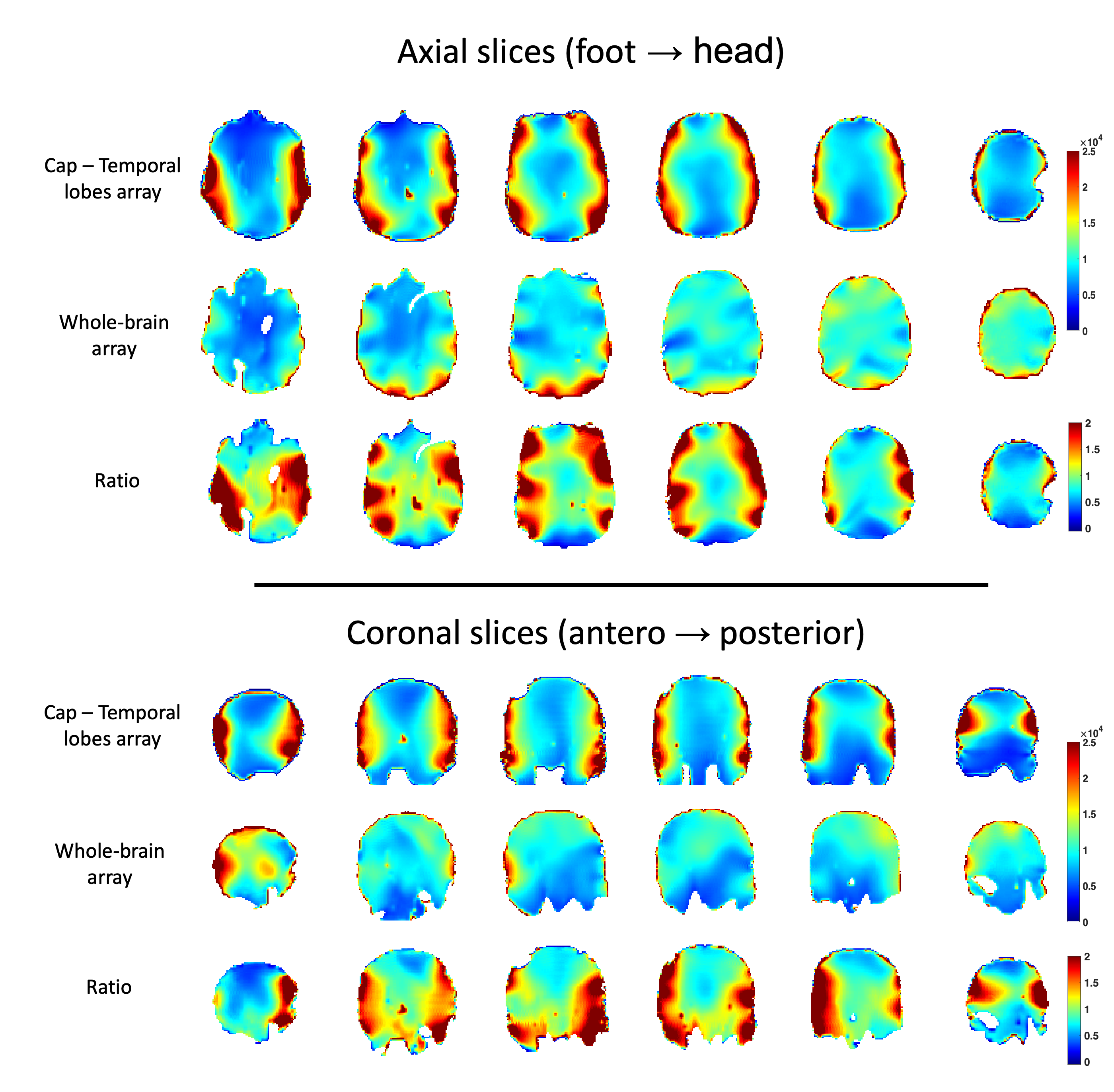
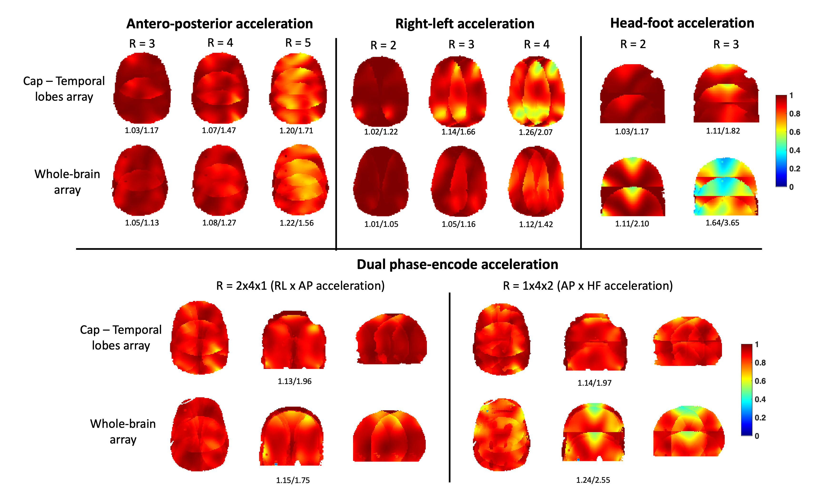
The measured mean noise correlation from the cap and the whole-brain arrays are 0.057 and 0.025, respectively (Figure 3). In the manually selected ROI highlighted in dashed black lines, the SNR is about 1.7 times higher for the cap in average, with up to a factor 4 enhancement in the periphery of the temporal lobes as compared to the whole brain receive array (Figures 3 and 4). From the shown g-maps (Figure 5), the right-left acceleration is difficult with the cap as it is barely segmented along this axis. The antero-posterior acceleration shows results close to the whole-brain receive array with a slight improvement in the vicinity of the temporal lobes. For head-foot acceleration, a significant improvement is shown since the whole-brain receive array has only two rows of resonators (and a patch).Conclusion
Our cap receive array demonstrated an improved SNR in the temporal lobes compared to a whole-brain receive array, of about a factor 1.7 in average. Since the loops can be freely positioned on the cap, other imaging modalities are possible depending on the region of interest (e.g., the frontal or occipital cortex). The first in-vivo studies are expected in 2024, pending regulatory approval.Acknowledgements
We would like to thank Edouard Chazel for his help during the preamplifiers conception, the cable mounting and the overall coil integration. We also would like to thank Nicolas Boulant for authorizing the adaptation of the first AROMA coil as transmit-only to assess our receive array (cf. project AROMA H2020 FET-Open #885876).References
1. C. Le Ster et al. Magnetic field strength dependent SNR gain at the center of a spherical phantom and up to 11.7T. In: Magnetic Resonance in Medicine 88.5 (2022), pp. 2131–2138. doi: 10.1002/mrm.29391.
2. A. Amadon et al. Preliminary SNR comparison between the 11.7T Iseult RF coil and its twin coil at 7T. In: Proceedings of the 32nd annual meeting of the ISMRM, Toronto, CA, 2023, Abstract 4076.
3. Gruber et al. A 128-Channel Receive Array for Cortical Brain Imaging at 7 T. In: Magnetic Resonance in Medicine (2023). doi: 10.1002/mrm.29798.
4. K. Ugurbil et al. Brain Imaging with Improved Acceleration and SNR at 7 Tesla Obtained with 64‐channel Receive Array. Magnetic Resonance in Medicine 82, no. 1 (July 2019): 495–509, doi: 10.1002/mrm.27695.
5. B. Zhang, D. K. Sodickson, and M. A. Cloos. A high-impedance detector-array glove for magnetic resonance imaging of the hand. In: Nature Biomedical Engineering 2.8 (Aug. 2018), pp. 570–577. doi: 10.1038/s41551-018-0233-y.
6. L. Nohava et al. Flexible Multi-Turn Multi-Gap Coaxial RF Coils: Design Concept and Implementation for Magnetic Resonance Imaging at 3 and 7 Tesla. In: IEEE Transactions on Medical Imaging 40.4 (Apr. 2021) pp. 1267–1278. doi: 10.1109/TMI.2021. 3051390.
7. M. Luong et al. A compact 16Tx-32Rx geometrically decoupled phased array for 11.7 T MRI. Proceedings of the 31st annual meeting of the ISMRM, London, UK, 2022, Abstract 707.
8. F. Mauconduit et al. Opening new horizons with the first human brain in vivo experiments at 11.7T. ISMRM 2024, Singapore.
9. D. Seeber, J. Jevtic and A. Menon. Floating shield current suppression trap. In: Concepts in Magnetic Resonance Part B: Magnetic Resonance Engineering 21B.1 (2004), pp. 26– 31. doi: 10.1002/cmr.b.20008.
10. S. Chu, V. Gras, F. Mauconduit, A. Massire, N. Boulant and S. Gunamony. Electromagnetic and RF pulse design simulation based optimization of an eight-channel loop array for 11.7T brain imaging. In: Magnetic Resonance in Medicine (2023). doi: 10.1002/mrm.29654.
11. M. A. Cloos et al. kT-points: Short three-dimensional tailored RF pulses for flip-angle homogenization over an extended volume. In: Magnetic Resonance in Medicine (2011). doi: 10.1002/mrm.22978.
12. P. B. Roemer, W. A. Edelstein, C. E. Hayes, S. P. Souza, and O. M. Mueller. The NMR Phased Array. In: Magnetic Resonance in Medicine. (1990) doi: 10.1002/mrm.1910160203.
13. K. P. Pruessmann, M. Weiger, M. B. Scheidegger and P. Boesiger. SENSE: sensitivity encoding for fast MRI. In: Magnetic Resonance in Medicine 42.5 (1999). PMID: 10542355.
Figures