1030
A 72-Channel Head Coil with an Integrated 16-Channel Field Camera for the Connectome 2.0 Scanner1Institute of Medical Physics and Radiation Protection, Mittelhessen University of Applied Sciences, Giessen, Germany, 2Athinoula A. Martinos Center for Biomedical Imaging, Massachusetts General Hospital and Harvard Medical School, Charlestown, MA, United States, 3Max Planck Institute for Human Cognitive and Brain Sciences, Leipzig, Germany, 4Department of Diagnostic and Interventional Radiology, University Hospital Marburg, Philipps University of Marburg, Marburg, Germany
Synopsis
Keywords: RF Arrays & Systems, RF Arrays & Systems
Motivation: Diffusion MRI utilizing ultra-high performance gradients for high b-value in vivo brain images still suffers from low SNR and increased eddy currents artifacts.
Goal(s): To construct a high-density coil array with an integrated field monitoring system. To enhance SNR and parallel image encoding, while capturing 3rd-order field dynamics.
Approach: Utilizing simulations, 3D printing technology, and radiofrequency electronics to construct a 72-channel head coil and incorporate a field monitoring system. Optimization of the combined system to operate jointly in a space-constraint MRI gradient coil environment.
Results: High-resolution, high b-value diffusion in vivo imaging with greatly minimized image artefacts.
Impact: The constructed 72-channel head coil along with the new Connectome 2.0 scanner will enable the investigation of new microstructure features and connectivity in the living human brain.
Introduction
Diffusion MRI technology has reached a remarkable turning point for mapping the connectivity pathways in the living human brain by utilizing powerful gradient coils1,2,3 with a performance of Gmax= 500mT/m on each axis, while being capable of slewing up to SRmax= 600T/m/s. This enables ultra-high b-value diffusion imaging of >30,000 s/mm2. However, these images continue to suffer from low SNR, while the strong and fast switching gradient pulses substantially increase the eddy current fields and Maxwell terms, further degrading image quality. To mitigate these limitations, the strong diffusion gradients must be paired with an advanced RF front-end design to enhance sensitivity and acquisition speed due to highly parallel detection, while allowing concomitant measurement of the spatiotemporal magnetic field dynamics. The aim of this study was to develop a 72-channel head array coil with an integrated 16-channel field monitoring system for the Connectome 2.0 scanner.Methods
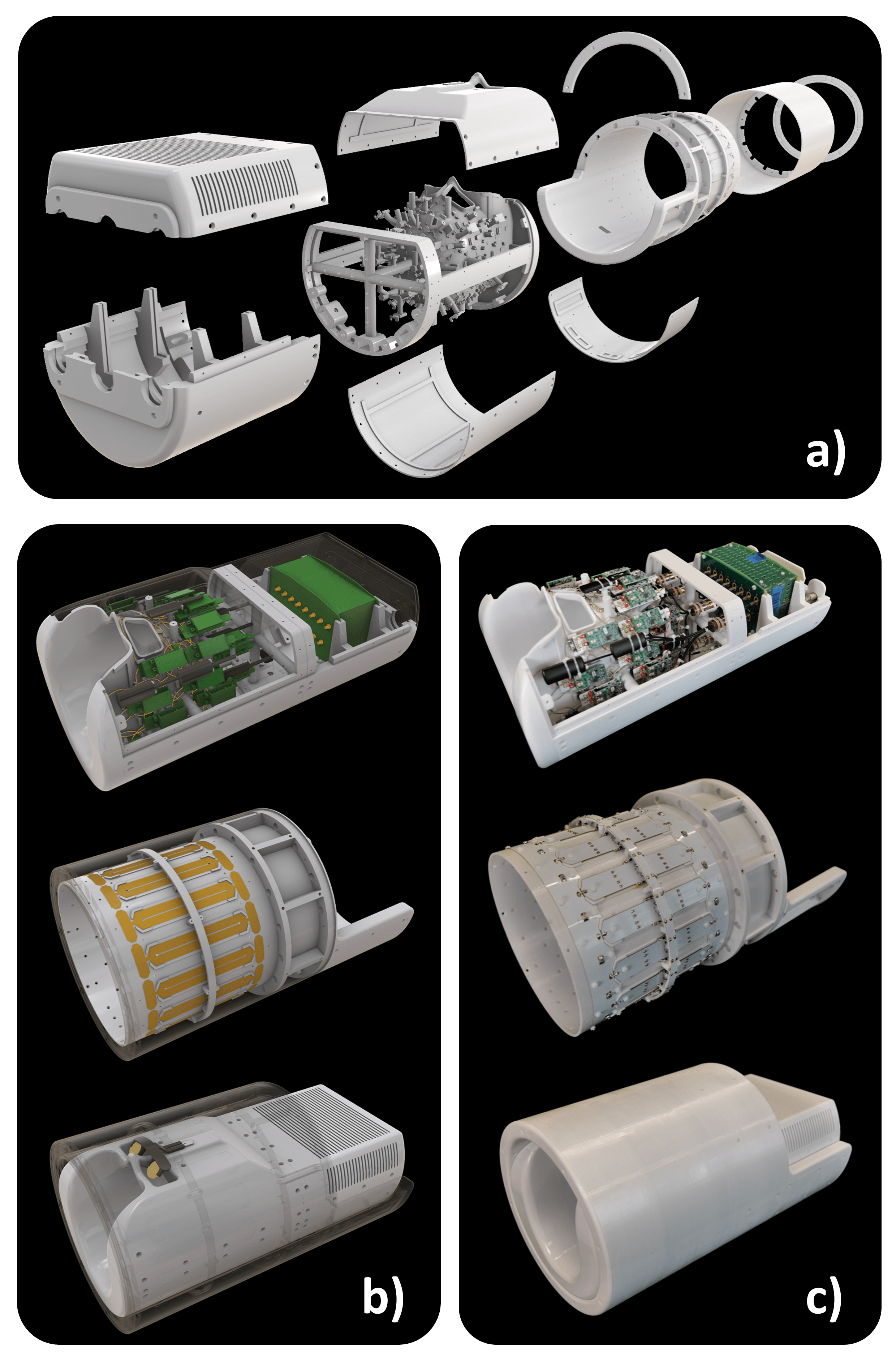
Rx coil: The head coil (Fig.1) was constructed on an anatomically shaped former. All coil parts were 3D printed in polycarbonate plastic. All adjacent coil elements were decoupled by critical overlap. Next-nearest neighboring elements used preamplifier decoupling.4 Pairs of coils were attached to a daughter board containing the matching network, active/passive detuning, and a twin preamplifier (Siemens Healthineers, Erlangen). Preamplifier outputs passed through cable traps to suppress common modes. Element tuning, active detuning, and preamplifier decoupling for each coil element were confirmed through RF bench measurements. Two high-density system plugs were incorporated, enabling all elements to be connected to the MRI scanner’s receive chain.Tx coil: Due to the multi-layer gradient coil design, the Connectome 2.0 scanner architecture requires a local transmit coil on the patient bed. Thus, the 72-channel receiver array was outfitted with a compact CP birdcage5 coil (band-pass design, actively tuned, 16 rungs, L=280mm, coil diam.: 318mm, shield diam.: 280mm). All copper parts of the coil and shielding were optimized for low eddy currents, which is crucial for operating Tx/Rx coil systems at high performance gradients.
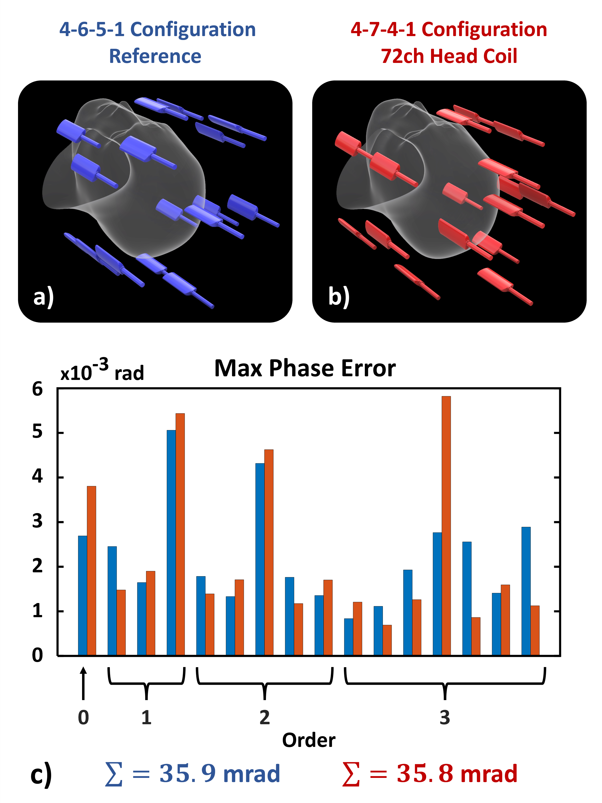
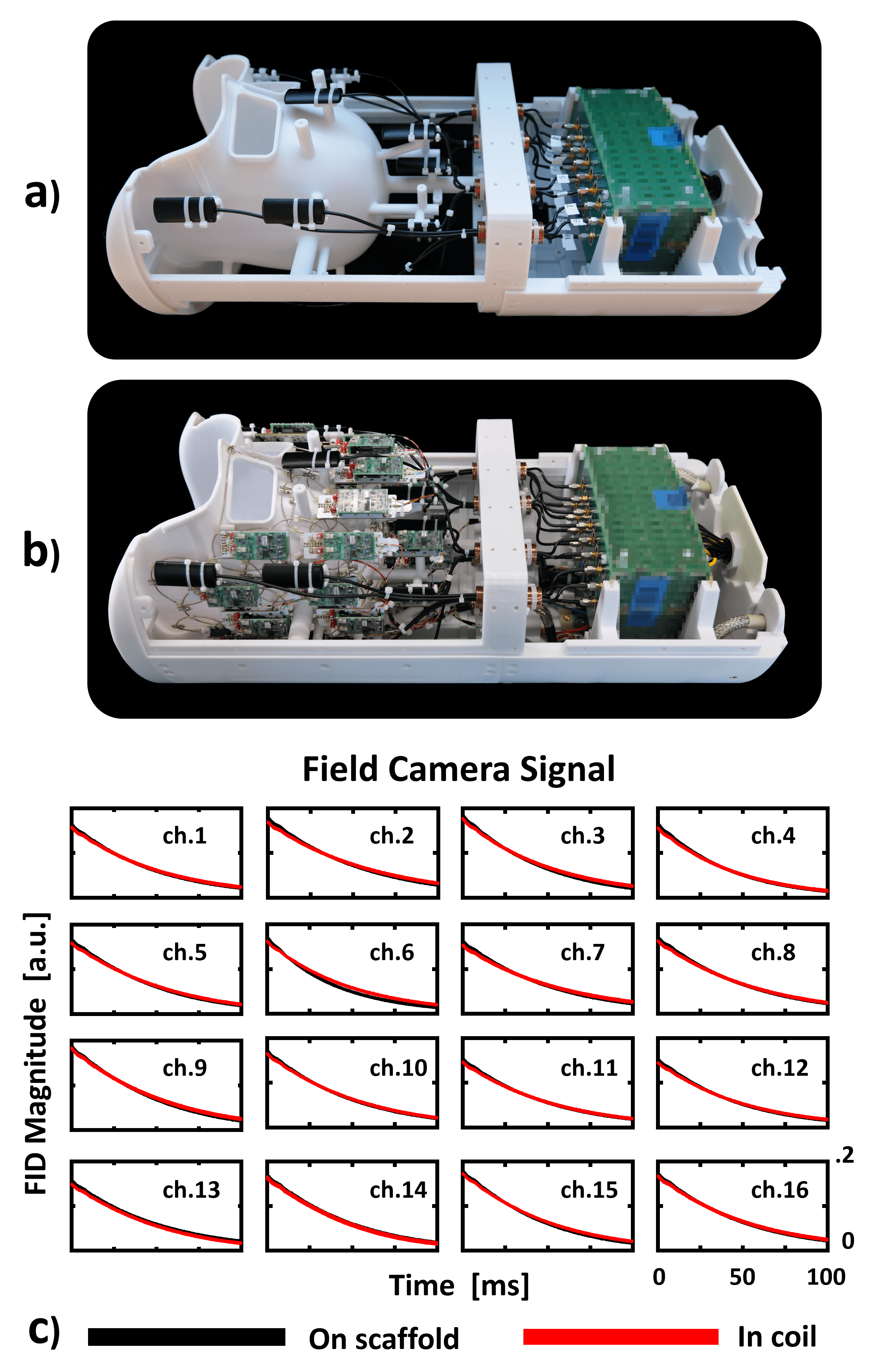
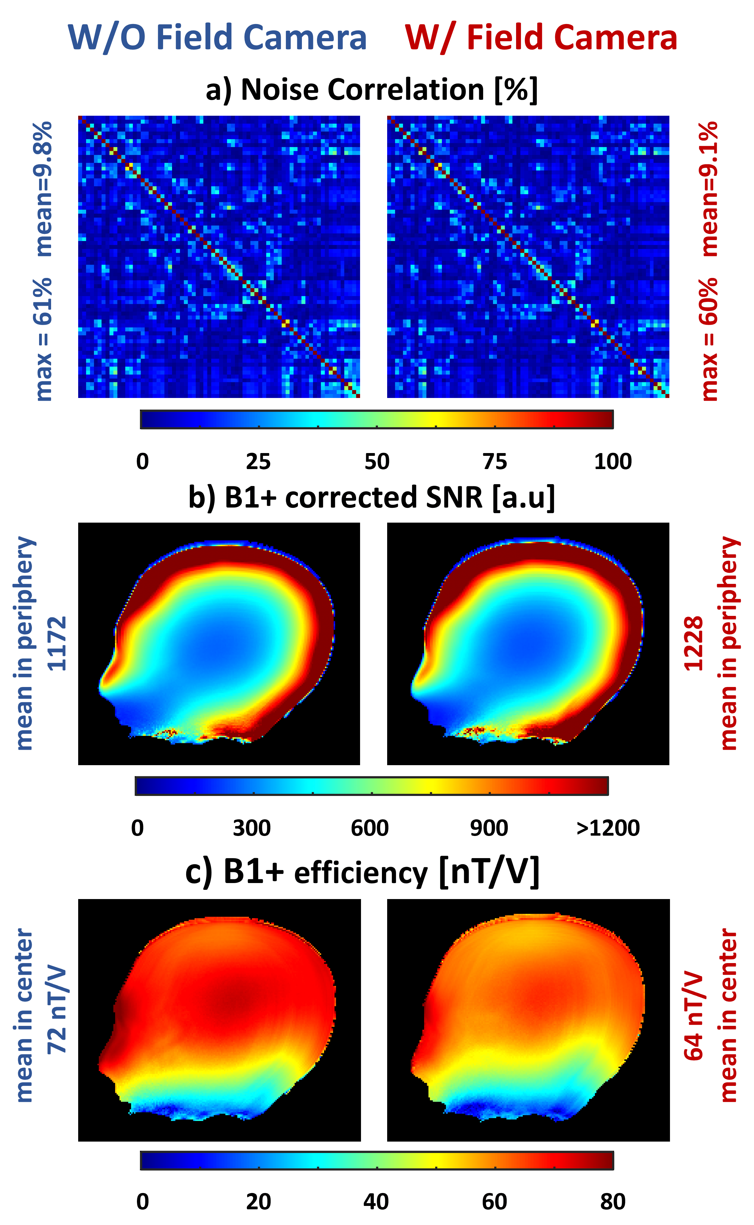
Field camera: 16 field camera probes (Skope Inc, Zurich) were distributed around the Rx coil, using an iterative simulation process to minimize the phase error, while taking into account the positioning constraints imposed by the numerous components of the high-density Rx array.6,7 Possible mutual interactions between the coil system and the field camera were investigated by comparing performance parameters with and without the field camera present (Fig.3).8,9,10,11 In both cases, the FID signals of the probes were determined by averaging the data from 10 measurements. Pixel-wise SNR and B1+ maps were measured with and without the field probes equipped.12,13
Data acquisition: Initial in vivo DWI brain data were taken from a healthy volunteer using a modified 2D PGSE-EPI sequence on the Connectome 2.0 scanner (MAGNETOM Connectom.X, Siemens Healthineers). The sequence featured trigger and synchronization pulses at the beginning for precise synchronization between measured field and k-space data. The multi-shell diffusion protocol included 30 directions at various b-values (1,000 to 12,000 s/mm²). The maximum gradient strength was 495 mT/m. The phase evolution expressed in the spherical harmonic basis was incorporated in the image encoding of a 3rd-order SENSE forward model.14
Results and Discussion
RF bench measurements: The QU/QL-ratio of the Rx elements was 178/49=3.6 at 123.25MHz. Adjacent neighboring coupling ranged from -10dB to -18dB, added with further -20dB via preamplifier decoupling. Tuned/detuned state showed >42dB isolation. The two quadrature ports of the birdcage coil showed an isolation of -18dB.Field camera integration: The FID signals of the field probes showed nearly identical signal decays with and without the coil system present (Fig3-c). Effects in the coil’s noise correlation and SNR performance were neglectable when the camera was installed (Fig4-a,b). The field camera, however, reduced the transmit efficiency of the birdcage coil, where a B1+ attenuation of -11% could be measured (Fig4-c), which is likely attributed to RF shielding effects of the field probes and their routed coaxial cables.
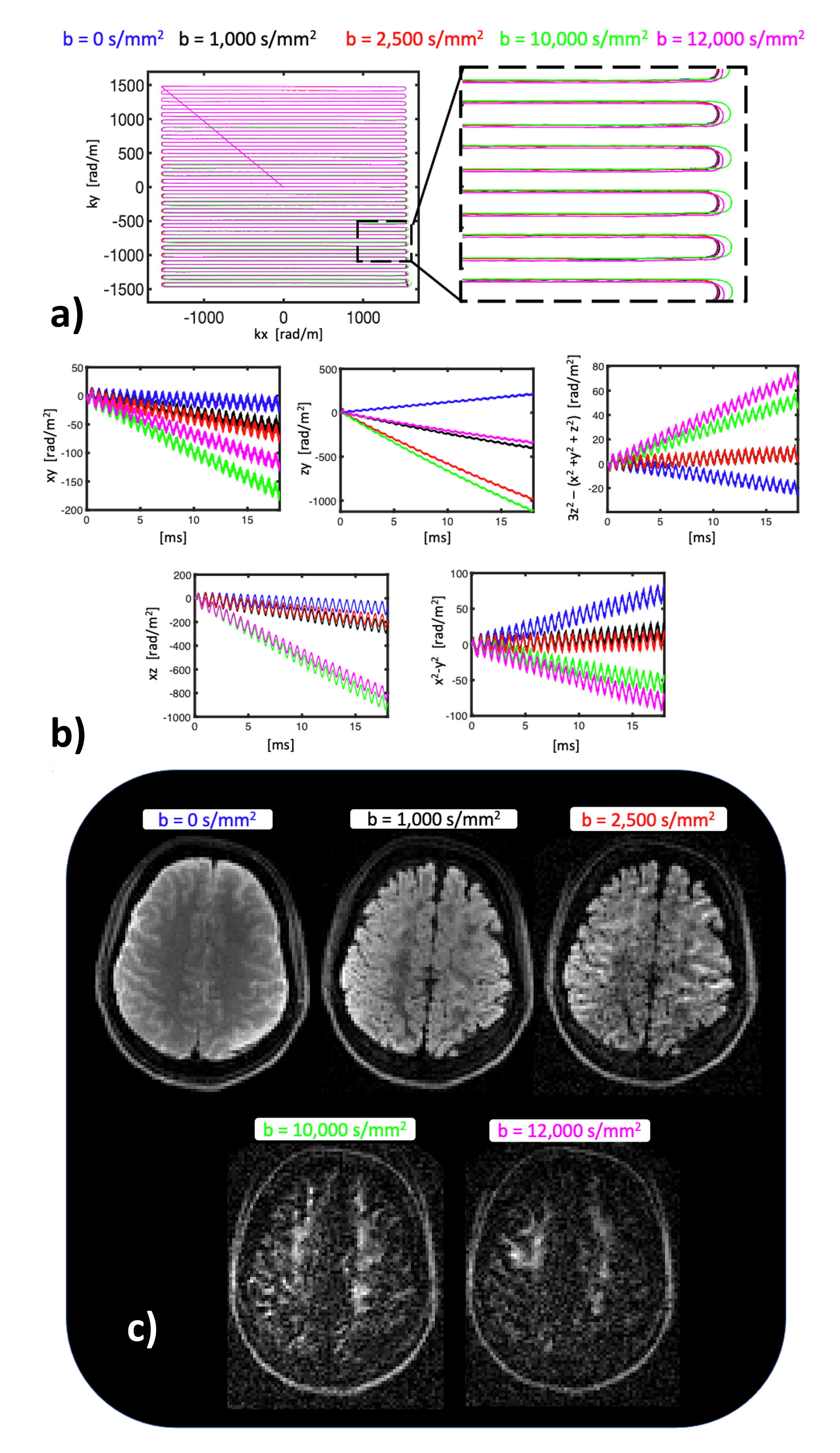
Measured phase coefficients: Eddy currents generated by ultra-high b-values and gradient strengths (495 mT/m) caused significant deviations in the phase coefficients compared to the b=0 data (Fig5-a,b).15
Diffusion imaging: When the 3rd-order SENSE reconstruction was performed with actual phase evolution, distortion and ghosting-free diffusion images with high b-values were obtained (Fig5-c).
Conclusion
A 72-channel head coil with an integrated 16-channel field camera was designed, constructed, and validated. The integration of the field camera into the coil was achieved without compromising the performance of either system. Initial in vivo measurements with concurrent field monitoring have shown artifact-free diffusion images with high b-values.Acknowledgements
This work was supported by the National Institute of Biomedical Imaging and Bioengineering of the National Institutes of Health under award number U01EB026996.We would like to thank Cameron Cushing, Paul Weavers, and Simon Gross for the valuable discussions regarding the field camera. Moreover, we would like to thank Andreas Potthast, René Gumbrecht, and Jasmine Fischer for the assistance in integrating the constructed coil into the Connectome 2.0 scanner.
References
1. Ramos-Llordén
G, Dietz P, Davids M, et al. Connectome 2.0: Performance evaluation and initial
in vivo human brain diffusion MRI results. Proceedings of the International
Society for Magnetic Resonance in Medicine. Submitted to ISMRM 2024.
2. Huang SY, Witzel
T, Keil B, et al. Connectome 2.0:
Developing the next-generation ultra-high gradient strength human MRI scanner
for bridging studies of the micro-, meso- and macro-connectome. Neuroimage 243 (2021):118530.
3. Setsompop K, Kimmlingen R, Eberlein E, et al. Pushing the limits of in vivo diffusion MRI
for the Human Connectome Project. NeuroImage 80 (2013): 220-233.
4. Roemer PB,
Edelstein WA, Hayes CE, et. al. The NMR phased
array. Magnetic Resonance in Medicine 16.2 (1990): 192-255.
5. Hayes CE,
Edelstein WA, Schenck JF, et.al. An efficient highly homogeneous radiofrequency
coil for whole-body NMR imaging at 1.5T. Journal of Magnetic Resonace 63.3
(1985): 622-628.
6. Barmet C, De
Zanche N, and Pruessmann KP. Spatiotemporal magnetic field monitoring for MR.
Magnetic Resonance in Medicine 60.1 (2008): 187-197.
7. Barmet C, Wilm BJ, Pavan M, et.al. Concurrent
higher-order field monitoring for routine head MRI: an integrated heteronuclear
setup. Proceedings of the 18th Annual Meeting of ISMRM, Vol. 216. (2010).
8. Lee Y, Kennedy
M, and Nagy Z. A custom-made design for integrating a magnetic field monitoring
system into a 32ch MRI head coil. Proceedings of the International Society for
Magnetic Resonance in Medicine. Virtual Conference, 2020.
9. Mahmutovic M, Scholz A, Kutscha N, et. al. A 64-channel brain array coil with an
integrated 16-channel field monitoring system for 3T MRI. Proceedings of the
29th Annual Meeting of ISMRM. Vol.623. (2021).
10. Gilbert KM, Dubovan PI, Gati JS, et.al.
Integration of an RF coil and commercial field camera for ultrahigh-field MRI.
Magnetic Resonance in Medicine 87.5 (2022):2551-2565.
11. Scholz A, Mahmutovic M, Alem M, et al. Design of a 64-Channel ex vivo
Brain Rx Array Coil with field monitoring and temperature control for DWI at
3T, Proceedings of the International Society for
Magnetic Resonance in Medicine 31 (2023): 0215.
12. Kellman P and McVeigh ER. Image
reconstruction in SNR units: A general method for SNR measurement. Magnetic
Resonance in Medicine 54.6 (2005): 1439-1447.
13. Insko EK and Bolinger L. Mapping of the
Radiofrequency Field. Journal of Magnetic Resonance, Series A 103 (1993):
82-85.
14. Wilm BJ, Barmet C, Pavan M, et. al. Higher
order reconstruction for MRI in the presence of spatiotemporal field
perturbations. Magnetic Resonance in Medicine. 65.6 (2011): 1690-1701.
15. Ramos-Llordén G, Park DJ, Kirsch JE, et. al. Eddy current-induced artifact correction in
high b-value ex vivo human brain diffusion MRI with dynamic field monitoring.
Magnetic Resonance in Medicine. 2023 Sep 27. doi: 10.1002/mrm.29873. Epub ahead
of print. PMID: 37753621.
Figures